Apparatus and Method For Assessing Transmurality of a Tissue Ablation

a tissue ablation and applicator technology, applied in the field of applicator and method for assessing tissue ablation transmurality, can solve the problems of traumatic operation, inability to restore normal cardiac hemodynamics, and inability to alleviate the patient's vulnerability, so as to improve the assessment of the responsive signal and improve the assessment of the transmurality

Inactive Publication Date: 2007-06-28
MAQUET CARDIOVASCULAR LLC
View PDF18 Cites 4 Cited by
  • Summary
  • Abstract
  • Description
  • Claims
  • Application Information

AI Technical Summary

Benefits of technology

[0020] Accordingly, by collectively analyzing this measured data, a surgeon may gauge whether an ablation procedure has been properly performed. Unlike the current transmurality assessment procedures, the present invention is capable of conducting measurements through the means of very simple and straight forward purse string openings leading to the interior of the organ and placement of various sensors therein.
[0024] In yet another aspect of the present invention, a method is provided for assessing the transmurality of an ablation lesion from a first surface of a targeted biological tissue to an opposed second surface thereof. The method includes placing a first sensor proximate a first surface of a target tissue and placing a second sensor proximate to a second opposing surface of the target tissue. Once the sensors are placed, a first signal is transmitted from the first sensor. The first signal propagates through at least a portion of the target tissue and a signal responsive to the first signal is received by the second sensor, the responsive signal being related to one or more of conduction time, conduction velocity, phase angle, and impedance through at least a portion of the targeted tissue, to determine the transmurality of the ablation lesion created or being created. The method further includes utilization of digital signal processing to better evaluate the measured responsive signal and, thus, better assess transmurality during or after the ablation lesion is being created.
[0027] In yet another aspect of the present, a method for treating medically refractory atrial fibrillation of the heart is provided. This method includes manipulating an antenna assembly of an ablation instrument into engagement with or substantially adjacent to a first surface of targeted cardiac tissue of the heart, and generating an electromagnetic field from the antenna assembly sufficiently strong to cause tissue ablation to the first surface to form an ablation lesion extending from the first surface toward an opposed second surface of the heart. The antenna assembly includes a first electrode attached thereto. In accordance with this aspect of the present invention, before, during or after generating, the method next includes piercing a needle member having an elongated shaft into cardiac tissue from the heart epicardial surface. The needle member includes at least a second electrode which is placed proximate to an endocardial surface of the heart. Next the method includes transmitting and receiving electrical signals to measure at least one of conduction time, conduction velocity, phase angle, and impedance through at least a portion of the targeted cardiac to determine the transmurality of the ablation lesion. The manipulating, generating, piercing and measuring events are repeated to form a plurality of strategically positioned ablation lesions and / or to divide the left and / or right atria to substantially prevent reentry circuits.

Problems solved by technology

However, these procedures have for the most part failed to restore normal cardiac hemodynamics, or alleviate the patient's vulnerability to thromboembolism because the atria are allowed to continue to fibrillate.
Further, strategically placed atrial incisions not only interrupt the conduction routes of the common reentrant circuits, but they also direct the sinus impulse from the sinoatrial node to the atrioventricular node along a specified route.
While this MAZE III procedure has proven effective in treating medically refractory atrial fibrillation and associated detrimental sequelae, this operational procedure is traumatic to the patient since this is an open-heart procedure and substantial incisions are introduced into the interior chambers of the Heart.
However, radio frequency energy has several limitations including the rapid dissipation of energy in surface tissues resulting in shallow “burns” and failure to access deeper arrhythmic tissues.
Another limitation of RF ablation catheters is the risk of clot formation on the energy emitting electrodes.
Such clots have an associated danger of causing potentially lethal strokes in the event that a clot is dislodged from the catheter.
It is also very difficult to create continuous long lesions with RF ablation instruments.
Ablation lesions of insufficient depth may enable currents to pass over or under the lesion, and thus be incapable of disrupting, or otherwise interrupting, the reentry circuits.
This, however, is often difficult to perform and / or assess since the cardiac tissue thickness varies with location and, further, varies from one individual to another.
Excessive thermal energy at the interface between the tissue and the ablation head, on the other hand, is detrimental as well.
Moreover, the tissue may adhere to the tip, resulting in tearing at the ablation site upon removal of the ablation instrument, or immediate or subsequent perforation may occur.
Thin walled tissues are particularly susceptible.
Since these variables may change constantly during the ablation procedure, and without overheating the tissues at the interface, it is often difficult to estimate the interface temperature and time of ablation to effect a proper transmural ablation, especially with deeper arrhythmic tissues.
This may be problematic since the measurement of such electrical properties can produce false indications with respect to transmurality of the ablation; a decrease in the change of impedance measured across the lesion indicative of transmurality, however, knowing there is insufficient energy applied to truly created a transmural lesion, as one example.

Method used


Image

  • Apparatus and Method For Assessing Transmurality of a Tissue Ablation
  • Apparatus and Method For Assessing Transmurality of a Tissue Ablation
  • Apparatus and Method For Assessing Transmurality of a Tissue Ablation

Examples


Embodiment Construction

[0039] While the present invention will be described with reference to a few specific embodiments, the description is illustrative of the invention and is not to be construed as limiting the invention. Various modifications to the present invention can be made to the preferred embodiments by those skilled in the art without departing from the true spirit and scope of the invention as defined by the appended claims. It will be noted here that for a better understanding, like components are designated by like reference numerals throughout the various Figures.

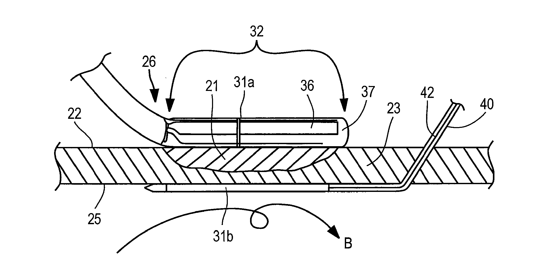
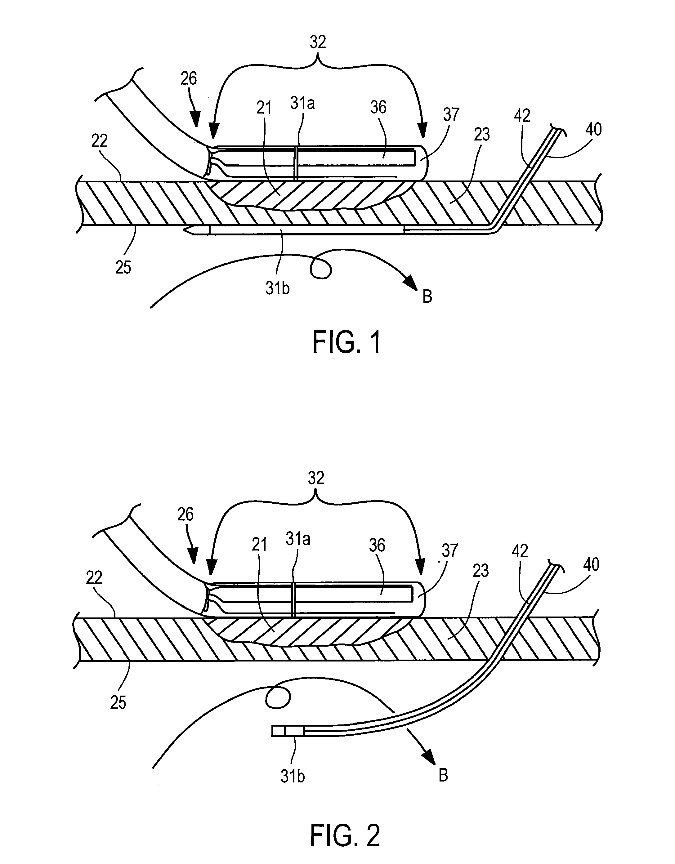
[0040] Turning now to the figures, a measurement or assessment instrument or device, generally designated 20 in FIGS. 4A, 4B, is provided to assess the transmurality of an ablation lesion 21 which extends from a first surface 22 of a targeted biological tissue 23 toward an opposed second surface 25 thereof. As will be described in greater detail below, these lesions are generally formed during surgical tissue ablation procedures ...


PUM


Abstract

An instrument assesses the transmurality of an ablation lesion from a first surface of a targeted biological tissue to an opposed second surface thereof. The instrument includes at least a first electrode operably attached to an ablation instrument adapted to engage a tissue first surface, and at least a second electrode adapted to engage a tissue second surface, the at least second electrode positioned generally opposed to the at least first electrode. Alternatively, the at least second electrode may be adapted to be placed within a chamber of an organ. These electrodes facilitate measuring at least one of conduction time, conduction velocity, phase angle, and impedance through at least a portion of the targeted tissue to determine the transmurality of the ablation lesion.

Description

RELATED APPLICATION [0001] This application claims the benefit under 35 U.S.C. § 120 as a continuation of U.S. patent application Ser. No. 10 / 369,887, filed Feb. 19, 2003, which claims priority under 35 U.S.C. § 120 to U.S. Provisional Application Ser. No. 60 / 358,215, filed Feb. 19, 2002, entitled TRANSMURALITY ASSESSMENT DEVICE, which applications are incorporated herein in their entireties by this reference thereto to form a part hereof.FIELD OF INVENTION [0002] This invention relates to tissue ablation instruments and lesion formations, and more particularly to apparatus and method for assessing tissue ablation transmurality. BACKGROUND OF THE INVENTION [0003] It is well documented that atrial fibrillation, either alone or as a consequence of other cardiac disease, continues to persist as the most common cardiac arrhythmia. According to recent estimates, more than two million people in the U.S. suffer from this common arrhythmia, roughly 0.15% to 1.0% of the population. Moreover,...

Claims


Application Information

Patent Timeline
Patent Type & Authority Applications(United States)
IPC IPC(8): A61B18/14A61B17/00
CPCA61B18/1492A61B2017/00026A61B2017/00243A61B2018/00351A61B2018/00702A61B2018/00875
Inventor CHAPELON, PIERRE-ANTOINEBERUBE, DANYNGUYEN, HIEPKALLEL, FAOUZI
Owner MAQUET CARDIOVASCULAR LLC
Why Eureka
Try Eureka