Method of forming planar lipid double membrane for membrane protein analysis and apparatus therefor

Active Publication Date: 2007-07-12
JAPAN SCI & TECH CORP
View PDF1 Cites 18 Cited by
  • Summary
  • Abstract
  • Description
  • Claims
  • Application Information

AI Technical Summary

Benefits of technology

[0011] Under such circumstances, it is an object of the present invention to provide a method of forming a planar lipid-bilayer me

Problems solved by technology

Therefore, the dead volumes are large and microscopic observation cannot be performed.
Additionally, in these methods, when a plurality of planar lipid-bilayer membranes are simultaneously formed in a channel by providing a plurality of apertures, adjacent apertures (planar lipid-bilayer membr

Method used


Image

  • Method of forming planar lipid double membrane for membrane protein analysis and apparatus therefor
  • Method of forming planar lipid double membrane for membrane protein analysis and apparatus therefor
  • Method of forming planar lipid double membrane for membrane protein analysis and apparatus therefor

Examples


first embodiment

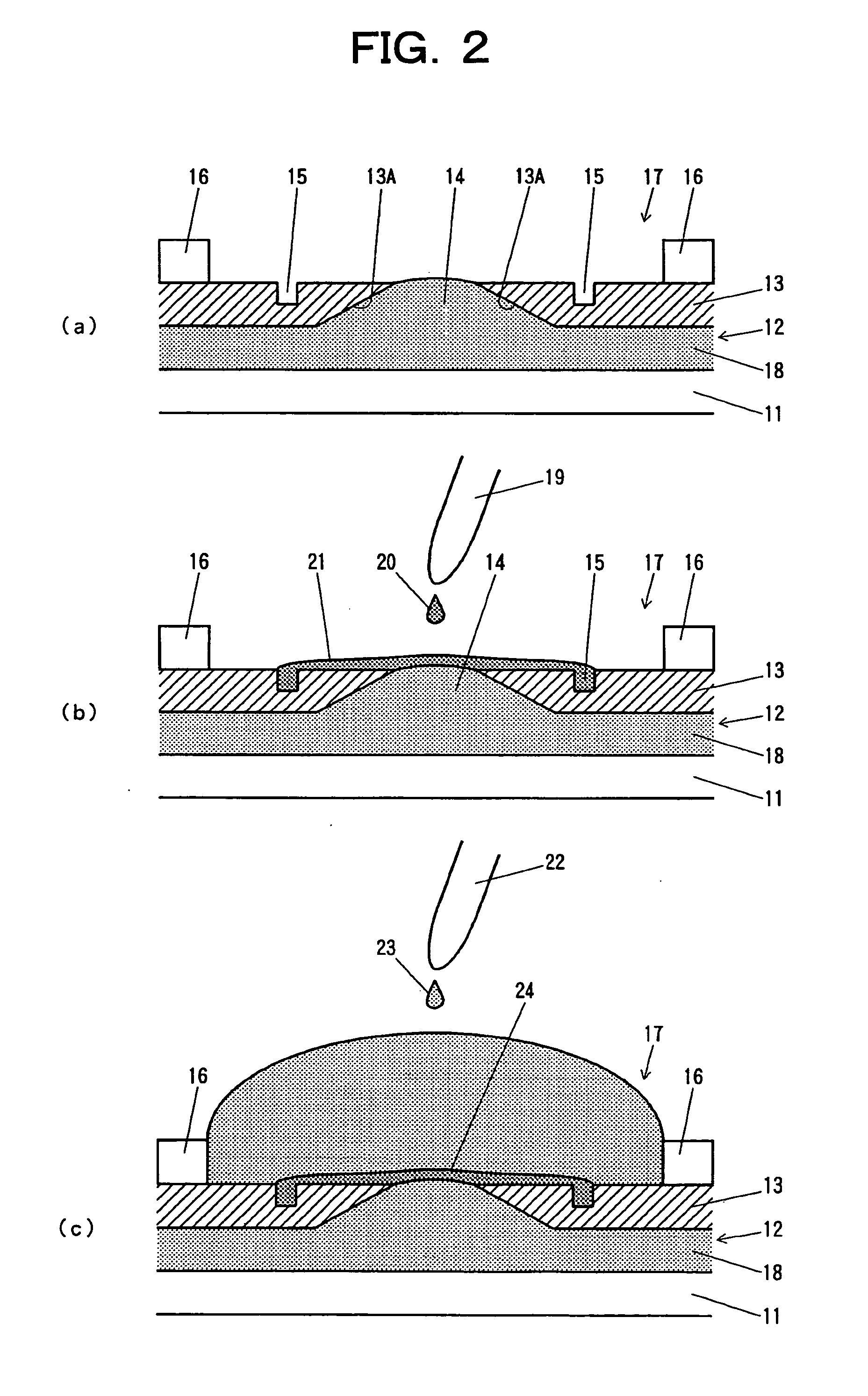
[0044]FIG. 2 is a schematic diagram of a device for forming a planar lipid-bilayer membrane according to a first embodiment of the present invention. FIG. 3 is a schematic diagram showing a lipid solution.

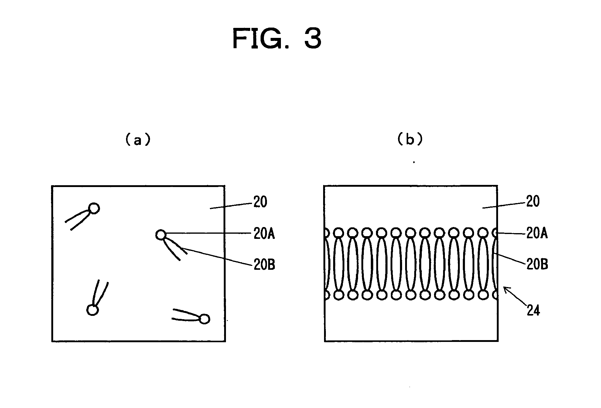
[0045] In FIG. 2, a reference numeral 11 denotes a glass substrate, a reference numeral 12 denotes a microchannel, a reference numeral 13 denotes a partition wall, a reference numeral 14 denotes an aperture (opening) provided to the partition wall 13, a reference numeral 15 denotes a liquid trap provided on the partition wall 13, a reference numeral 17 denotes a chamber defined by a well 16, a reference numeral 18 denotes a buffer solution which fills the microchannel 12 and the aperture (opening) 14, a reference numeral 19 denotes a microinjection device (microinjector), a reference numeral 20 denotes a lipid solution applied as a droplet from the microinjection device 19, a reference numeral 21 denotes a layer of the lipid solution, a reference numeral 22 denotes a microinjectio...

second embodiment

[0053]FIG. 4 is a schematic diagram of a device for forming a planar lipid-bilayer membrane according to a second embodiment of the present invention.

[0054] In a second embodiment, in addition to the components in the first embodiment, a first thin-film electrode 25 is provided on the glass substrate 11 of the microchannel 12 and a second thin-film electrode 26 is provided on the partition wall 13 within the chamber 17 defined by the well 16. Since the thin-film electrodes 25 and 26 are independently provided in the chamber 17 defined by the well 16, the membrane potential and current can be measured.

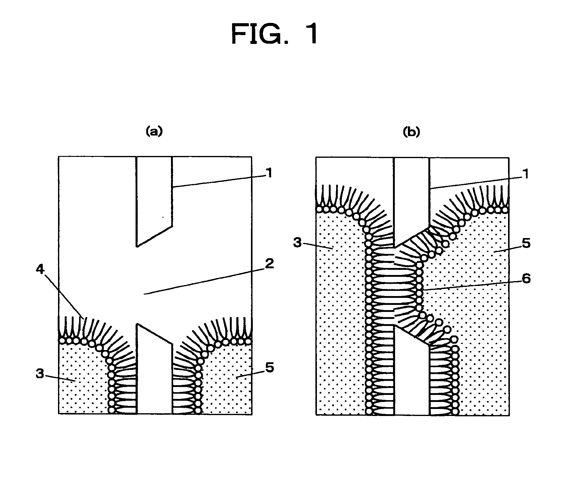
[0055] In order to incorporate a membrane protein to be analyzed into the planar lipid-bilayer membrane 24 formed at the interface of the buffer solution 18, a spherical vesicle (liposome) of the same lipid bilayer is used.

[0056] As shown in FIG. 5, a liposome 31 containing Alamethicin 32 which is a channel protein is prepared. Droplets of the liposome is mixed in the buffer solution...

third embodiment

[0057]FIG. 6 is a schematic diagram of a device for forming a planar lipid-bilayer membrane according to a third embodiment of the present invention.

[0058] In this embodiment, the device is provided with a channel 12A connecting to the liquid trap 15. Therefore, it is possible to control the thickness of the layer of the lipid solution remaining on the interface of the buffer solution 18. In other words, when a thick lipid solution layer 21 is formed by the lipid solution 20 remaining on the interface of the buffer solution 18, the thickness of the lipid solution layer 21 can be decreased by sucking the excess of the lipid solution 20 through the channel 12A connecting to the liquid trap 15. Reversely, when a thin lipid solution layer 21 is formed by the lipid solution 20 remaining on the interface of the buffer solution 18, the thickness of the lipid solution layer 21 can be increased by pushing the lipid solution 20 back through the channel 12A connecting to the liquid trap 15.


PUM


Abstract

To provide a method of forming a planar lipid-bilayer membrane for membrane protein analysis, by which downsizing, simplifying, and multichanneling of a device therefor are achieved. A planar lipid-bilayer membrane 24 is formed by filling a microchannel 12 with a buffer solution 18, the microchannel 12 disposed under a horizontal partition wall 13 having an aperture 14; applying a small amount of a lipid solution 20 as a droplet on the aperture 14 filled with the buffer solution 18 to thereby form a thin layer 21 of the lipid solution in a chamber, the chamber 17 being formed at a position corresponding to the aperture 14 and provided with a liquid trap 15 inside the chamber; and applying a buffer solution 23 as a droplet to the chamber 17 from the upper side of the chamber.

Description

TECHNICAL FIELD [0001] The present invention relates to methods of forming planar lipid-bilayer membranes for membrane protein analysis and devices therefor. The planar lipid-bilayer membranes are used in fields such as biotechnology, biochips, membrane protein analysis, drug discovery screening, and biosensors. BACKGROUND ART [0002] As typical methods for producing planar lipid-bilayer membranes used in analysis of membrane proteins such as ion channels, painting method and Langmuir-Blodgett method (LB method) are conventionally known. Both methods are of forming a planar lipid-bilayer membrane in an aperture opened in a Teflon (registered trademark) sheet in a chamber filled with a buffer solution. The aperture has a size of several-hundred microns. In the painting method, a lipid solution is applied to the aperture with a brush. The LB method utilizes the fact that a monomolecular layer of a lipid molecule is formed on the surface of a solution. In this method, a planar lipid-bil...

Claims


Application Information

Patent Timeline
IPC IPC(8): C12M1/34G01N1/28B01J19/00B01L3/00G01N33/48
CPCB01J2219/00317B01J2219/00585B01J2219/00599B01J2219/00653B01L2400/0472B01L3/5027B01L3/5088B01L2300/0819B01J2219/00725
Inventor TAKEUCHI, SHOJI
Owner JAPAN SCI & TECH CORP
Why Eureka
Try Eureka