Ocular fluid markers

a technology of ocular fluids and markers, applied in the field of analysis and monitoring of ocular fluids, can solve the problems of major unmet early diagnosis and preventive treatment of these disorders

Inactive Publication Date: 2007-09-27
GEORGE MASON INTPROP INC
View PDF8 Cites 11 Cited by
  • Summary
  • Abstract
  • Description
  • Claims
  • Application Information

AI Technical Summary

Benefits of technology

[0009] Cystoid Macular Edema (CME) is a type of edema of the macula that causes retinal damage and occurs in a wide variety of ocular disorders. There is intense interest among practitioners and pharmaceutical companies to develop treatments for CME. Understanding the ocular fluid proteome profile of patients with CME will provide new opportunities in prevention and treatment.
[0011] Tools are needed to track the pharmacokinetics of intraocular drugs delivered directly or indirectly and track ocular / systemic drug partition between vitreous and serum. Thereby drug development and modification will be greatly facilitated providing significant value to the rapidly growing industry of developing drugs for retinal disorders.

Problems solved by technology

While photocoagulation surgery and vitrectomy are highly effective in reducing vision loss, early diagnosis and preventative treatments for these disorders remain a major unmet clinical need.

Method used


Image

  • Ocular fluid markers
  • Ocular fluid markers
  • Ocular fluid markers

Examples


Embodiment Construction

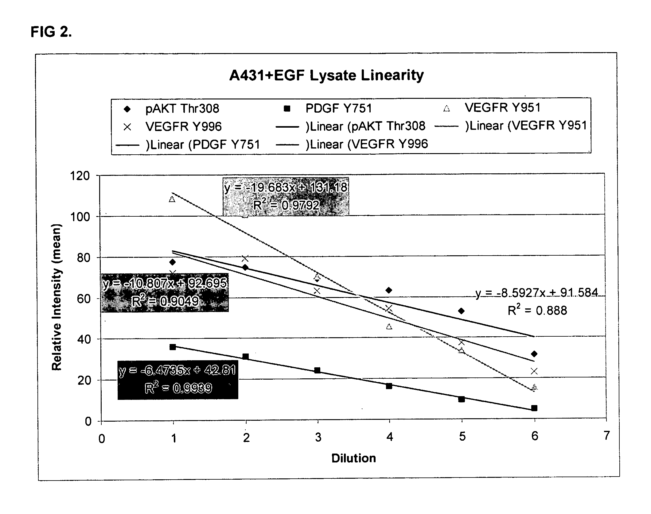
[0045] In the forgoing and in the following examples, all temperatures are set forth uncorrected in degrees Celsius (° C.) and, all parts and percentages are by weight, unless otherwise indicated.

Experimental Procedure
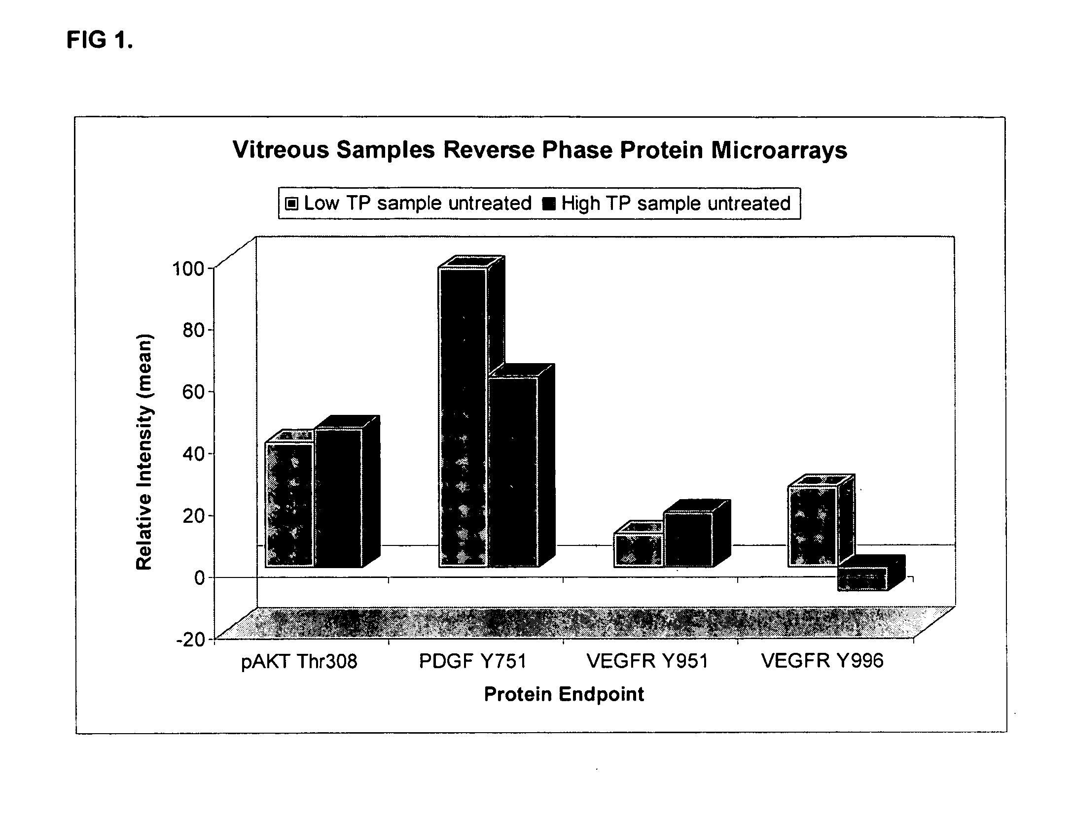
Vitreous Sampling:

Pars Plana Vitrectomy

[0046] All vitreous samples were obtained prior to the vitrectomy portion of the surgery. The surgery would have been done regardless of participation in the study. The patient was prepped and draped in the usual sterile fashion. Prior to the vitrectomy portion of the study, a minute amount of vitreous (approximately 0.1 ml) was obtained in a sterile TB syringe through the pars plana. The vitreous sample was then frozen at −20° C. or −80° C. for storage and subsequent analysis of the vitreous proteome. There was no additional risk to the patient in addition to that incurred from the surgery alone.

Autopsy Study

[0047] At an unrestricted consented autopsy, 2-8 ml of clear vitreous gel were extracted by inserting in the late...


PUM

PropertyMeasurementUnit
electrophoresisaaaaaaaaaa
mass spectroscopyaaaaaaaaaa

Abstract

The present invention relates to the analysis and monitoring of ocular fluids for determining the physiological state of an organism, to monitor drug efficacy and dynamics, for early disease detection, as well as to certain molecular markers and fingerprints of identified molecules or molecule fragments in such analysis.

Description

CROSS-REFERENCE TO RELATED APPLICATIONS [0001] This application claims the benefit of earlier-filed U.S. Provisional Application Ser. No. 60 / 762,499, filed Jan. 27, 2006, which is incorporated herein by reference in its entirety.FIELD OF THE INVENTION [0002] The present invention relates to the analysis and monitoring of ocular fluids for determining the physiological state of an organism, e.g., to monitor drug efficacy and dynamics, for early disease or other physiological state detection, as well as to monitor / quantitate certain molecular markers and fingerprints identified in such analysis. SUMMARY OF THE INVENTION [0003] This invention relates to, e.g., a method of characterizing the physiological state of the eye, comprising detecting the presence or absence in vitreous fluid of one or more polypeptides, or fragments thereof; a method of characterizing the physiological state of the eye, comprising detecting the presence or absence in vitreous fluid of one or more biomarker att...

Claims


Application Information

Patent Timeline
Patent Type & Authority Applications(United States)
IPC IPC(8): G01N33/53C12Q1/48G06F19/00A61M31/00
CPCC12Q1/485G01N2800/164G01N33/6842G01N33/68Y02A90/10G01N33/48
Inventor LIOTTA, LANCE A.ZHOU, WEIDONGESPINA, VIRGINIAPETRICOIN, EMANUEL
Owner GEORGE MASON INTPROP INC
Why Eureka
Try Eureka