Esophagus imaging enhancement device

a technology of enhancing device and esophagus, which is applied in the field of medical and therapeutic systems, can solve problems such as damage to the esophagus, and achieve the effect of enhancing the imaging quality of the esophagus and enhancing the imaging quality of the organ

Active Publication Date: 2007-09-27
BIOSENSE WEBSTER INC
View PDF23 Cites 46 Cited by
  • Summary
  • Abstract
  • Description
  • Claims
  • Application Information

AI Technical Summary

Benefits of technology

[0007] In some medical procedures, such as cardiac ablation, it is desirable to enhance the imaging quality of an organ. For example, when performing ablation in

Problems solved by technology

Catheter-based cardiac procedures such as cardiac ablation may sometimes cause damage to the esophagus

Method used


Image

  • Esophagus imaging enhancement device
  • Esophagus imaging enhancement device
  • Esophagus imaging enhancement device

Examples


Embodiment Construction

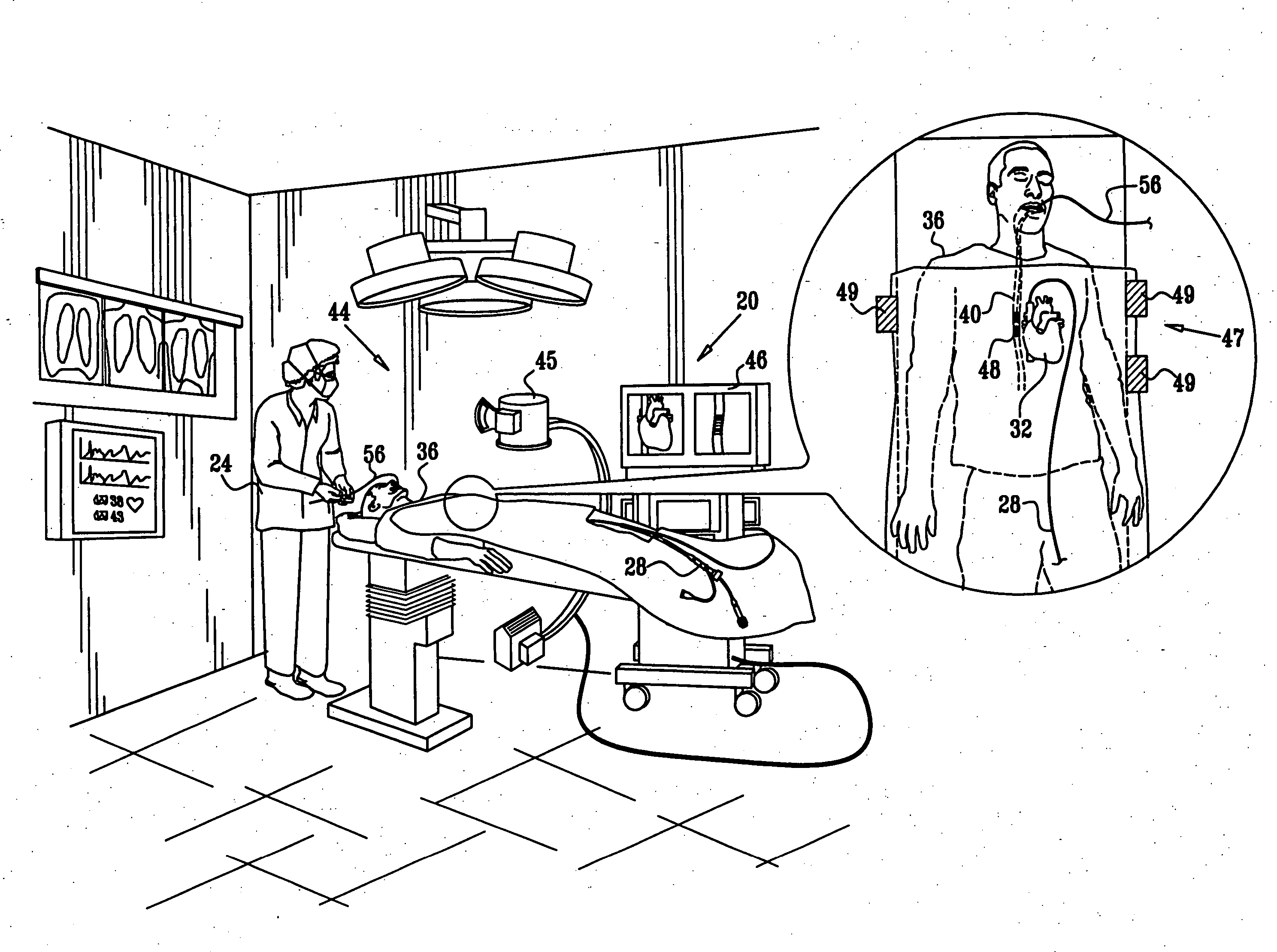
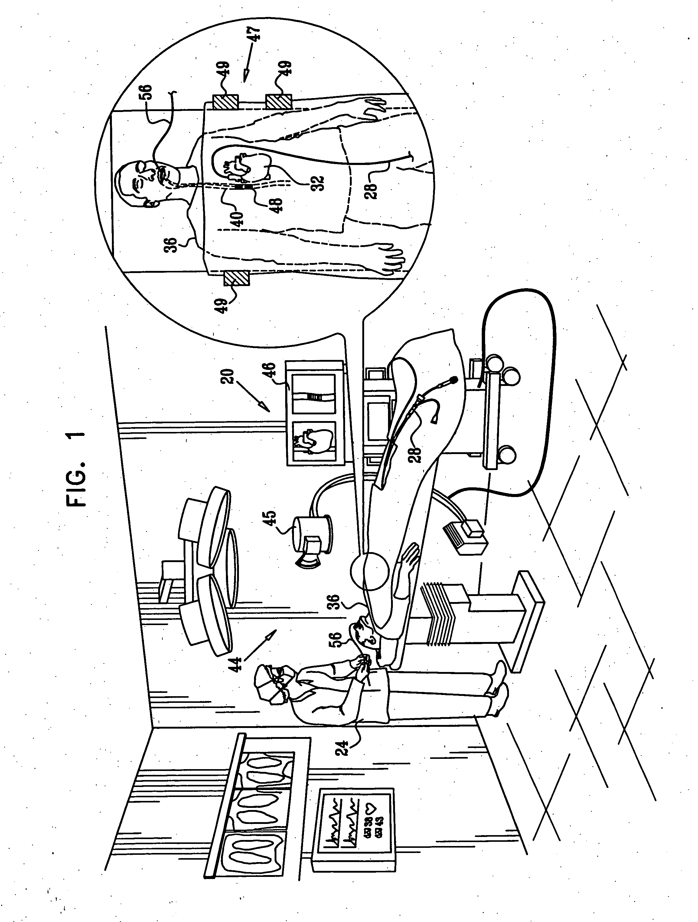
[0043]FIG. 1 is a, schematic, pictorial illustration of a system 20 for performing cardiac ablation, in accordance with an embodiment of the present invention. A physician 24 inserts a catheter 28 into a heart 32 of a patient 36 in order to perform a cardiac ablation procedure. Catheter 28 typically comprises an ablation electrode, which applies concentrated RF energy to selected spots on the endocardium (the inner surface of the heart), as is known in the art.

[0044] In some cases, parts of an esophagus 40 of the patient may overlap, or be adjacent to, parts of heart 32, and in particular the posterior part of the left atrium and the coronary sinus. Because of this proximity, the ablation procedure may cause thermal damage to the esophagus, sometimes resulting in its perforation.

[0045] In order to prevent damage from being caused to the esophagus, a visualization system 44 provides the physician with an image of at least part of the patient's body, typically comprising the heart a...


PUM


Abstract

A device for enhancing visualization of an esophagus when imaged by a visualization system includes a structure including material that is clearly visible when imaged by the visualization system. The structure is arranged, when inserted into the esophagus, to expand so as to conform to an inner surface of the esophagus in order to enhance a visualization quality of the surface when imaged by the visualization system. A cord attached to the structure is arranged to extend out of the esophagus so as to assist a removal of the device from the esophagus.

Description

FIELD OF THE INVENTION [0001] The present invention relates generally to medical and therapeutic systems, and particularly to methods and devices for enhancing the imaging quality of organs during therapeutic procedures. BACKGROUND OF THE INVENTION [0002] Catheter-based cardiac procedures such as cardiac ablation may sometimes cause damage to the esophagus, since sections of the esophagus often pass in close proximity to the left atrium and coronary sinus. The anatomical proximity of the left atrium to the esophagus is described, for example, by Lemola et al., in “Computed Tomographic Analysis of the Anatomy of the Left Atrium and the Esophagus: [0003] Implications for Left Atrial Catheter Ablation,” Circulation, (110:24), Dec. 14, 2004, pages 3655-60, which is incorporated herein by reference. The proximity of the coronary sinus to the esophagus is described, for example, by Tsao et al., in “Anatomic Proximity of the Esophagus to the Coronary Sinus: Implication for Catheter Ablatio...

Claims


Application Information

Patent Timeline
IPC IPC(8): A61B5/05
CPCA61B5/06A61B5/061A61B5/4233A61B2562/164A61B6/12A61B8/0833A61B5/6885A61B17/00A61B1/273
Inventor PORATH, JOSHUASCHWARTZ, YITZHACK
Owner BIOSENSE WEBSTER INC
Why Eureka
Try Eureka