Optical Vascular Function Imaging System and Method for Detection and Diagnosis of Cancerous Tumors

Inactive Publication Date: 2007-12-13
SRI INTERNATIONAL
View PDF11 Cites 15 Cited by
  • Summary
  • Abstract
  • Description
  • Claims
  • Application Information

AI Technical Summary

Benefits of technology

[0011] During the process of angiogenesis, tumors develop abnormal vasculature, and as a result, cancerous tissue is often hypoxic, a condition that can be observed with hemoglobin oxygenation measurements. The present inventi

Problems solved by technology

Unfortunately, X-ray mammography is less effective at detecting cancer in younger women's breasts, which a

Method used


Image

  • Optical Vascular Function Imaging System and Method for Detection and Diagnosis of Cancerous Tumors
  • Optical Vascular Function Imaging System and Method for Detection and Diagnosis of Cancerous Tumors
  • Optical Vascular Function Imaging System and Method for Detection and Diagnosis of Cancerous Tumors

Examples


Example

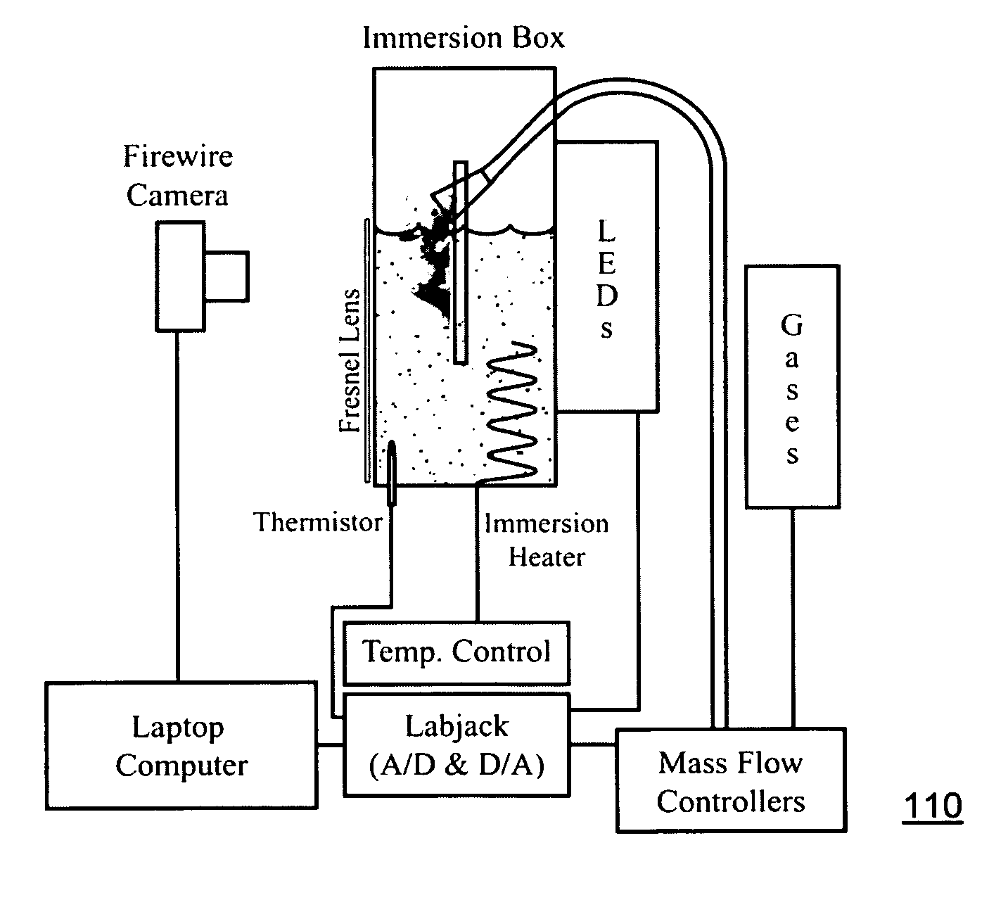
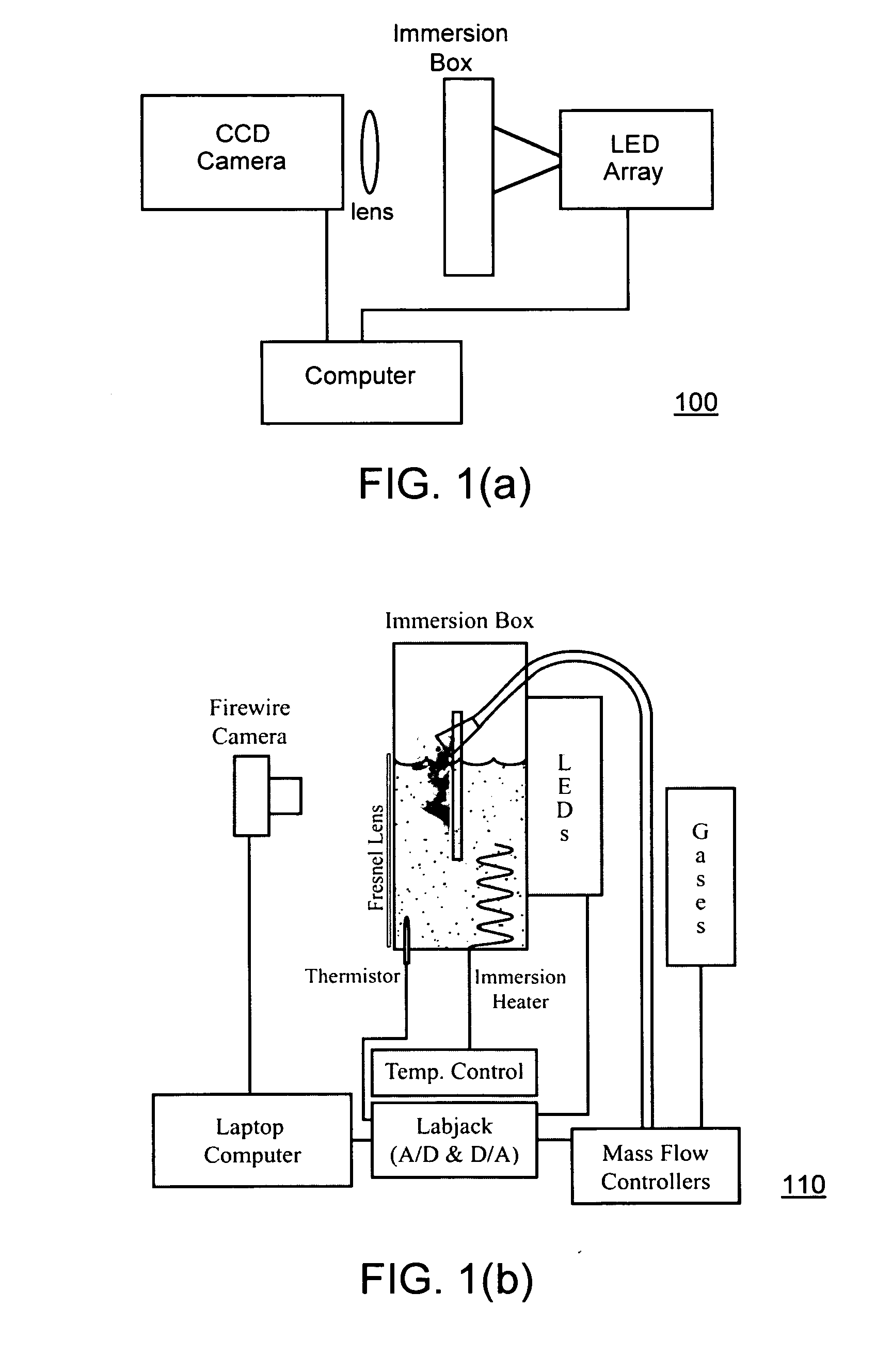
[0033] A primary goal of the invention is to develop reliable and yet inexpensive technology to improve sensitivity and specificity (lower false-negative and false-positive rates) for early breast cancer detection and diagnosis. We have achieved this goal with enhanced functional (physiological) optical imaging using a new type of contrast based on the unusual vascular function of tumors (atypical oxygenation improvement, atypical vasoactivity, and blood pooling).

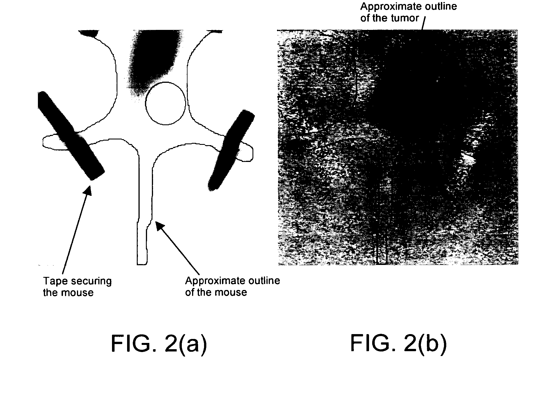
[0034] Another goal is to improve imaging through dense breasts where X-ray mammography is less successful. We have been investigating this differential vasoactive optical imaging (DVOI) approach in animal model studies. That work has demonstrated strong contrast between cancerous and noncancerous tissue during differential imaging in rodents in association with inhalation of O2 / CO2 gas mixtures.

[0035] The contrast achieved by DVOI results from the vasculature in tumors and can arise from atypical oxygenation improvement,...


PUM


Abstract

An in vivo optical imaging system and method of identifying unusual vasculature associated with the angiogenic vasculature in tumors. An imaging system acquires images through the breast. Benign, noninvasive oxygen and carbon dioxide are used as vasoactive agents and administered by inhalation to stimulate vascular changes. Images taken before and during inhalation are subtracted. An optical vascular functional imaging system monitors abnormal vasculature through optical measurements on oxy- and deoxy-hemoglobin during inhalation of varying levels of O2 and CO2. The increase in contrast between tumor (cancerous) and normal (noncancerous) tissue is dramatic, facilitating accurate early detection of cancerous tumors and improving sensitivity and specificity (lower false negative and false positive rates). The invention is useful in mammography, dermatology, prostate imaging and other optically accessible areas.

Description

CROSS-REFERENCE TO RELATED APPLICATIONS [0001] This application is a 371 filing claiming priority to PCT Application PCT / US05 / 03090 filed Jan. 21, 2005, which claims priority to U.S. Provisional Patent Application No. 60 / 538,765, filed Jan. 23, 2004, the entire content of which is incorporated herein by reference.STATEMENT REGARDING FEDERALLY SPONSORED RESEARCH OR DEVELOPMENT [0002] This invention was made with Government support under Grant DAMD17-02-1-0570 awarded by the U.S. ARMY MEDICAL RSCH. ACQUISITION ACTIVITY. The Government has certain rights in this invention.BACKGROUND OF THE INVENTION [0003] 1. Field of the Invention [0004] This invention relates generally to medical imaging systems and methods. More particularly, it relates to an innovative optical vascular functional imaging technology with significantly improved image quality, sensitivity and specificity, particularly useful in early detection and diagnosis of cancerous tumors such as breast cancer. [0005] 2. Descript...

Claims


Application Information

Patent Timeline
IPC IPC(8): A61K49/00A61B5/06
CPCA61B5/0091A61B5/4312A61B5/14551
Inventor FARIS, GREGORY
Owner SRI INTERNATIONAL
Why Eureka
Try Eureka