
Clip device and protective cap, and methods of using the protective cap and clip device with an endoscope for grasping tissue endoscopically

a technology of endoscopy and clip device, which is applied in the field of clip devices, can solve the problems of insufficient strength of conventional haemostatic devices to cause permanent hemostasis, inability to deliver via incision using rigid shafted instruments, and inability to continue bleeding for many patients, etc., and achieve the effect of improving healthcar

Inactive Publication Date: 2007-12-20
WILSONCOOK MEDICAL
View PDF99 Cites 176 Cited by
  • Summary
  • Abstract
  • Description
  • Claims
  • Application Information

AI Technical Summary

Benefits of technology

[0017] The present invention relates to medical devices and methods for grasping tissue using an endoscope flexible distal insert portion and for improving healthcare to individuals undergoing endoscopic procedures.

Problems solved by technology

If left untreated, this common and serious condition sometimes can lead to fatal results.
While this problem has prompted a few endoscopic therapeutic approaches to achieve hemostasis, such as injecting sclerosing agents and using contact thermo-coagulation techniques, such approaches occasionally allow continued bleeding for many patients.
One of the problems associated with using conventional haemostatic devices, however, is that they can only be delivered using rigid shafted instruments via incision or trocar cannula.
Moreover, none of the conventional haemostatic devices are strong enough to cause permanent hemostasis.
While U.S. Pat. No. 5,766,189 describes one proposed solution and shows a device that gastroenterologists may use to deliver a detachable clip having a pair of open arms that close about a blood vessel, one problem with using this clip device and other similar clip devices having only two arms is that gastroenterologists need to rotate the clip in order to grasp properly the area to be clipped.
Rotating the operating wire through the bends of the tube(s) used to deliver the clip, however, often hinders or complicates the procedure.
Another problem often encountered with using conventional clip devices is the difficulty in securing the clip device to the delivery apparatus prior to reaching the target area within the patient.
Turning to another problem to be solved, gastroenterologists often encounter blood or other bodily fluids that obscure the surgical site when using conventional clip devices.
These fluids increase the difficulty in properly positioning the device and grasping the area to be clipped.
For example, when attempting to clip a bleeding vessel, blood typically fills the gastroenterologists' working space, thereby impeding the gastroenterologists' ability to locate and / or clip the vessel.
In addition to the need for a separate catheter, the procedure often results in delays in clipping the vessel, because additional time is needed for inserting and positioning the catheter.
One problem with these conventional through-the-scope clip delivery apparatus is that the delivery apparatus (e.g., clip-wire assembly, holding tube, hollow coil sheath, and outer tubular sheath distal end) may be too large to be placed through smaller endoscope working channels.
Another problem is that two coaxial tubes are needed, and still another problem is that the holding tube must have an inner diameter that is large enough to attach to the outside of the hollow coil sheath, and the outer diameter of the outer tubular sheath needs to be greater than the outer diameter of the holding tube.
Yet another problem is that the conventional coaxial tubes are prone to kinking, buckling, bending, and / or bowing that result from passing the delivery apparatus through the working channel of an endoscope, pulling the wire proximally, or sliding one tube relative to the other tube.

Method used


Image

  • Clip device and protective cap, and methods of using the protective cap and clip device with an endoscope for grasping tissue endoscopically
  • Clip device and protective cap, and methods of using the protective cap and clip device with an endoscope for grasping tissue endoscopically
  • Clip device and protective cap, and methods of using the protective cap and clip device with an endoscope for grasping tissue endoscopically

Examples


Embodiment Construction

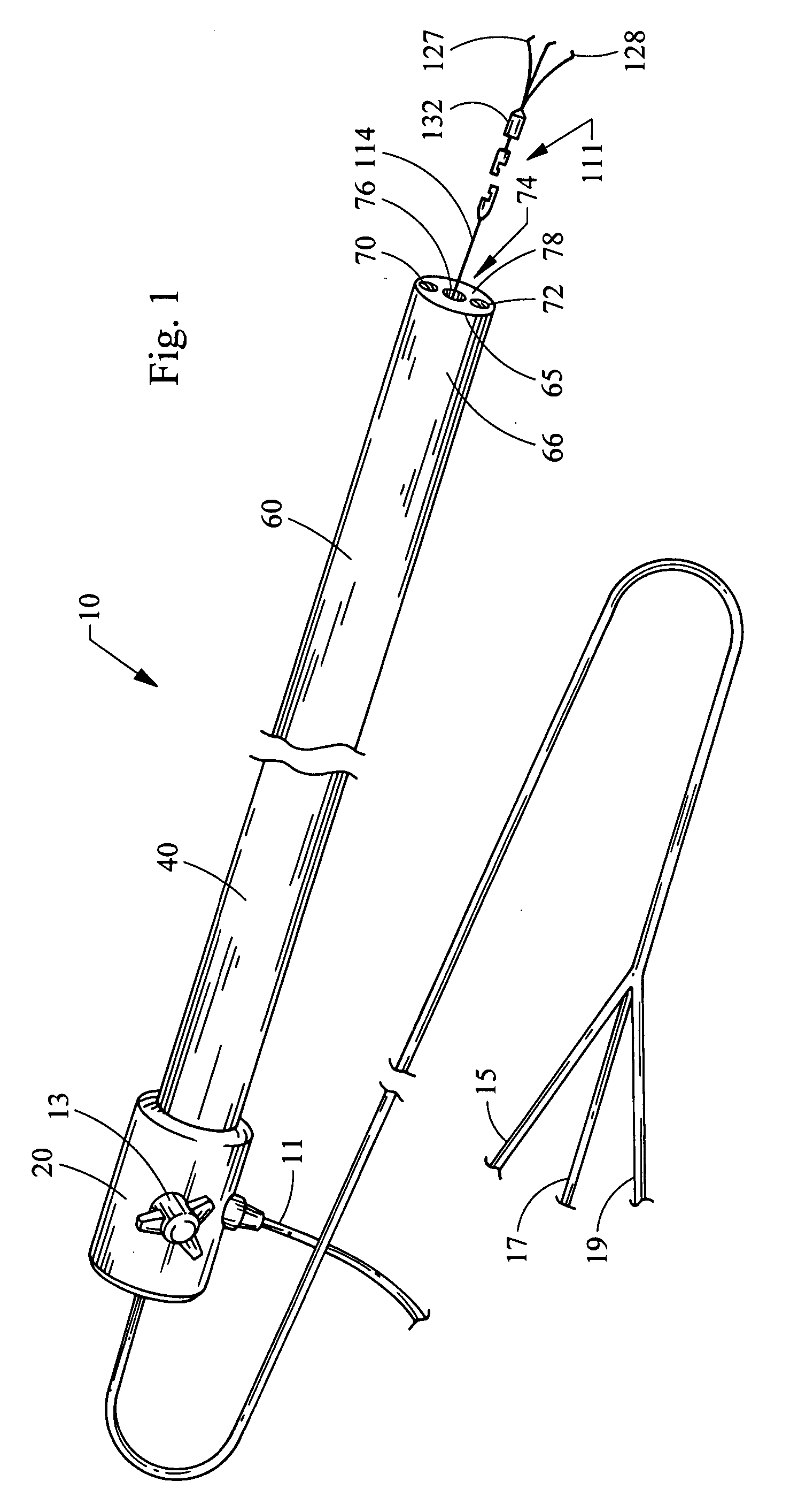
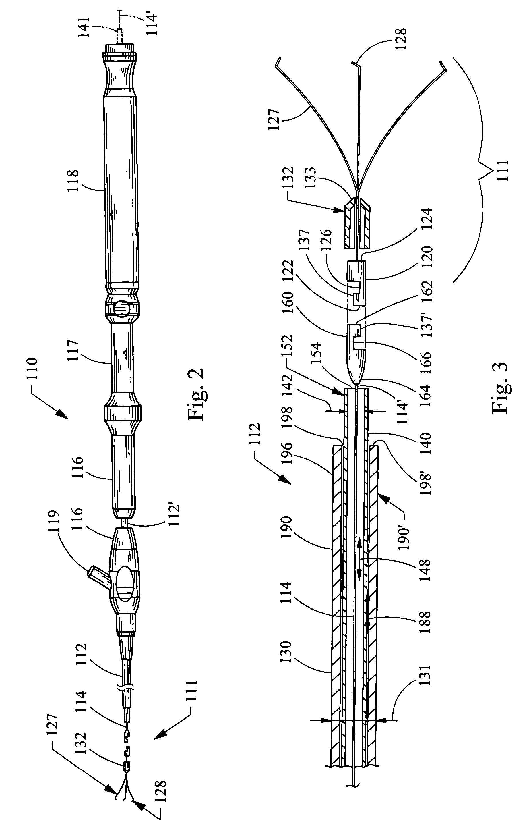
[0029] Although not limited in its scope or applicability, the present invention relates generally to devices and methods of grasping tissue endoscopically. More particularly, the invention relates to a clip and protective cap device, and methods of using a protective cap and a clip device with an endoscope, to grasp tissue endoscopically for performing hemostasis, tissue marking, endoscopic mucosal resection, tissue ligation, and a number of other applications related to colorectal medical procedures or gastrointestinal bleeding and the like.

[0030] For the purpose of promoting an understanding of the principles of the invention, the following provides a detailed description of embodiments of the invention as illustrated by the drawings as well as the language used herein to describe various aspects of the invention. The description is not intended to limit the invention in any manner, but rather serves to enable those skilled in the art to make and use the invention. As used herei...


PUM


Abstract

A clip device delivery apparatus for hemostasis has an introducer tube, such as an endoscope or sheath having a distal end portion and working channel receiving an operating wire detachably securing a clip device comprising tissue engaging arms and a sliding ring for closing the arms. A protective cap having a tube mounting section and a distal section having a receiving chamber define a passageway therebetween, the mounting section being mounted to the introducer tube distal end portion and the distal section receiving tissue and clip arms to close about tissue. The operating wire and clip device are inserted within the working channel. Methods of grasping tissue are provided using the introducer tube introduced endoscopically into a patient, tissue suctioned and clip arms deployed into tissue receiving chamber, sliding ring moved distally to close the clip arms about the tissue, and the clip detached from the operating wire.

Description

RELATED APPLICATIONS [0001] The present patent document claims the benefit of the filing date under 35 U.S.C. §119(e) of United States Provisional Patent Application filed on Mar. 10, 2006 entitled, “Clip Device and Protective Cap, and Methods of Using the Protective Cap and Clip Device with an Endoscope for Grasping Tissue Endoscopically,” and having an application Ser. No. 60 / 780,994, the disclosure of which is hereby incorporated by reference in its entirety.TECHNICAL FIELD [0002] The present invention relates to a clip device and more specifically to a protective cap and clip device for delivering a clip that can be used to cause hemostasis of blood vessels along the gastrointestinal tract, or that can be used as an endoscopic tool for holding tissue and the like. The invention also relates to methods of grasping tissue endoscopically. More particularly, the invention relates to methods of using a protective cap and a clip device with an endoscope to grasp tissue endoscopically ...

Claims


Application Information

Patent Timeline
IPC IPC(8): A61B17/128
CPCA61B1/012A61B17/122A61B2017/308A61B17/1285A61B17/1227
Inventor SOETIKNO, ROY M.SURTI, VIHAR C.
Owner WILSONCOOK MEDICAL
Learn More