Method and system for detecting electrophysiological changes in pre-cancerous and cancerous tissue and epithelium

a technology of pre-cancerous and cancerous tissue and epithelium, applied in the field of abnormal or cancerous tissue detection, can solve the problems of high mortality rate, invasive, high cost, and/or uncomfortable procedures,

Inactive Publication Date: 2008-01-10
EPI SCI LLC
View PDF35 Cites 62 Cited by
  • Summary
  • Abstract
  • Description
  • Claims
  • Application Information

AI Technical Summary

Benefits of technology

[0055] In yet another embodiment the method further comprises the steps of: analyzing the electrical parameter, and controlling the ultrasound application based on results of the analysis. In a specific embodiment the step of controlling comprises deriving an impedance value based on the electrical parameter and modifying or discontinuing application of the ultrasound when a control condition is reached, t...

Problems solved by technology

Difficulty in detecting abnormal pre-cancerous or cancerous tissue before treatment options become non-viable is one of the reasons for the high mortality rate.
Detecting of the presence of abnormal or cancerous tissues is difficult, in part, because such tissues are largely located deep within the body, thus requiring expensive, complex, invasive, and/or uncomfortable procedures.
For this reason, the use of detection procedures is often restricted until a patient is experiencing symptoms related to the abnormal tissue.
Many forms of cancers or tumors, however, require extended periods of time to attain a detectable size (and thus to produce significant symptoms or signs in the patient).
It is often too late for effective treatment by the time the detection is performed with currently available diagnostic modalities.
Both are fraught with problems of inaccuracy.
Clinical breast examinations are limited because lesions less than one cm are usually undetectable and larger lesions may be obscured by diffuse nodularity, fibrocystic change, or may be too deep in the breast to enable clinical detection.
Accordingly, mammography and clinical breast examination have relatively poor specificity in diagnosing breast cancer.
Therefore many positive mammographic findings or lesions detected on clinical breast examination ultimately prove to be false positives resulting in physical and emotional trauma for patients.
However, such techniques are unable to determine whether a solid mass, or calcifications are benign or malignant.
Its high cost and low specificity limit its general applicability for diagnosing and screening for breast cancer.
Nuclear imaging with Positron Emission Tomogaphy (PET) has a lower sensitivity for small lesions, but is limited by cost.
The disadvantage of this and similar systems is that the DC electrical properties of the epithelium are not considered.
Another disadvantage of the above referenced system is that the frequency range is not defined.
Another disadvantage with the above referenced system is that the topography of altered impedance is not examined.
Another disadvantage of the above referenced methods is that they do not probe the specific conductive pathways that are altered during the malignant process.
This approach relies on the differences in conductivity and impedivity between different tissue types and relies on data acquisition and image reconstruction algorithms which are difficult to apply clinically.
Another disadvantage with using EIT to diagnose breast cancer is the inhomogeneity of breast tissue.
Empirical measurements, however, are difficult to interpret and use in diagnosis.
There are se...

Method used


Image

  • Method and system for detecting electrophysiological changes in pre-cancerous and cancerous tissue and epithelium
  • Method and system for detecting electrophysiological changes in pre-cancerous and cancerous tissue and epithelium
  • Method and system for detecting electrophysiological changes in pre-cancerous and cancerous tissue and epithelium

Examples


example 1

Breast Cancer

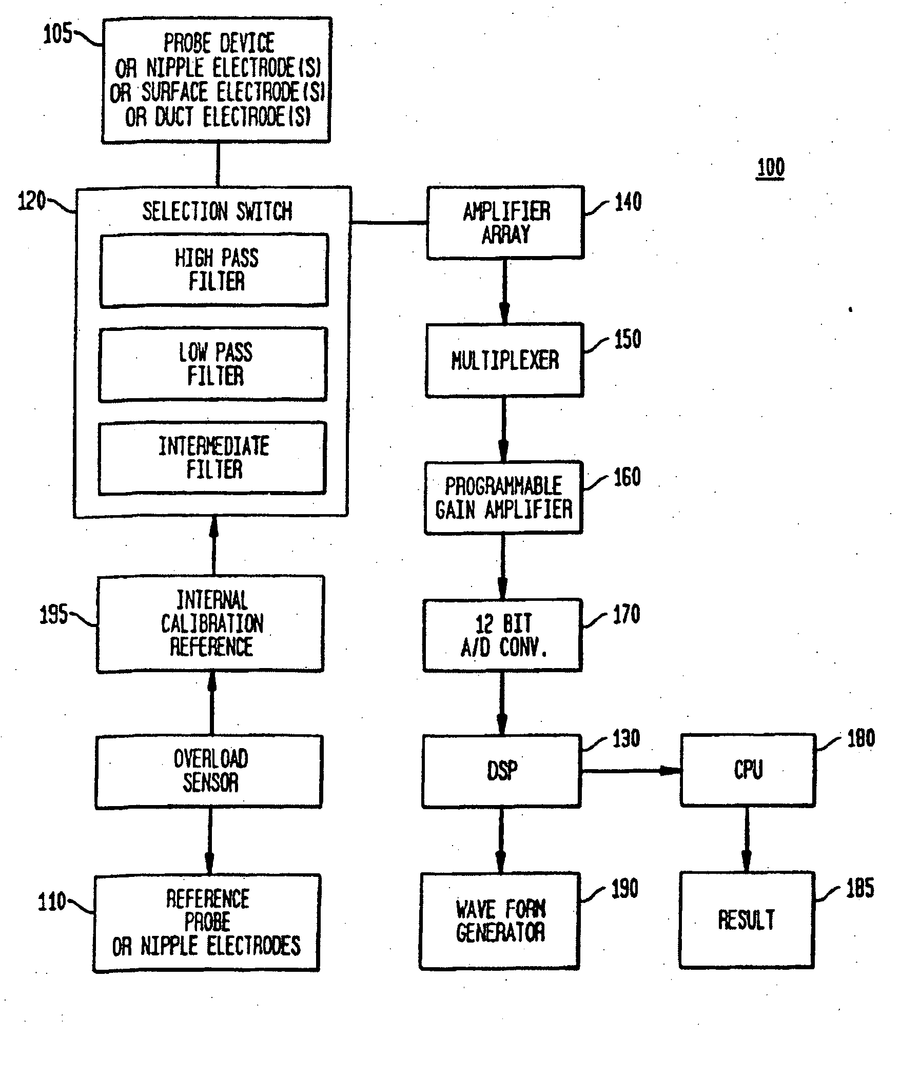
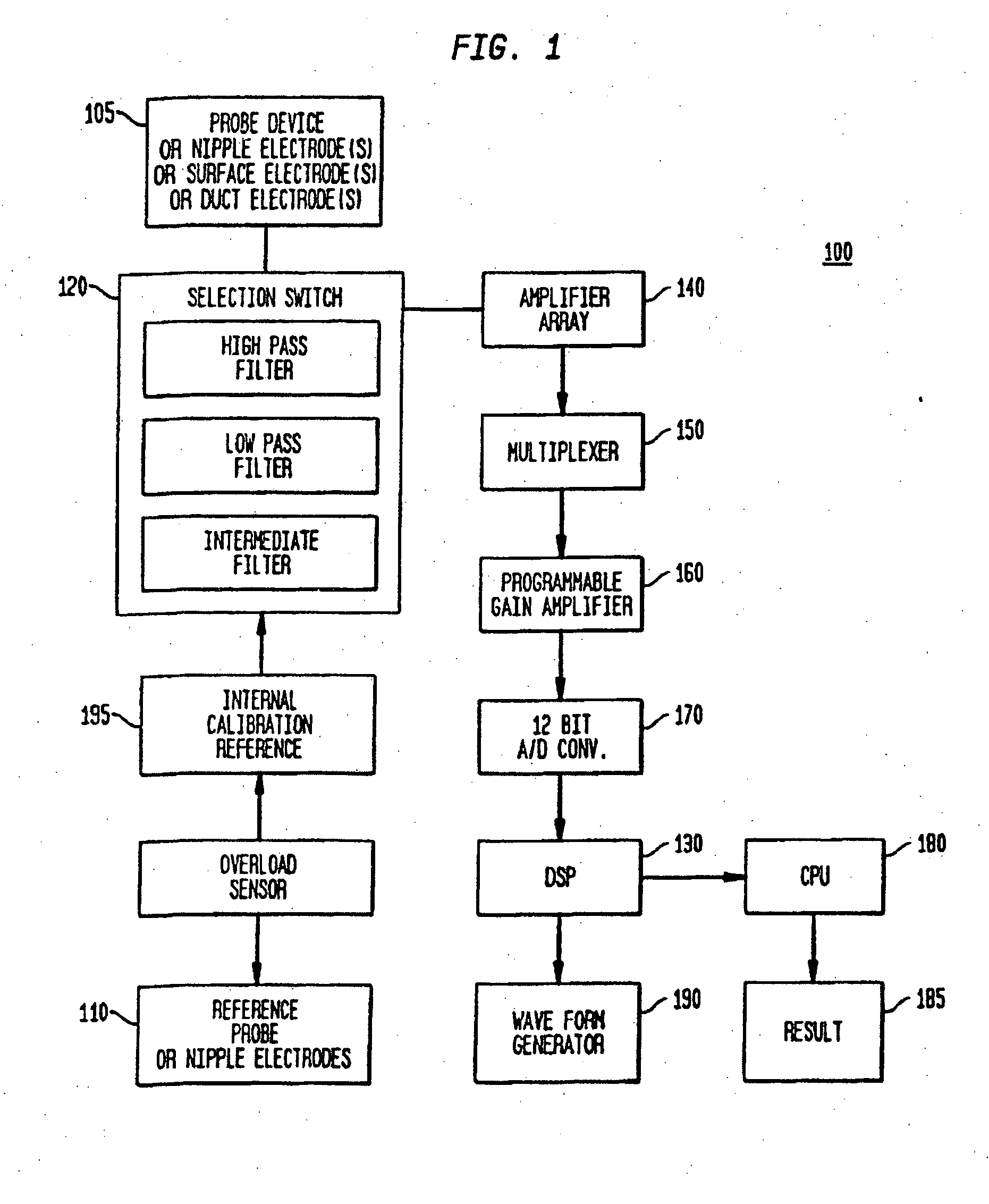
[0223] As mentioned above, impedance and DC electrical potential have been used separately at the skin's surface to diagnose breast cancer. Neither of these methods measures the ductal transepithelial DC or AC electrical properties of the breast. This significantly reduces the accuracy of the approach, because the origins of breast cancer are within the ductal epithelium, and not the surrounding breast stroma. Accuracy is further improved when the transepithelial measurements of impedance and DC potential are combined. The use of pharmacological and / or hormonal agents in combination with impedance or DC electrical potential measurements, provide a more effective method for detecting abnormal pre-cancerous or cancerous breast tissue.

[0224] Breast cancer develops within a background of disordered proliferation, which primarily affects the terminal ductal lobular units (TDLUs). The TDLUs are lined by epithelial cells, which maintain a TEP (transepithelial potential). In ...

example 2

Chemopreventative and Therapeutic Use

[0248] In addition to the ionic, pharmacologic, and hormonal agents described above, the system and method of the present invention may be used with cancer preventative and therapeutic agents and treatments. Specifically, electrical measurement of altered structure and function provides a method for evaluating a patient's response to the drugs without requiring a biopsy and without waiting for the cancer to further develop. Patients who respond to a given chemopreventative or therapeutic agent would likely show restoration of epithelial function to a more normal state. Patients who do not respond would show minimal change or may even demonstrate progression to a more advanced stage of the disease. This system and method, thus, may be used by either clinicians or drug companies in assessing drug response or by clinicians in monitoring the progress of a patient's disease and treatment, or monitoring the process of carcinogenesis (cancer developmen...

example 3

Electrophysiological Changes in Other Epithelia

[0249] The examples illustrated by FIGS. 12 and 13 were performed in human colon specimen removed at the time of surgery. Based on in vitro studies in breast epithelial tissues, similar changes in human ductal epithelium that can be measured in vivo are expected.

[0250]FIG. 12 demonstrates the short circuit current (Isc) of human colonic epithelium ex-vivo. The figure demonstrates the time course along the x-axis while varying the potassium gradient across the tissue. The potassium permeability of the apical membrane of human colonic mucosa (PKa) was determined in surgical specimens of controls and grossly normal-appearing mucosa obtained 10-30 cm proximal to colorectal adenocarcinomas. The mucosa was mounted in Ussing chambers and the basolateral membrane resistance and voltage were nullified by elevating the K+ in the serosal bathing solution. The apical sodium (Na+) conductance was blocked with 0.1 mM amiloride. This protocol reduce...


PUM


Abstract

Methods and systems are provided for determining a condition of an organ, or epithelial or stromal tissue, for example in the human breast. The methods incorporate sonophoresis, the application of ultrasonic energy, in order to condition tissue for testing and enhance test measurements. A plurality of electrodes are used to measure surface and transepithelial electropotential and impedance of breast tissue at one or more locations and at several frequencies, particularly very low frequencies. An agent may be introduced into the region of tissue to enhance electrophysiological characteristics. Pressure, drugs and other agents can optionally be applied for enhanced diagnosis. Tissue condition is determined based on the electropotential and impedance profile at different depths of the epithelium, stroma, tissue, or organ, together with an estimate of the functional changes in the epithelium due to altered ion transport and electrophysiological properties of the tissue. Devices for practicing the disclosed methods are also provided.

Description

CROSS-REFERENCE TO RELATED APPLICATIONS [0001] This application is a continuation-in-part of and claims the benefit of the filing date of U.S. patent application Ser. No. 11 / 409,144 filed Apr. 21, 2006, which claimed the benefit of the filing date of U.S. Provisional Patent Application No. 60 / 673,448 filed Apr. 21, 2005, the disclosures of which are hereby incorporated herein by reference.BACKGROUND OF THE INVENTION [0002] The present invention relates generally to the detection of abnormal or cancerous tissue, and more particularly, to the detection of changes in the electrophysiological characteristics of abnormal or cancerous tissue and to changes in those electrophysiological characteristics related to the functional, structural and topographic (the interaction of shape, position and function) relationships of the tissue during the development of malignancy. These measurements are made in the absence and presence of pharmacological and hormonal agents to reveal and accentuate th...

Claims


Application Information

Patent Timeline
IPC IPC(8): A61B5/053
CPCA61B5/0048A61B5/0536A61B5/411A61B5/6834A61B5/418A61B5/4312A61B5/415A61B5/0053A61B5/0055
Inventor DAVIES, RICHARD J.
Owner EPI SCI LLC
Why Eureka
Try Eureka