
Removable Filter Apparatus and Endoscope Apparatus

Inactive Publication Date: 2008-01-31
OLYMPUS CORP
View PDF10 Cites 21 Cited by
  • Summary
  • Abstract
  • Description
  • Claims
  • Application Information

AI Technical Summary

Benefits of technology

[0013] As a result, it is possible to decide whether no fluorescence is detected because cancer cells do not exist, or whether no fluorescence is detected because the fluorescer has not spread. Therefore, it is easy to detect cancer cells and so on.
[0047] According to the endoscope apparatus of the present invention, it is possible to replace the removable filter apparatus according to the wavelengths of the fluorescences to be observed. Therefore, an advantage is provided in that it is possible to easily observe fluorescences of different wavelengths with a single endoscope apparatus. In addition, an advantage is provided in that it is possible to easily perform simultaneous observation of fluorescences having different wavelengths.

Problems solved by technology

Therefore, there is a problem in that it is not possible to determine whether no fluorescence is detected because cancer cells do not exist, or whether no fluorescence is detected because the fluorescer has not spread; therefore, it is not possible to easily detect cancer cells and so on.

Method used


Image

  • Removable Filter Apparatus and Endoscope Apparatus
  • Removable Filter Apparatus and Endoscope Apparatus
  • Removable Filter Apparatus and Endoscope Apparatus

Examples


first embodiment

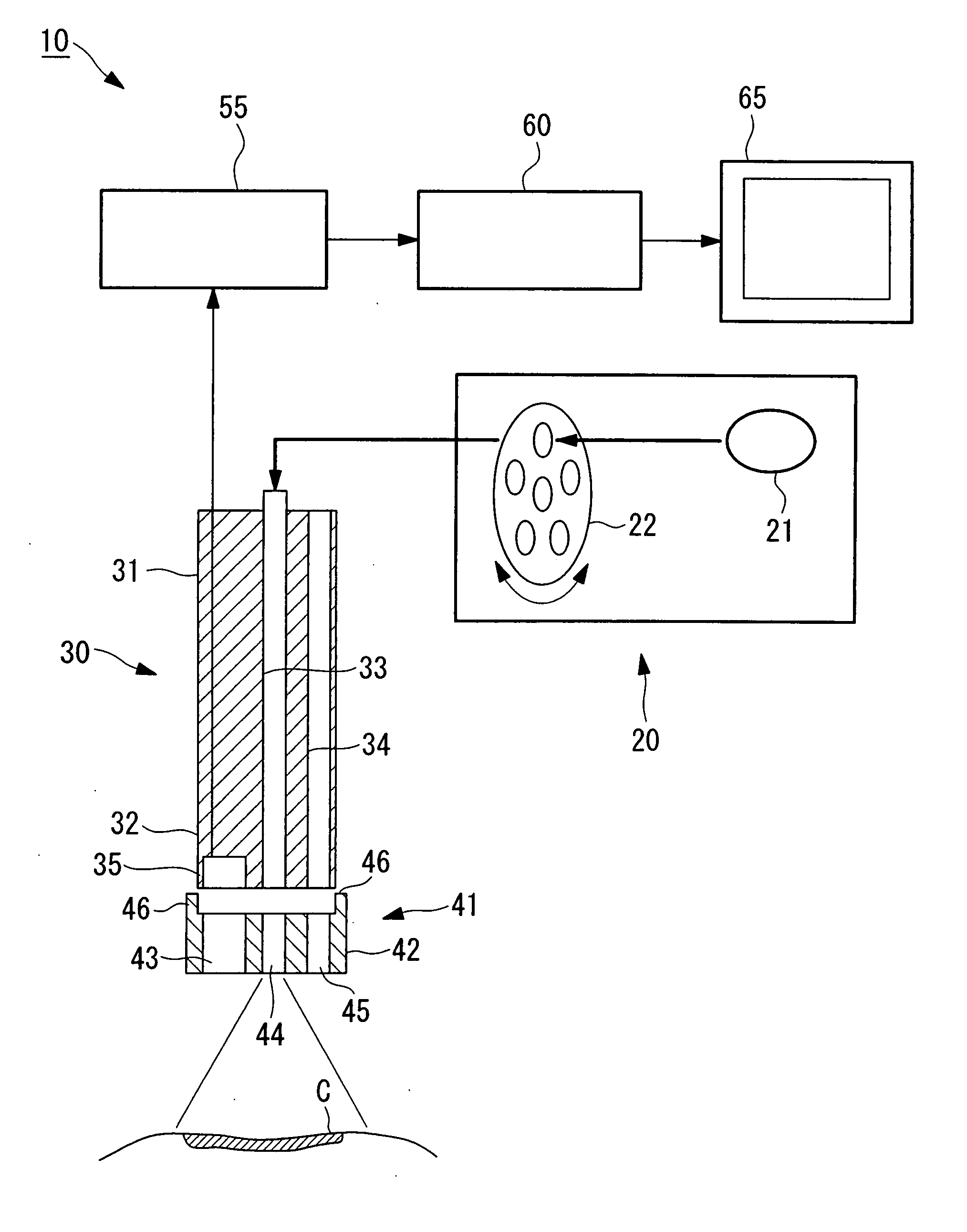
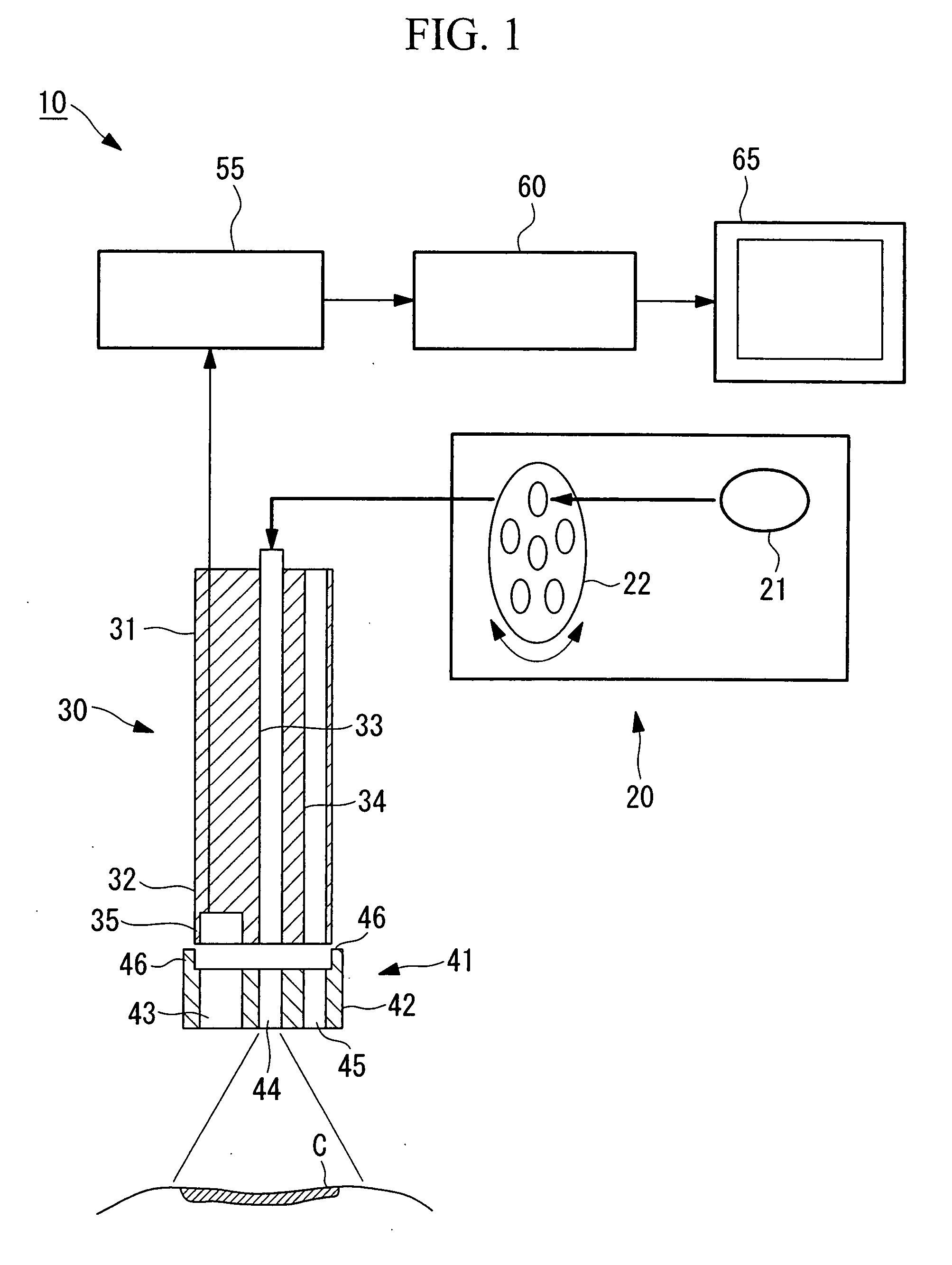
[0060] A first embodiment of the present invention will be described below with reference to FIGS. 1 to 6.

[0061]FIG. 1 is a diagram showing the overall configuration of an endoscope apparatus according to a first embodiment of the present invention for diagnosing lesions using fluorescence.

[0062] As shown in FIG. 1, the endoscope apparatus 10 is principally formed of a light source unit (projection unit) 20 for emitting visible light, excitation light, and so on; an endoscope (inserting portion) 30 for insertion into the body cavity of a living organism to form an image of a lesion (biological tissue) C; a camera control unit 55 for converting the image signal formed by the endoscope 30 into a video signal; an image processor 60 for processing the video signal to make it easier to recognize the lesion C and a normal region; and a monitor 65 for displaying the output from the image processor 60.

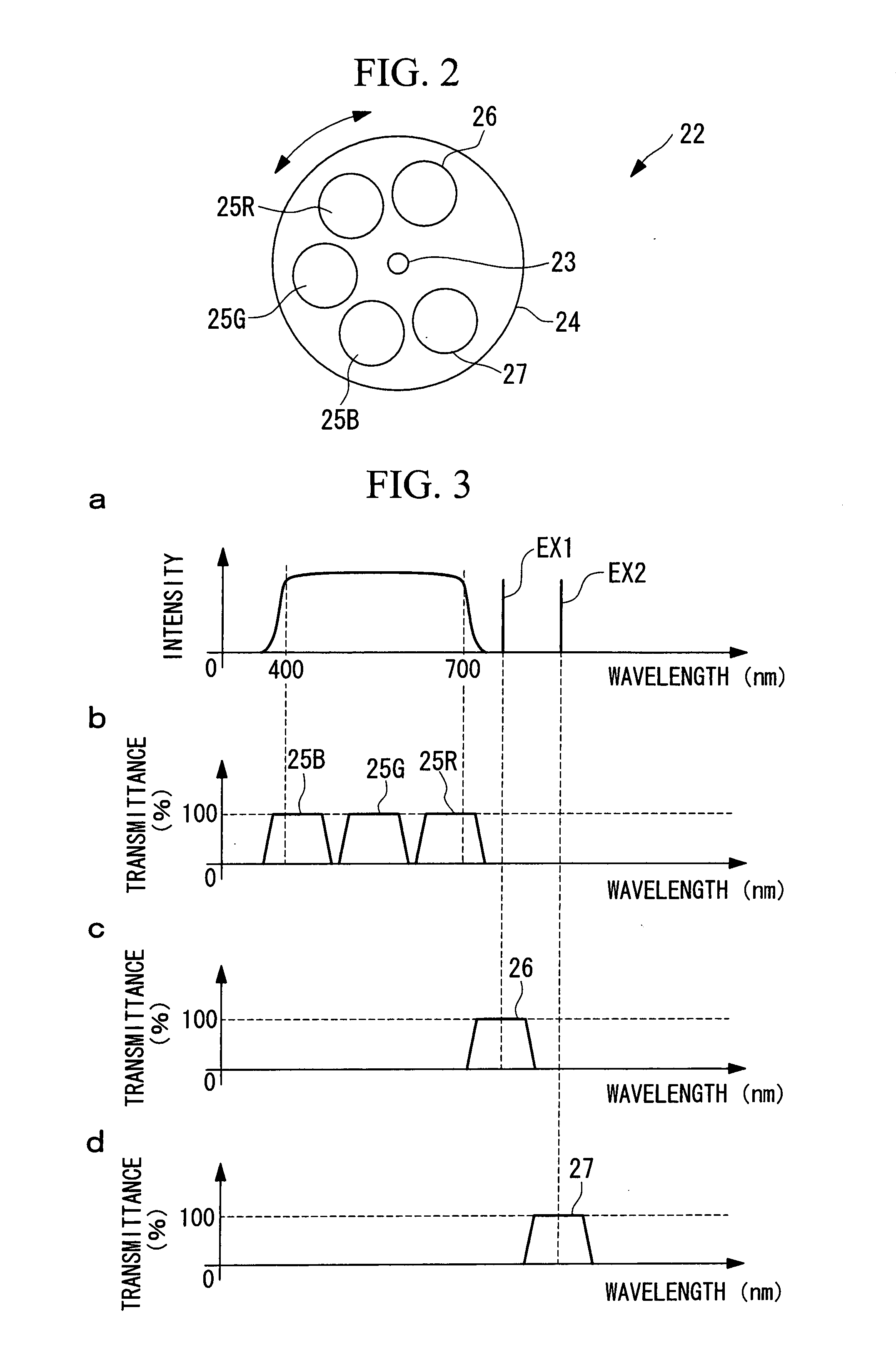
[0063]FIG. 2 is a plan view of a filter turret 22 provided in the light source unit 20 ...

second embodiment

[0099] Next, a second embodiment of the present invention will be described with reference to FIGS. 7 and 8.

[0100] The basic configuration of the endoscope apparatus of this embodiment is the same as that of the first embodiment, but the configuration of the endoscope is different from that of the first embodiment. Therefore, only the vicinity of the endoscope in this embodiment will be described using FIGS. 7 and 8, and a description of the light source apparatus and so on will be omitted.

[0101]FIG. 7 is an enlarged cross-sectional view of an tip portion, which is inserted into a body cavity, of the endoscope apparatus according to this embodiment.

[0102] Elements which are identical to those of the first embodiment are assigned the same reference numerals, and a description thereof shall be omitted.

[0103] As shown in FIG. 7, an endoscope (inserting portion) 130 of the endoscope apparatus 110 is mainly formed of a scope main body 31 and a cap (removable filter apparatus) 141 whi...

third embodiment

[0113] Next, a third embodiment of the present invention will be described with reference to FIG. 9.

[0114] The basic configuration of the endoscope apparatus according to this embodiment is the same as that of the second embodiment, but the configuration of the filter portion differs from that of the second embodiment. Therefore, in this embodiment, only the vicinity of the filter portion will be described using FIG. 9, and a description of the light source apparatus and so on will be omitted.

[0115]FIG. 9 is an enlarged cross-sectional view of the tip portion, which is inserted into the body cavity, of the endoscope apparatus according to this embodiment.

[0116] The same reference numerals are assigned to elements which are identical to those of the second embodiment, and a description thereof shall be omitted.

[0117] As shown in FIG. 9, the endoscope 230 of the endoscope apparatus 210 mainly includes a scope main body 31 and a cap (removable filter apparatus) 241 which is attache...


PUM


Abstract

A removable filter apparatus and an endoscope using the same which can easily select and transmit fluorescences of different wavelengths are provided. There is provided a removable filter apparatus 1 which is attached, in an attachable and removable manner, to a tip 32 of an inserting portion 30 of an endoscope apparatus including a projection unit 33 for emitting light towards biological tissue and a solid-state image-acquisition device 35 for detecting return light from the biological tissue, the removable filter apparatus 1 comprising a plurality of filters 50a and 50b for transmitting fluorescences of different wavelengths among the return light from the biological tissue and for blocking excitation light which generates the fluorescences.

Description

TECHNICAL FIELD [0001] The present invention relates to a removable filter apparatus of a video endoscope apparatus for observing fluorescences of a plurality of wavelengths and to an endoscope apparatus using the same. BACKGROUND ART [0002] In the related art, there are known techniques for observing autofluorescence from an organism or fluorescence of a substance injected into an organism to diagnose diseases in biological tissue, such as degeneration and cancer. [0003] For example, if the biological tissue is irradiated with excitation light, fluorescence with a longer wavelength than the excitation light is generated. Substances which produce fluorescence in response to excitation light are also present inside organisms, and it is becoming clear that there are relationships between those substances and diseases. It is also known that there are fluorescers having fluorescent pigments which attach to cancer cells. Because more of these fluorescers than normal cells gather in cance...

Claims


Application Information

Patent Timeline
IPC IPC(8): A61B1/06
CPCA61B1/00096A61B1/00101A61B1/00186A61B5/0084A61B1/0646A61B5/0071A61B1/043
Inventor XIE, TIANYU
Owner OLYMPUS CORP
Learn More