
Methods for Diagnosis and Treatment of Cancer

a cancer and cancer technology, applied in the field of cancer diagnosis and treatment, can solve the problems of limiting the utility of tracking the response of patients to therapy, and affecting the treatment effect of patients

Inactive Publication Date: 2008-03-13
MINERVA BIOTECH
View PDF16 Cites 23 Cited by
  • Summary
  • Abstract
  • Description
  • Claims
  • Application Information

AI Technical Summary

Benefits of technology

[0028]Taking advantage of understanding the biochemistry of the state of MUC1 on normal cells versus cancer cells, it is possible to detect cancer cells by assaying for binding of a ligand to the MGFR portion of MUC1 to determine the distribution of the MUC1 on the cell surface. Uniform distribution indicates cancerous cells. Thus, in one aspect, the invention is directed to a method for diagnosing cancer cells, comprising: (i) contacting a population of cells with a MGFR specific ligand bound to a signal generating label; and (ii) assaying for binding of the ligand to MGFR on a membrane, in which presence of uniform signal on the membrane on cell surface when contacted with the ligand indicates that the cells are cancerous.

Problems solved by technology

Unfortunately, the mechanism that goes awry in CML represents only a small percentage of human cancers.
Failure to characterize a cancer according to molecular signatures prior to treatment could cause the patient more harm than good.
However, because a diagnostic test or tracking test must detect elevated levels of shed MUC1, the variable number of antibody epitopes makes it impossible to discriminate between elevated levels and increased antibody binding because that person's MUC1 contains a greater number of tandem repeat units.
This variability in the number of repeat units from person to person introduces variability into the test and thus limits its utility for tracking a patient's response to therapy and prevents its use as a diagnostic.

Method used


Image

  • Methods for Diagnosis and Treatment of Cancer
  • Methods for Diagnosis and Treatment of Cancer
  • Methods for Diagnosis and Treatment of Cancer

Examples


example 1

[0214]A tumor from a patient suspected of having cancer is biopsied. The tissue specimen is assessed using the standard methods known to those skilled in the art to enable an assessment of whether or not the specimen is cancerous. The tissue specimen is then characterized as to whether it is a MUC1-positive or negative cancer using methods of the invention. The patient is diagnosed as having a MUC1-positive cancer. The physician prescribes a treatment that includes an agent(s) that inhibits MUC1. Preferred treatments are with agents that bind to the MGFR portion of the MUC1 receptor and / or inhibit its cleavage. Especially preferred are compounds disclosed in Tables 2-5, and in particular, compound numbers 28, 118, 107, 109, 125, 173, 182, 184, 185, and 188.

example 2

Antibody Production

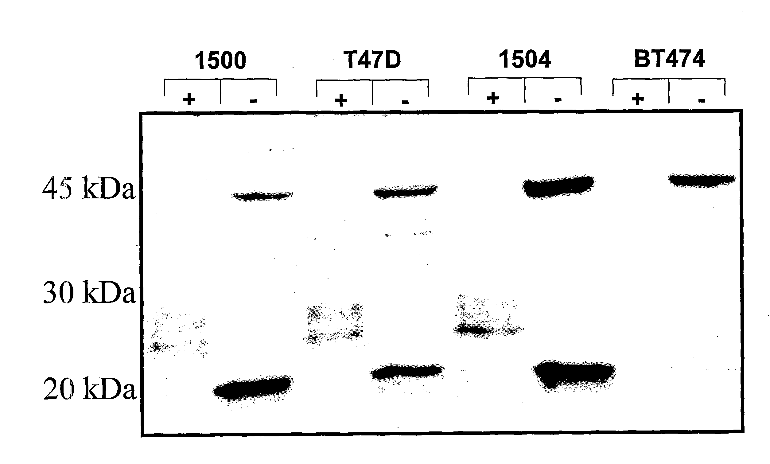
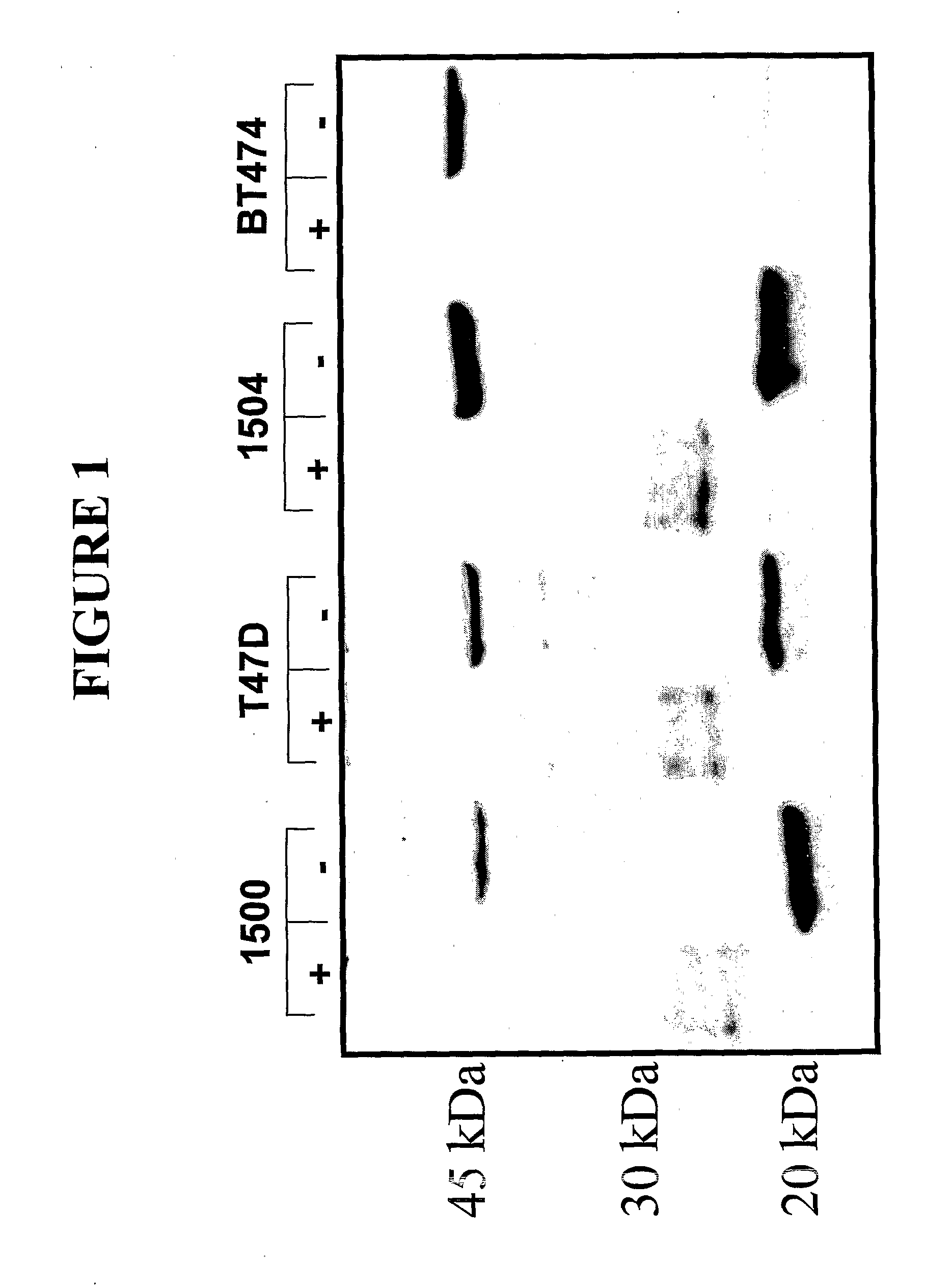
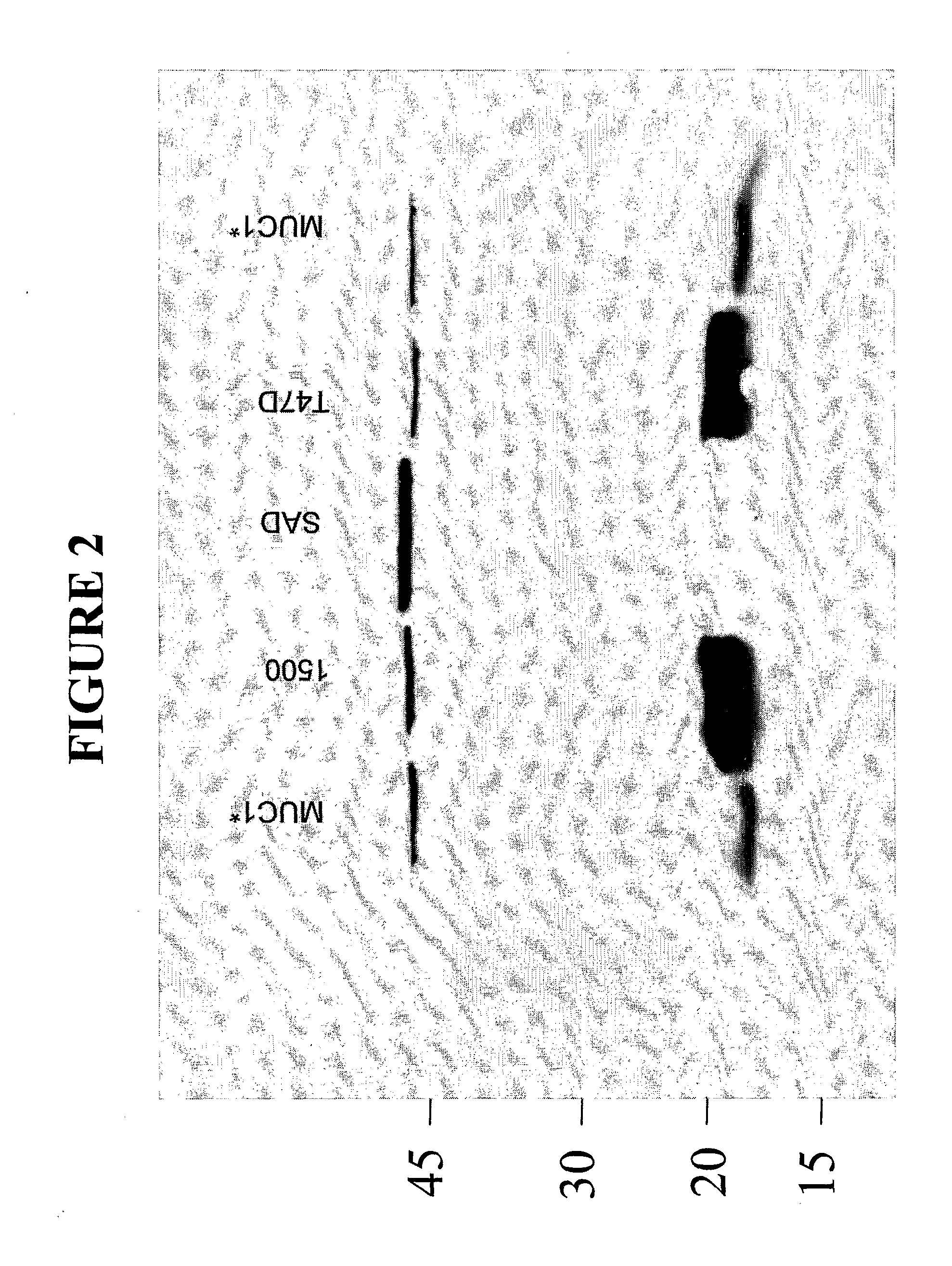
[0215]This example demonstrates that antibodies raised against the MGFR region of the MUC1 receptor, specifically bind to the form of the MUC1 receptor that is predominantly expressed on cancer cells. In this example it is shown that inventive antibodies raised against the MGFR bind to a MUC1 species that is expressed on cancer cells, wherein virtually all of the tandem repeat units have been cleaved and shed from the cell surface leaving the MGFR attached to the cell surface. Inventive antibodies were raised against the PSMGFR portion of the MUC1 receptor, in particular nat-PSMGFR or var-PSMGFR shown in Table 1 using standard methods of antibody production. Rabbit polyclonal antibodies were produced and purified by column chromatography in which the immunizing peptide was attached to the chromatography column beads. The antibodies, anti-nat-PSMGFR and anti-var-PSMGFR, were shown to specifically and sensitively bind to the immunizing peptide, MUC1 extracted from t...

example 3

MUC1-Positive Breast Cancers

[0216]Formalin fixed, paraffin embedded anonymous tissue specimens were separately tested for reactivity to two antibodies that recognize different epitopes on the MUC1 receptor: 1) a rabbit polyclonal antibody, anti-PSMGFR, that binds to the PSMGFR portion of the MUC1 receptor that remains attached to the cell surface after receptor shedding; and 2) a commercially available mouse monoclonal, VU4H5 (Santa Cruz) that binds to a sequence in the tandem repeat section of the receptor. Both antibodies were at 1 ug / ml. Protocols and treatments that the specimens were subjected to were identical for both antibodies. Contiguous sections from the same tissue specimen block were separately probed with either VU4H5 or anti-PSMGFR so that nearly identical sections were compared. As controls, like specimens were also probed with irrelevant mouse and rabbit antibodies. One section from each block was stained with hemotoxin and eosin (H&E) to aid in assessing tumor grad...


PUM

PropertyMeasurementUnit
temperatureaaaaaaaaaa
chemical stabilityaaaaaaaaaa
structureaaaaaaaaaa

Abstract

The present application discloses methods of detecting and treating cancer which express MUC1 aberrantly.

Description

CROSS-REFERENCE TO RELATED APPLICATIONS[0001]The present application is a continuation-in-part application of U.S. patent application Ser. No. 09 / 996,069, filed Nov. 27, 2001, which claims the benefit of priority to U.S. Provisional Patent Application Ser. Nos. 60 / 253,361, filed Nov. 27, 2000; 60 / 256,027, filed Dec. 15, 2000; 60 / 258,157, filed Dec. 22, 2000; 60 / 259,615, filed Jan. 3, 2001; 60 / 260,186, Jan. 5, 2001; 60 / 266,169, filed Feb. 2, 2001; 60 / 289,444, filed May 7, 2001; 60 / 266,929, filed Feb. 6, 2001; 60 / 278,093, filed Mar. 23, 2001; 60 / 294,887, filed May 31, 2001; and 60 / 298,272, filed Jun. 14, 2001. The present application is also a continuation-in-part application of U.S. patent application Ser. No. 10 / 237,150, filed Sep. 5, 2002, which claims the benefit of priority to U.S. Provisional Patent Application Ser. Nos. 60 / 317,302, filed Sep. 5, 2001 and 60 / 376,732, filed May 1, 2002. The present application is also a continuation-in-part application of U.S. patent application ...

Claims


Application Information

Patent Timeline
Patent Type & Authority Applications(United States)
IPC IPC(8): A61K31/517A61K31/341A61K31/437A61P35/00G01N33/53A61K31/55A61K31/496
CPCA61K31/495B82Y30/00A61K31/517A61P35/00A61P43/00
Inventor BAMDAD, CYNTHIA C.
Owner MINERVA BIOTECH
Learn More