

Methods for the identification of polypeptide antigens associated with disorders involving aberrant cell proliferation and compositions useful for the treatment of such disorders
a polypeptide antigen and aberrant cell technology, applied in the field of polypeptide antigen identification of aberrant cell proliferation disorders and compositions useful for the treatment of such disorders, to achieve the effect of limited general toxicity
- Summary
- Abstract
- Description
- Claims
- Application Information
AI Technical Summary
Problems solved by technology
Method used
Image

Examples
example 1
6.1. Example 1
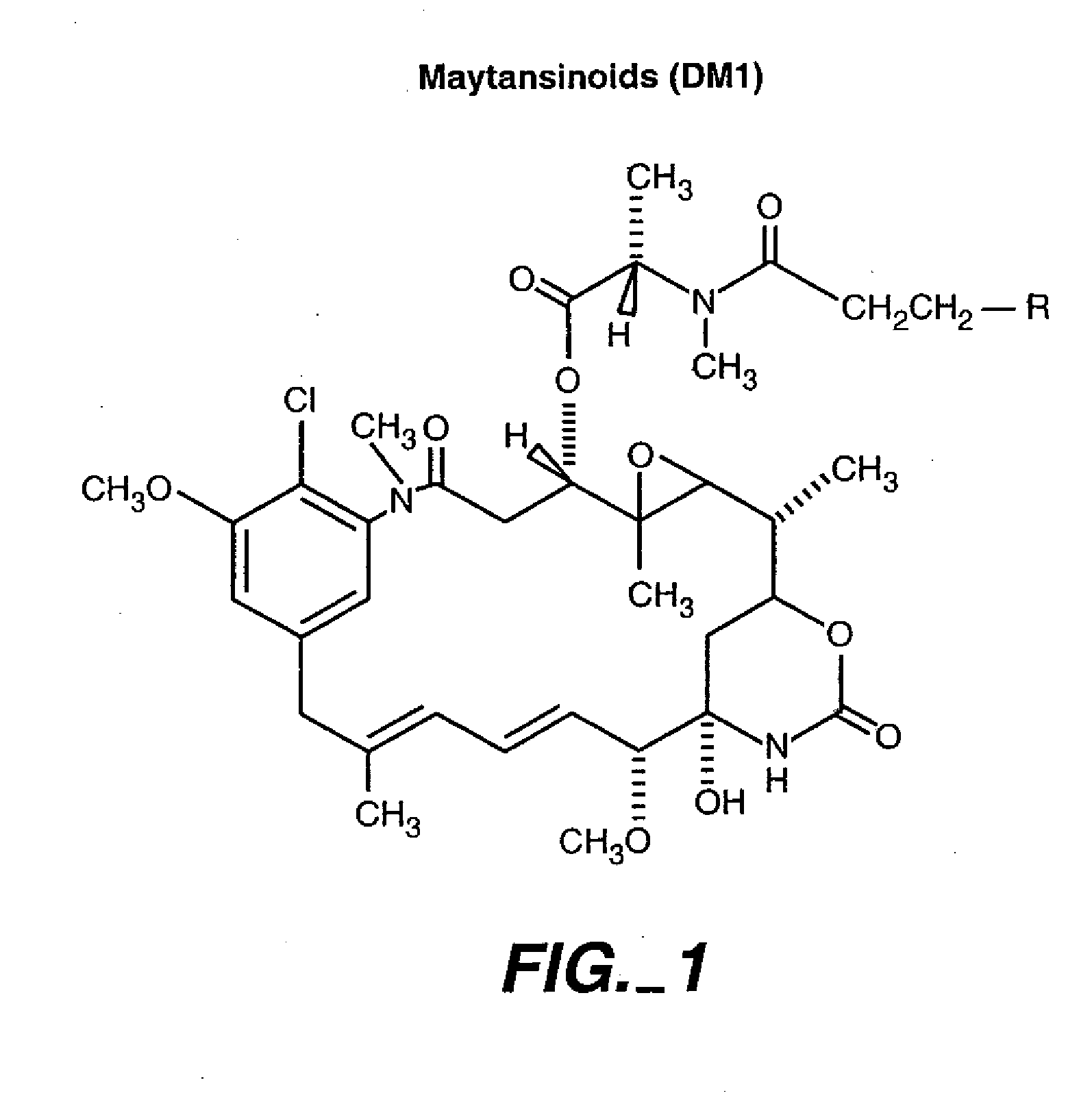
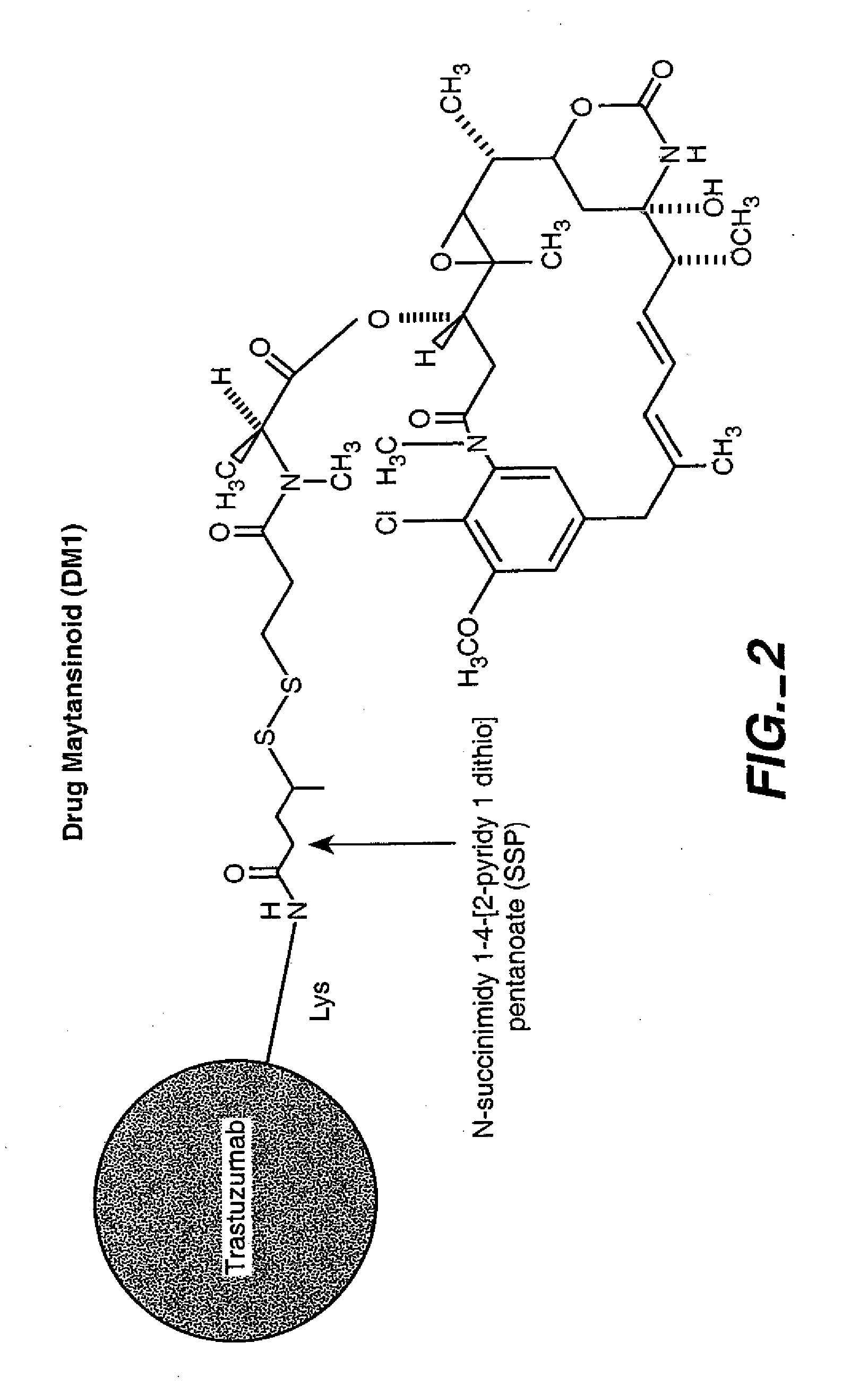
HERCEPTIN®-DM1 Conjugates
[0286] 6.1.1. Purification of HERCEPTIN®
[0287] HERCEPTIN® (huMAb4D5-8, rhuMAb HER2, U.S. Pat. No. 5,821,337) (1 vial containing 440 mg antibody) was dissolved in 50 mL MES buffer (25 mM MES, 50 mM NaCl, pH 5.6). The sample was loaded on a cation exchange column (Sepbarose S, 15 cm×1.7 cm) that had been equilibrated in the same buffer. The column was then washed with the same buffer (5 column volumes). HERCEPTIN® was eluted by raising the NaCl concentration of the buffer to 200 mM. Fractions containing the antibody were pooled, diluted to 10 mg / mL, and dialyzed into a buffer containing 50 mm potassium phosphate, 50 mM NaCl, 2 mM EDTA, pH 6.5.
[0288] 6.1.2. Modification of HERCEPTIN® with SPP
[0289] The purified HERCEPTIN® antibody was modified with N-succinimidyl-4-(2-pyridylthio)pentanoate (SPP) to introduce dithiopyridyl groups. The antibody (376.0 mg, 8 mg / mL) in 44.7 mL of 50 mM potassium phosphate buffer (pH 6.5) containing NaCl (50 mM) an...
example 2
6.2. Example 2
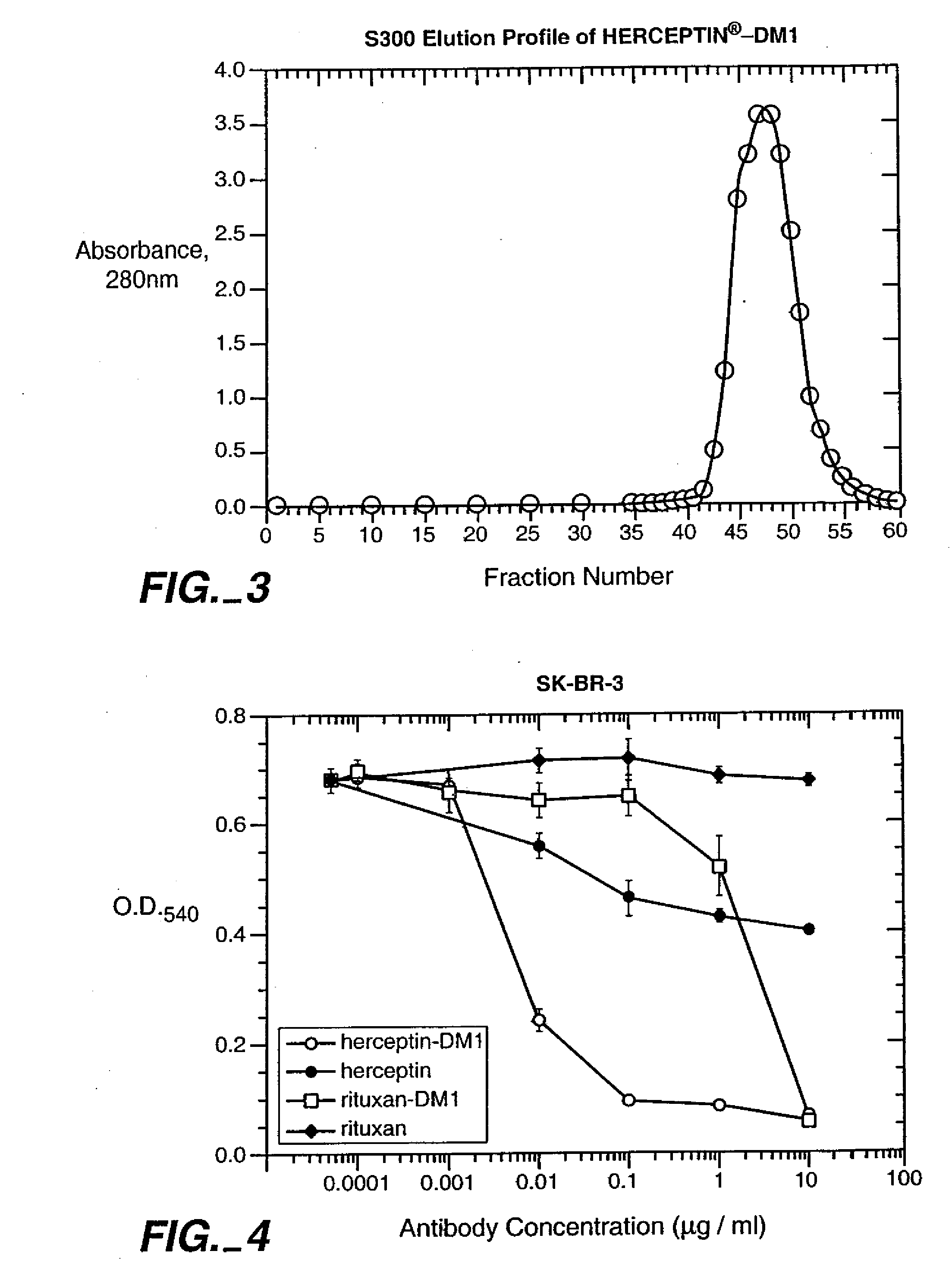
Lack of Toxicity with HERCEPTIN®-DM1 Conjugates
[0295] The following experiment demonstrates the lack of in vivo toxicity associated with HERCEPTIN®-DM1 conjugates.
[0296] 6.2.1. Experimental Design HERCEPTIN®-DM1 was administered to young adult female cynomolgus monkeys (Macaca fascicularis; Primate Products, Inc., Miami Fla.) once weekly for four weeks. The average weight of the monkeys was three kilograms (range from 2.7 to 3.4 kilograms). A total of eight monkeys, divided into four groups of two monkeys each, were utilized for the study. The dosages of HERCEPTIN®-DM1 tested were 2, 10 and 30 mg / kg. A control group received vehicle only (an aqueous buffer (pH 5.0) containing sodium succinate (10 mM), sucrose (100 mg / ml) and TWEEN™ 20 (0.1%)) at the same dose volume as administered to the treated animals. The monkeys were analyzed for various toxicities, including, but not limited to, neurotoxicity and cardiotoxicity. Table 2, below, more particularly gives the detai...
example 3
6.3. Example 3
HERCEPTIN®-DM1 Conjugates are not Toxic to Normal Human Cells or to Growth-Arrested Cells
[0325] The following experiment demonstrates the lack of toxicity associated with HERCEPTIN®-DM1 conjugates to normal human cells and to growth-arrested cells.
[0326] 6.3.1. Experimental Design Normal human mammary epithelial cells (HMEC), small airway-epithelial cells (SAEC) and adult epidermal keratinocytes (NHEK) were obtained from Clonetics / BioWhittaker (San Diego, Calif.). Human hepatocytes were obtained from In Vitro Technologies (Baltimore, Md.). SK-BR-3 human breast carcinoma cells were from The American Type Culture Collection (Rockville, Md.). Culture media used were: MEGM (mammary epithelial cell growth media), SAGM (small airway epithelial cell growth media) and KGM (keratinocyte growth media), all from Clonetics / BioWhittaker; and hepatocyte incubation media (In Vitro Technologies). SK-BR-3 cells were cultured in high glucose DMEM:Ham's F-12 (50:50) supplemented with 1...
PUM
| Property | Measurement | Unit |
|---|---|---|
| molecular weight | aaaaa | aaaaa |
| concentration | aaaaa | aaaaa |
| time | aaaaa | aaaaa |
Abstract
Description
Claims
Application Information