Method And Device For Analyzing The Retinal Vessels By Means Of Degital Images

a retinal vessel and degital image technology, applied in image analysis, image enhancement, instruments, etc., can solve the problems of high incidence of subjective systematic and random errors, time-consuming, and inability to identify individual diagnoses, so as to improve the ability to discriminate, reduce manual effort, and save time

Active Publication Date: 2008-05-08
IMEDOS INTELLIGENTE OPTISCHE SYST DER MEDIZIN & MESSTECHNIK GMBH
View PDF17 Cites 5 Cited by
  • Summary
  • Abstract
  • Description
  • Claims
  • Application Information

AI Technical Summary

Benefits of technology

[0010]Therefore, it is the primary object of the invention to enhance the ability to discriminate between healthy vessel states and at-risk vessel states while reducing manual effort

Problems solved by technology

Because of the high measurement uncertainty with a method-related standard deviation over a number of classification limits in determining the parameters and because of the high inter-individual variability, individual follow-ups and the comparison of an individual A/V (PSH) ratio to a healthy test group have only a limited ability to discriminate between healthy individuals and

Method used


Image

  • Method And Device For Analyzing The Retinal Vessels By Means Of Degital Images
  • Method And Device For Analyzing The Retinal Vessels By Means Of Degital Images

Examples


Embodiment Construction

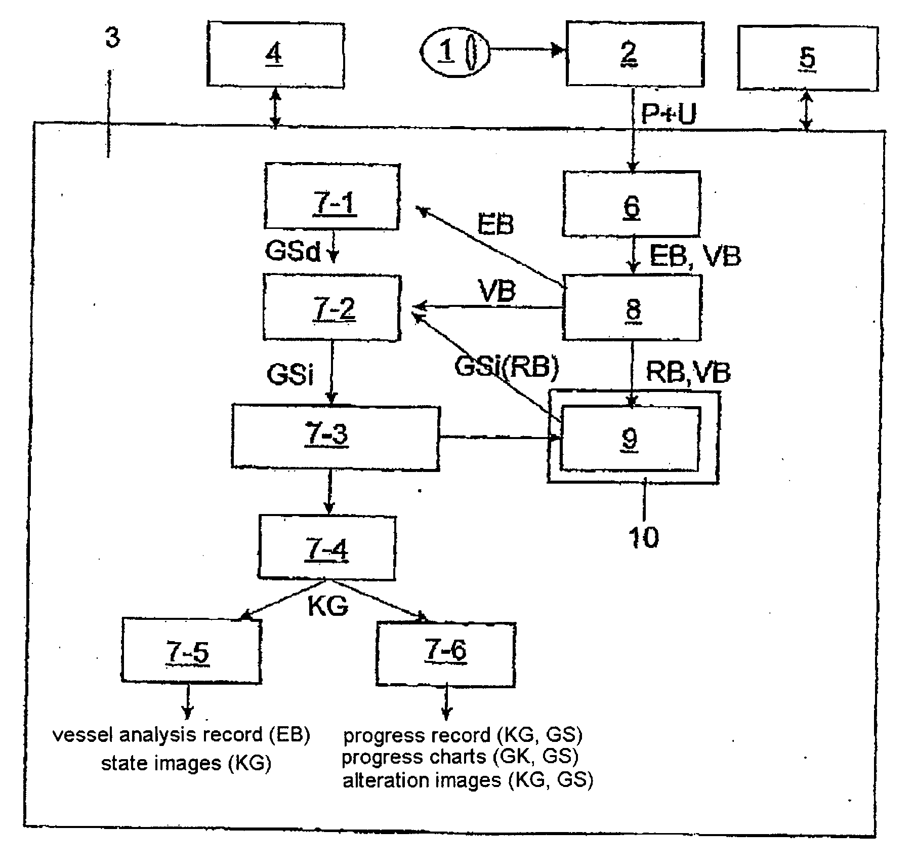
[0012]A series of adjoining vessel segments is selected along the vessel portions, and a vessel segment diameter, assignment to a vessel portion, the type of vessel distinguished by artery or vein and the associated image coordinates are determined for every vessel segment and are stored and archived by vessel segment together in a data set with reference to the evaluated image so that they can be accessed for subsequent comparison measurements. By means of comparison measurements in comparison images of the same eye recorded at offset times, the vessel diameters are automatically determined in the comparison image for vessel segments identical to a selected reference image, wherein the correlation of the vessel segment to the vessel type and vessel portion is adopted intact from the reference image. The data sets relating to the vessel segment in the comparison images are supplemented by reference to the reference image and a displacement vector of the vessel segment between the re...


PUM


Abstract

It is the object of a method and apparatus for retinal vessel analysis based on digital images to enhance the ability to discriminate between healthy vessel states and at-risk vessel states while reducing manual effort and saving time in order to allow individual vascular risk, particularly stroke risk, to be determined in a more reliable manner and with fewer subjective systematic and random errors. The vessel segment diameter, type of vessel and the image coordinates are determined for a series of adjoining vessel segments along vessel portions in a measurement zone surrounding the papilla and are stored by vessel segment with reference to the evaluated image, to a reference image recorded with a time offset, and to a displacement vector that is determined for the vessel segment between the reference image and an evaluated comparison image. Comparison measurements are carried out only on identical vessel segments already measured in the reference image. The correlation of vessel segments to vessel portions and to vessel type is adopted intact from the reference image. The stored data sets for the vessel segments of the reference image and comparison images provide a progression of coordinate-oriented vessel segment diameters for all measured vessel segments as basis for determining parameters and presenting them in a spatially resolved progression, e.g., in progress images.

Description

CROSS-REFERENCE TO RELATED APPLICATIONS[0001]This application claims priority of International Application No. PCT / DE2005 / 001680, filed Sep. 21, 2005 and German Application No. 10 2004 046 141.4, filed Sep. 21, 2004, the complete disclosures of which are hereby incorporated by reference.BACKGROUND OF THE INVENTION[0002]a) Field of the Invention[0003]The invention is directed to a method for the analysis of retinal vessels based on digital images in which a ring-shaped measurement zone containing vessel portions which proceed from a central vessel and are to be measured and which differ in type between arteries and veins is arranged around the papilla, a vessel diameter being determined for each of these vessel portions, and parameters are calculated from the determined vessel diameters of the vessel portions.[0004]The invention is further directed to an apparatus for the analysis of retinal vessels based on digital images containing a mydriatic or nonmydriatic retinal camera for rec...

Claims


Application Information

Patent Timeline
IPC IPC(8): G06K9/46
CPCA61B3/12G06T7/0012G06T2207/30041G06T7/602G06T7/2033G06T7/246G06T7/62
Inventor VILSER, WALTHARD
Owner IMEDOS INTELLIGENTE OPTISCHE SYST DER MEDIZIN & MESSTECHNIK GMBH
Why Eureka
Try Eureka