Portable orthovoltage radiotherapy

a radiotherapy and orthovoltage technology, applied in the field of ocular disorders using targeted photon energy, can solve the problems of central vision loss, macular degeneration, and contains several critical structures, such as the lens and the optic nerve, that can be damaged,

Active Publication Date: 2008-06-19
CARL ZEISS MEDITEC INC
View PDF99 Cites 10 Cited by
  • Summary
  • Abstract
  • Description
  • Claims
  • Application Information

AI Technical Summary

Benefits of technology

[0009]Retinal radiotherapy trials have shown stabilized or improved visual acuity without any significant toxicity. Radiation has also been shown to dry up neovascular membranes in patients and stabilize vision. However, due to limitations in the treatment of macular degeneration using radiotherapy including localization of the region to be treated as well as specific application of the radiation to the region to be treated, retinal radiotherapy often irradiates the entire retina, which is both unnecessary and possibly harmful.
[0012]In some embodiments, the radiation treatment system is used concomitantly with laser therapy. That is, rather than using a laser solely for pointing the x-ray device to the ocular target of choice, the laser is used for both pointing and therapy. In these embodiments, the laser preferably includes at least one additional energy or wavelength suitable for therapy of an ocular structure. The x-ray is preferably applied to the same region as the laser so as to prevent excessive scarring around the laser therapy.
[0014]In certain embodiments, a device using a treatment planning system is disclosed for providing targeted radiotherapy to specific regions of the eye. The treatment planning system integrates physical variables of the eye as well as disease variables from the physician to direct the x-ray system to deliver therapy to the ocular structures. The device applies narrow beams of radiation from one or more angles to focus radiation to a targeted region in or on the eye. In certain embodiments, the device may focus radiotherapy to structures of the posterior eye, such as the retina. In certain embodiments, the device may focus radiotherapy to structures of the anterior region of the eye, such as the sclera. The treatment planning system allows for planning of the direction of the beam entry into the eye at different points along the sclera. The unique anatomy of each individual is integrated into the treatment planning system for accurate targeting, and in some examples, automated positioning of the x-rays of the device.

Problems solved by technology

Macular degeneration is the leading cause of central vision loss in people over the age of fifty years.
The inflammatory process can result in direct destruction of the retina or destruction via formation of neovascular membranes which leak fluid and blood into the retina, quickly leading to scarring.
For example, the eye contains several critical structures, such as the lens and the optic nerve, that can possibly be damaged by excessive exposure to radiation.
The application of external beam therapy is limited by devices and methodologies used to apply the therapy.
These devices and methodologies are older radiation technologies used to treat conditions such as tumors anywhere in the body and were not developed specifically for ocular radiation therapy.
In addition, logistics are difficult as far as patient recruitment and administration of treatments because such treatment devices are borrowed from and displace oncologic therapies.
However, due to limitations in the treatment of macular degeneration using radiotherapy including localization of the region to be treated as well as specific application of the radiation to the region to be treated, retinal radiotherapy often irradiates the entire retina, which is both unnecessary and possibly harmful.
A major limitation of this treatment is that it requires invasive procedures involving partial removal of the vitreous fluid of the posterior chamber of the eye to place the brachytherapy probe.
In addition, it cannot be fractionated because of the invasiveness required to deliver it.
Furthermore, it would be difficult to apply this therapy to patients who do not yet have vision loss because of the potential complications from the procedure.
Procedures such as trabeculoplasty, trabeculotomy, canaloplasty, laser iridotomy, placement of shunts, and other procedures all suffer from a short-lived effect because of scar formation as a result of the surgical trauma.
Anti-inflammatory drugs appear to offer a palliative and / or preventative solution to the chronic scarring that occurs after these procedures; however, the drugs have to be given several times per day and are associated with their own side effect profile such as seepage into unwanted regions of the eye.
This scarring is caused by trauma from the surgery, proliferation of lens cells, and material incompatibility.

Method used


Image

  • Portable orthovoltage radiotherapy
  • Portable orthovoltage radiotherapy
  • Portable orthovoltage radiotherapy

Examples


Embodiment Construction

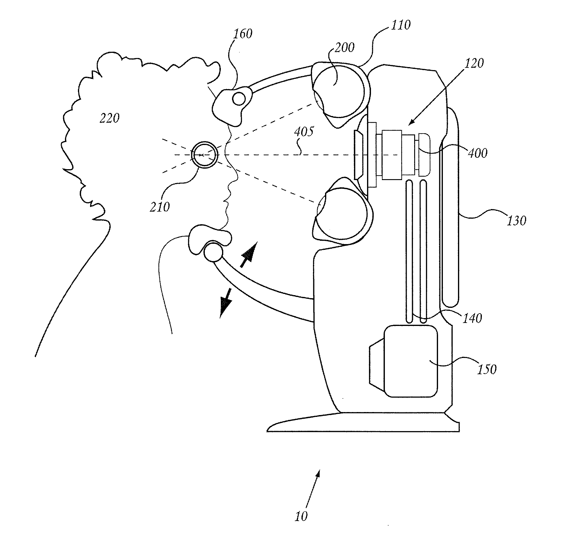
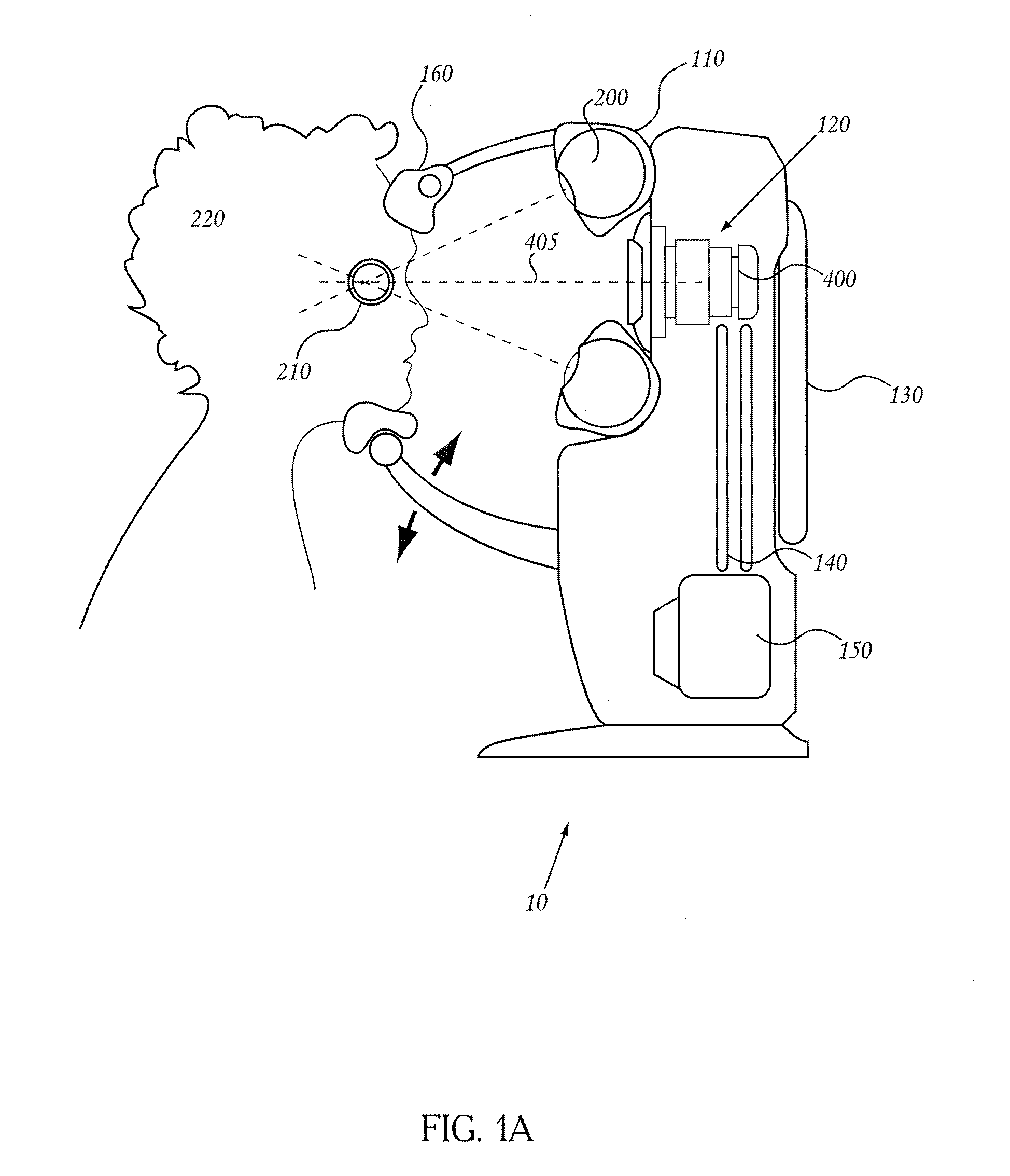
[0067]The present embodiments include systems and methods for treating a human eye with radiotherapy. Some embodiments described below relate to systems and methods for treating macular degeneration of the eye using radiotherapy. For example, in some embodiments, systems and methods are described for use of radiotherapy on select portions of the retina to impede or reduce neovascularization of the retina. Some embodiments described herein also relate to systems and methods for treatment of glaucoma or control wound healing using radiotherapy. For example, in some embodiments, systems and methods are described for use of radiotherapy on tissue in the anterior chamber following glaucoma surgery, such as trabeculoplasty, trabeculotomy, canaloplasty, and laser iridotomy, to reduce the likelihood of postoperative complications. In other embodiments, systems and methods are described to use radiotherapy to treat drusen, inflammatory deposits in the retina that are thought to lead to visio...


PUM


Abstract

A portable orthovoltage radiotherapy system is described that is configured to deliver a therapeutic dose of radiation to a target structure in a patient. In some embodiments, inflammatory ocular disorders are treated, specifically macular degeneration. In some embodiments, the ocular structures are placed in a global coordinate system based on ocular imaging. In some embodiments, the ocular structures inside the global coordinate system lead to direction of an automated positioning system that is directed based on the ocular structures within the coordinate system.

Description

RELATED APPLICATIONS[0001]This application claims priority benefit of U.S. Provisional Application No. 60 / 933,220, filed Jun. 4, 2007, entitled, “PORTABLE ORTHOVOLTAGE RADIOTHERAPY”; U.S. Provisional Application No. 60 / 922,741, filed Apr. 9, 2007, entitled, “RADIATION THERAPY SYSTEM FOR THE TREATMENT OF MACULAR DEGENERATION”; U.S. Provisional Application No. 60 / 869,872, filed Dec. 13, 2006, entitled, “XRAY TREATMENT SYSTEM”; U.S. Provisional Application No. 60 / 862,210, filed Oct. 19, 2006, entitled, “METHODS AND DEVICE FOR NON-INVASIVE ROBOTIC TARGETING OF INFLAMMATORY LESIONS USING RADIATION”; U.S. Provisional Application No. 60 / 862,044, filed Oct. 18, 2006, entitled, “METHODS AND DEVICES FOR NON-INVASIVE ROBOTIC TARGETING OF RETINAL LESIONS”; and U.S. Provisional Application No. 60 / 829,676, filed Oct. 16, 2006, entitled, “METHODS AND DEVICES TO APPLY FOCUSED ENERGY TO BODY REGIONS”; the entirety of each of which are incorporated herein by reference.BACKGROUND[0002]1. Field of the ...

Claims


Application Information

Patent Timeline
Patent Type & Authority Applications(United States)
IPC IPC(8): A61N5/10A61B5/00
CPCA61N5/1017A61N5/103A61N2005/1091A61N2005/105A61B6/506A61F9/009A61N2005/1056A61B2017/306A61N5/1049A61F9/007A61N5/10A61B5/00
Inventor GERTNER, MICHAEL
Owner CARL ZEISS MEDITEC INC
Why Eureka
Try Eureka