
Integrated endoscope and accessory treament device

Active Publication Date: 2008-06-19
CR BARD INC
View PDF102 Cites 18 Cited by
  • Summary
  • Abstract
  • Description
  • Claims
  • Application Information

AI Technical Summary

Benefits of technology

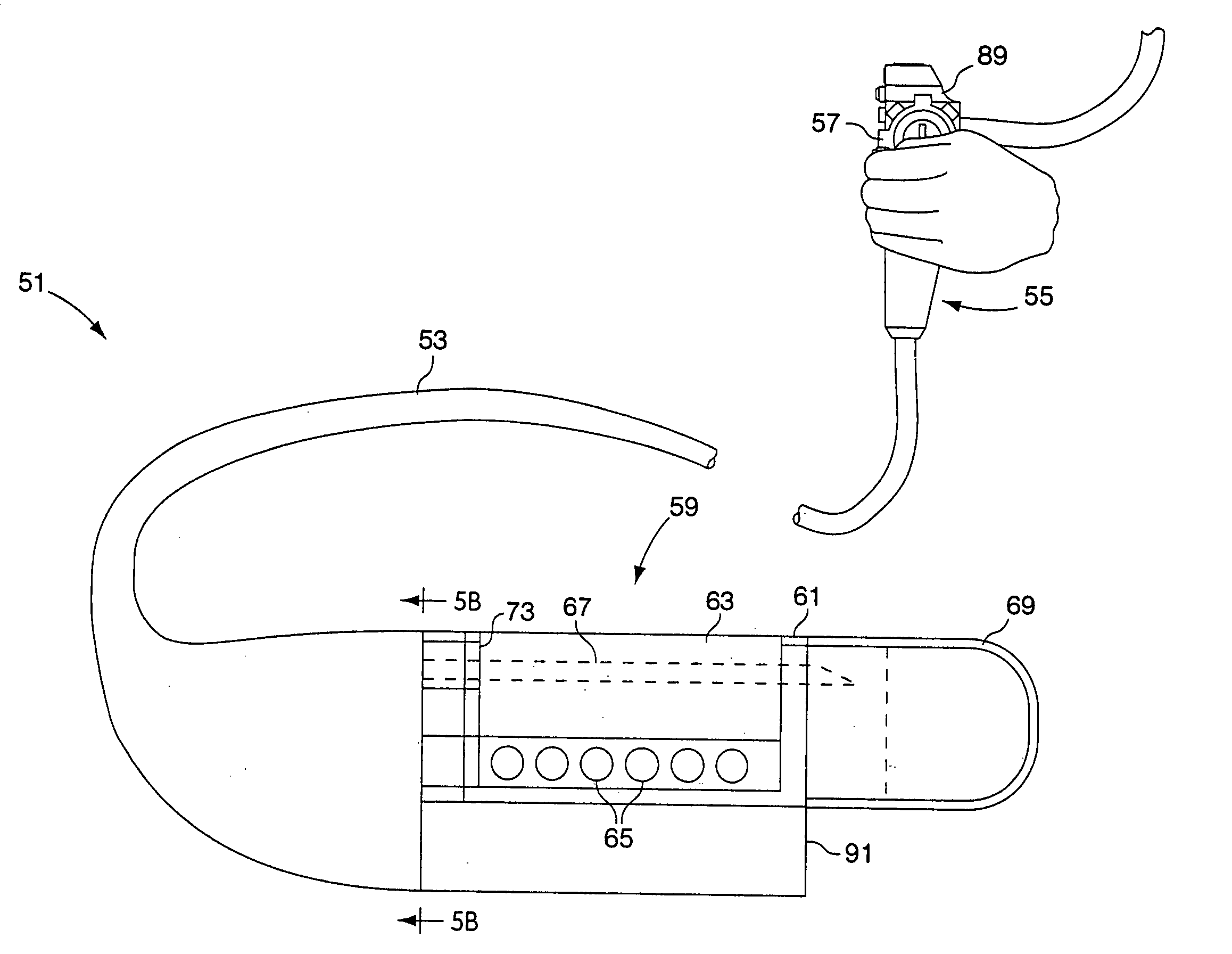
[0006]The present invention provides an integrated endoscope and treatment accessory that is secure, reliable and easy to operate accurately. The device comprises an endoscope shaft configured similarly to conventional endoscope shafts and may be provided with a working channel, a channel for optical fibers for viewing capability and light transmission as well as auxiliary channels to provide viewing lens cleaning or additional working channel capability. The endoscope shaft further provides along its length space for the operative control elements of the integrated treatment device placed at the distal end of the endoscope. The treatment device may consist of a housing in which separate, movable components are arranged to carry out the specified medical treatment or procedure. Elements extending through the shaft of the endoscope such as cables, wires, or fluid pressure serve to operate the components of the accessory. Because the accessory is integrated into the endoscope, the viewing elements can be specifically arranged to provide viewing capability of the location of the accessory where treatment will be carried out on tissue. Additionally, with control elements for the accessory being integrated into the endoscope shaft, separate open working channels can be left free to receive other devices or instruments in the overall diameter of the endoscope shaft need not be increased dramatically by an externally applied accessory control sheath or catheter.
[0007]The integrated endoscope and accessory may employ any type of treatment device presently known to be applicable to an endoscope as a separately attached accessory. Specifically, the integrated accessory may comprise forceps, a mechanical or electrical cutting element, suturing device, band ligating device or other tissue manipulating mechanism. The housing for the accessory is integrated into the shaft of the endoscope near its distal end appropriately positioned along the shaft for the treatment that will be carried out. The control mechanisms for operating the accessory may be similar to conventional specific accessories but will extend through spaces that are specifically formed through the endoscope to accommodate them. At the proximal end of the endoscope, an integrated control mechanism can be employed adjacent the endoscope distal end control mechanisms so that a user can operate both sets of controls easily. Alternatively, the accessory controls can be integrated with the endoscope controls for ease of use. Further the accessory controls can be positioned at any convenient location along the length of the endoscope where the physician would ordinarily grasp the endoscope shaft to control its movement in the patient.
[0010]It is another object of the invention to provide a method for using an integrated endoscope and medical treatment device accessory that reduces the number of steps and instruments required to complete an endoscopic procedure

Problems solved by technology

First, the separate accessory may limit viewing capability through the distal end of the endoscope when it is attached so as to extend distally from the distal face of the endoscope.
Second, there is always a risk that the accessory will become detached from the endoscope while in the patient, compromising the procedure and presenting problems for safely removing the detached accessory from the patient.
Third, with various manufacturers producing endoscopes and accessories of differing diameters, mounting a particular accessory to an endoscope can be problematic if their diameters are not compatible.
Fourth, the control mechanisms for operating the accessory must extend through existing working channels in the endoscope interfering with or prohibiting introduction of additional accessories or instruments through the endoscope during the procedure.
Also, the accessory controls may be awkward to mount and operate in conjunction with the endoscope as the endoscope was not originally designed to accommodate such additional controls.

Method used


Image

  • Integrated endoscope and accessory treament device
  • Integrated endoscope and accessory treament device
  • Integrated endoscope and accessory treament device

Examples


Embodiment Construction

[0038]The present invention provides an endoscope with an integrated medical device treatment accessory at its distal end. The devices contemplated for integration with the endoscope include tissue apposition devices, forceps or tissue cutting instruments. Several embodiments of the integrated endoscope employing tissue apposition devices are presented below.

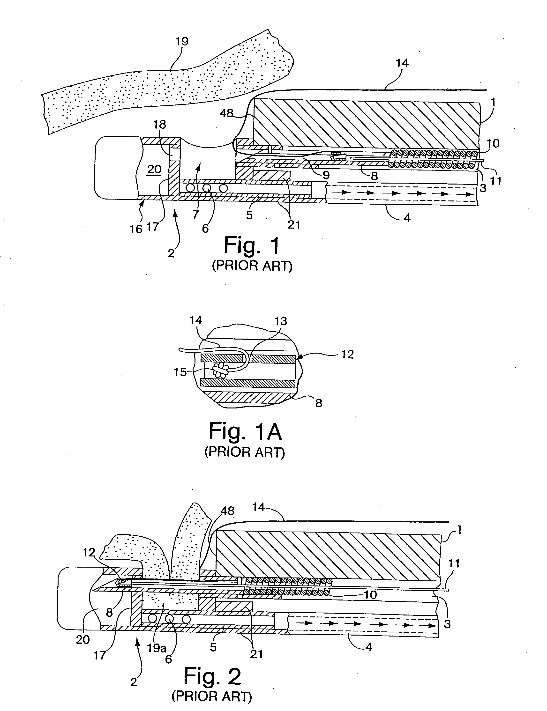
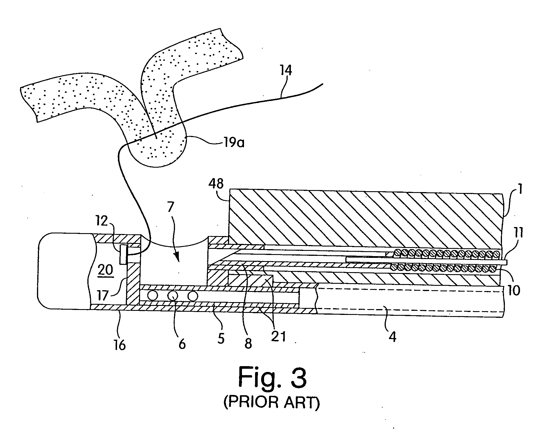
[0039]In one embodiment of the integrated endoscope, the tissue apposition device as disclosed in U.S. Pat. No. 5,792,153 may be employed. The '153 patent is incorporated by reference herein in its entirety. A brief explanation of the configuration and operation of the prior art tissue apposition device is presented with reference to the prior art FIGS. 1-3.

[0040]FIGS. 1-3 depict a prior art endoscopic suturing device disclosed in U.S. Pat. No. 5,792,153. FIG. 1 shows the distal end of a flexible endoscope 1, on which a sewing device 2 is attached. The endoscope is provided with a viewing channel, which is not shown, but which t...


PUM


Abstract

An integrated endoscope and accessory treatment device is provided. The device employs a medical treatment device at its distal end that is combined with the endoscope as a complete system. The treatment device is controlled by integrated elements at the proximal end of the endoscope. The treatment devices may include tissue apposition devices, tissue cutting devices, forceps and others.

Description

RELATED APPLICATIONS[0001]This application is a continuation of U.S. patent application Ser. No. 10 / 658,135, filed Sep. 8, 2003, which claims the benefit of U.S. Provisional Application No. 60 / 408,556, filed on Sep. 6, 2002, the subject matter of which is related to the disclosure document filed at the U.S. Patent and Trademark Office on Sep. 7, 2000, and assigned Disclosure Document No. 479631.FIELD OF THE INVENTION[0002]This invention relates to endoscopes and devices and methods for carrying out medical treatments using an endoscope.BACKGROUND OF INVENTION[0003]Viewing endoscopes permit remote treatment of internal locations within a patient by accessing those locations through a natural body lumen avoiding the need for surgery in some cases. The advantages of using an endoscope to treat internal maladies of the human body has led to the development of various endoscopic accessory treatment devices that can be fastened to the distal end of the endoscope to carry out mechanical ma...

Claims


Application Information

Patent Timeline
IPC IPC(8): A61B17/04A61BA61B1/00A61B1/12A61B17/30A61B19/00
CPCA61B1/00087A61B1/018A61B17/0469A61B2017/306A61B17/0643A61B19/5212A61B2017/00296A61B17/0482A61B90/361
Inventor AZNOIAN, HAROLD M.COSTA, RICHARD L.DIMITRIOU, JOHN E.GAMBALE, RICHARD A.LUKIN, PETER J.PAGE, EDWARD C.SILVA, SEAN J.ZELONIS, DAVID T.
Owner CR BARD INC
Learn More