3D Segmentation of the Colon in MR Colonography

a colon and 3d technology, applied in the field of image processing, can solve the problems of higher noise level of mr images, lower resolution of ct images, and difficult image post-processing and image analysis tasks, and achieve the effect of improving image quality, improving image quality, and improving image quality

Inactive Publication Date: 2008-08-07
SIEMENS MEDICAL SOLUTIONS USA INC
View PDF5 Cites 10 Cited by
  • Summary
  • Abstract
  • Description
  • Claims
  • Application Information

AI Technical Summary

Problems solved by technology

But MR images have higher noise levels and lower resolutions than CT images, which makes the tasks of image post-processing and im

Method used


Image

  • 3D Segmentation of the Colon in MR Colonography
  • 3D Segmentation of the Colon in MR Colonography
  • 3D Segmentation of the Colon in MR Colonography

Examples


Embodiment Construction

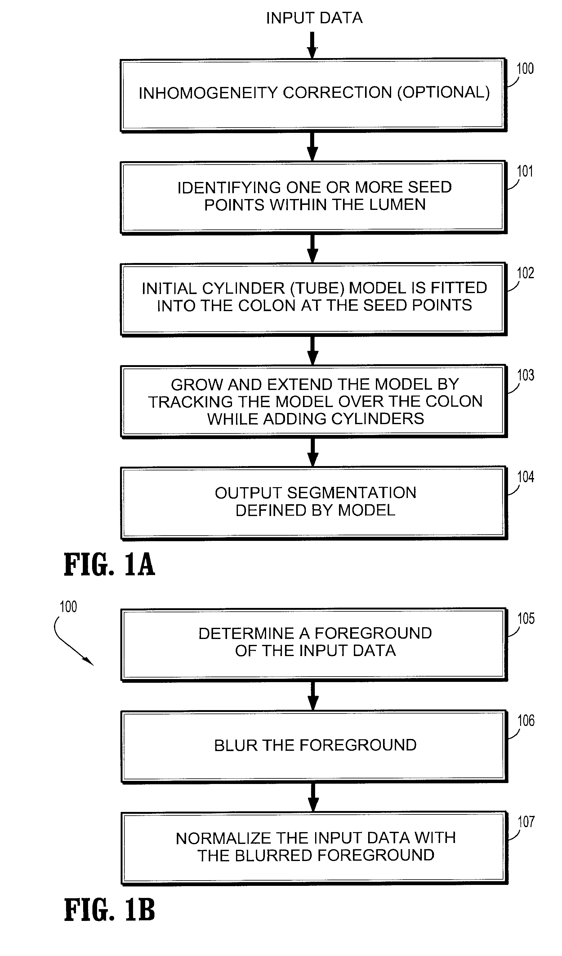
[0026]According to an embodiment of the present disclosure, a system and method for 3D segmentation in MR colonography segments the colon in 3D MR scans of the human abdomen. Results of the segmentation may facilitate polyp detection and classification tasks for both bright and dark lumen cases.

[0027]The segmentation is model-based to overcome limitations in imaging dark lumen areas. The model is tube-shaped to direct the segmentation and tracking. Voxel intensities and intensity gradients in the local region are the main image forces considered.

[0028]It should be understood that the methods described herein are applicable to applications other than colonography, and may be used for other tube-like structures.

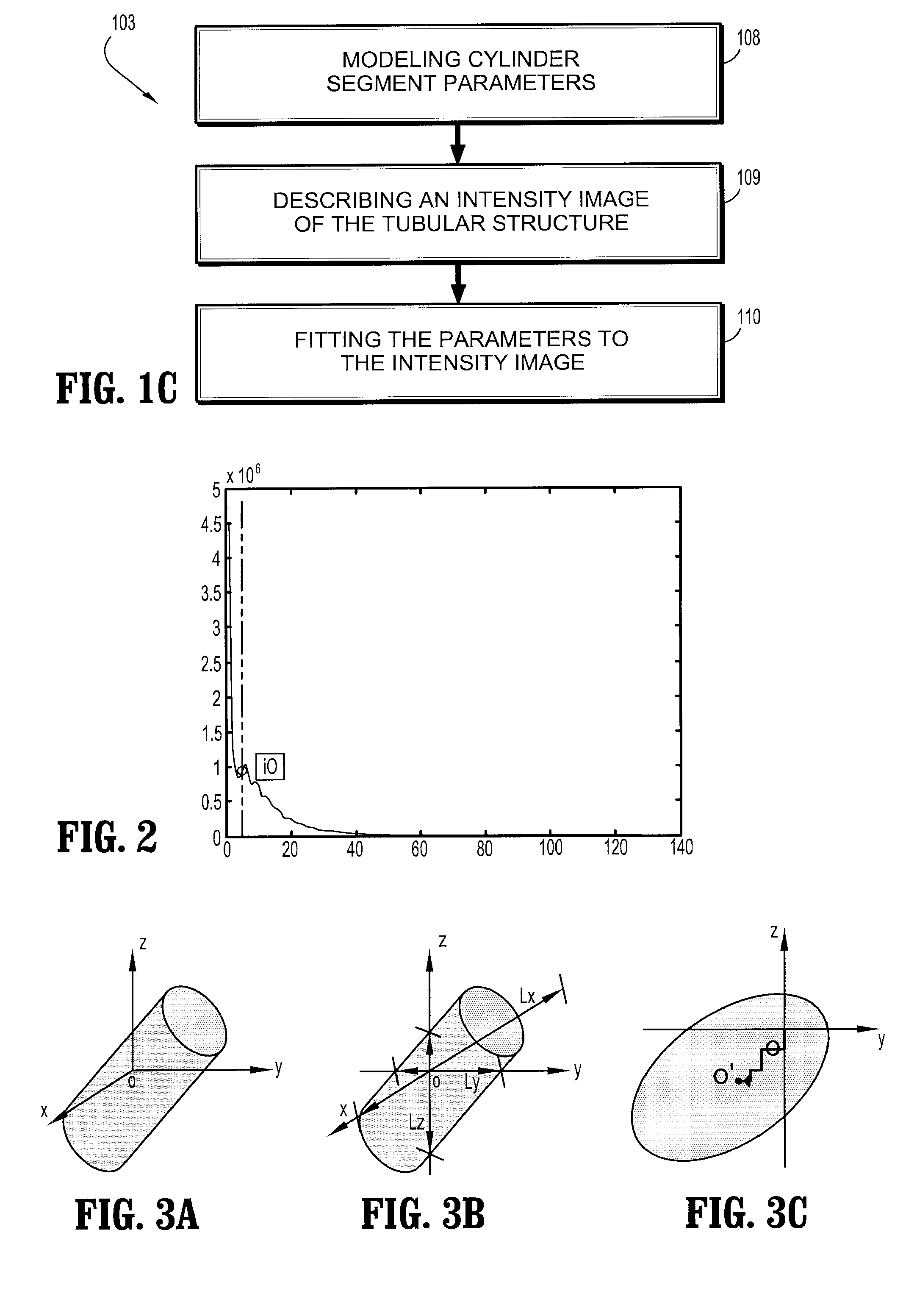
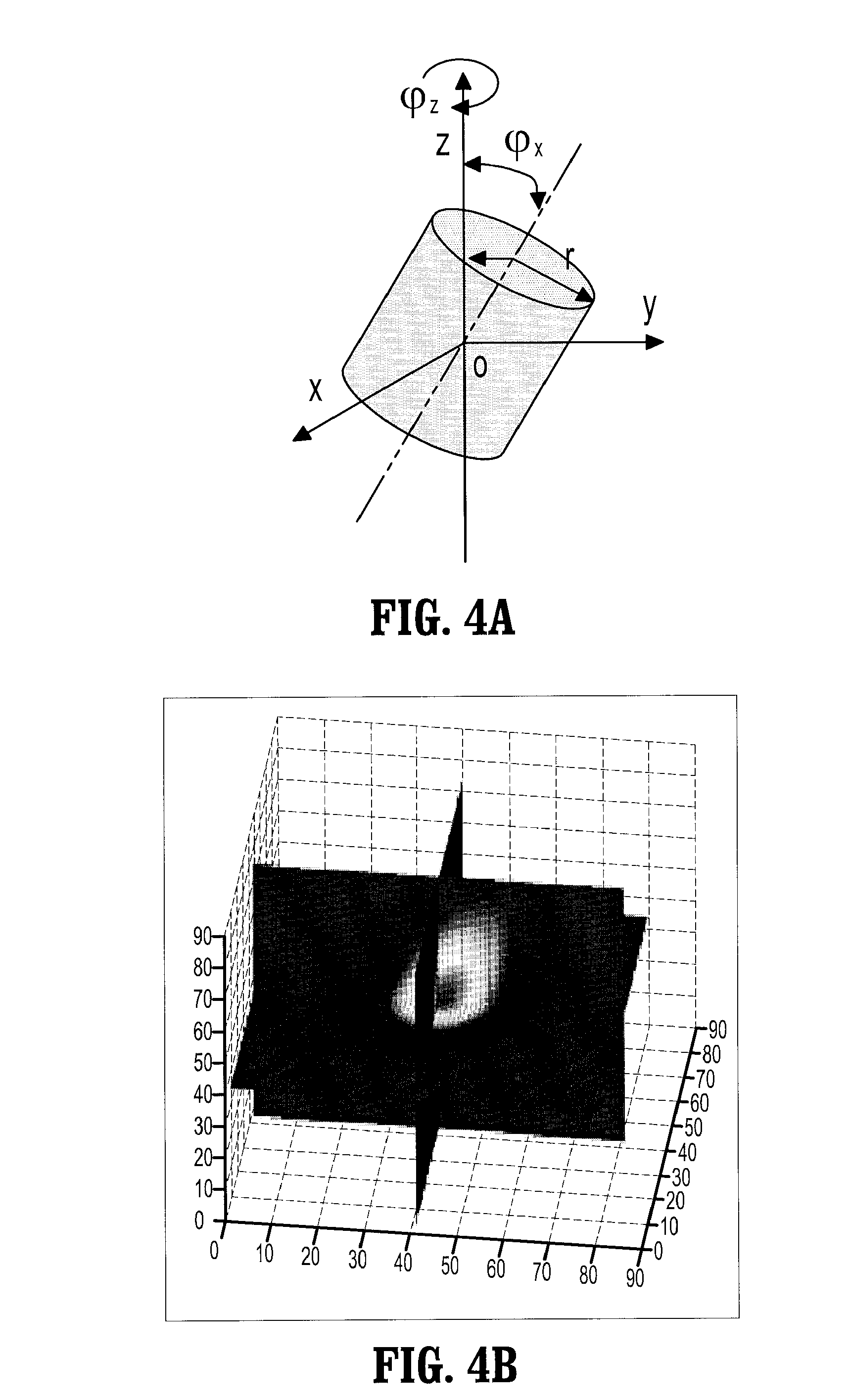
[0029]Referring to FIG. 1A, a method for segmentation includes identifying one or multiple seed points within the lumen 101, for example, as determined by an expert or by a computer program. An initial cylinder (tube) model is fitted into the colon at the seed point(s) as an in...


PUM


Abstract

A method for segmenting a tubular structure includes providing a three-dimensional image containing the tubular structure, providing at least one seed point within the tubular structure, fitting an initial cylinder into the tubular structure at the seed point, adding cylinder segments to the initial cylinder in forward and backward directions within the three-dimensional image of the tubular structure by tracking the cylinder model over the tubular structure, modeling each cylinder segment using parameters to account for a three-dimensional orientation, a radius, a length and a curvature, describing an intensity image of the tubular structure using a second model to represent an edge and intensity distribution of the tubular structure, fitting the parameters as the cylinder segment is added to the intensity image, and outputting cylinder segments as a segmentation of the tube.

Description

CROSS-REFERENCE TO RELATED APPLICATIONS[0001]This application claims the benefit of Provisional Application No. 60 / 888,393 filed on Feb. 6, 2007 in the United States Patent and Trademark Office, the contents of which are herein incorporated by reference in its entirety.BACKGROUND OF THE INVENTION[0002]1. Field of the Invention[0003]The present disclosure relates to image processing, and more particularly to a system and method for 3D segmentation in MR colonography.[0004]2. Description of Related Art[0005]MR colonography is a new technology for the accurate detection of colonic polyps. This technology is less painful for patients than conventional colonoscopy, which leads to better patient participation in screening programs of colorectal cancer. MR colonography is also considered safer than CT colonography, because MR imaging does not expose the patient to radiations. But MR images have higher noise levels and lower resolutions than CT images, which makes the tasks of image post-pr...

Claims


Application Information

Patent Timeline
IPC IPC(8): A61B5/00
CPCG06T7/0012G06T7/0083G06T7/0089G06T2207/30032G06T2207/20101G06T2207/20116G06T2207/20168G06T2207/10088G06T7/12G06T7/149Y10S128/922
Inventor QIAN, ZHENWOLF, MATTHIASBOGONI, LUCA
Owner SIEMENS MEDICAL SOLUTIONS USA INC
Why Eureka
Try Eureka