Automatic 3D segmentation and cortical surfaces reconstruction from t1 MRI

a technology of cortical surfaces and 3d segmentation, applied in the field of medical imaging, can solve problems such as requiring significant processing and generally requiring more than twenty hours

Active Publication Date: 2016-10-06
SONY CORP
View PDF0 Cites 11 Cited by
  • Summary
  • Abstract
  • Description
  • Claims
  • Application Information

AI Technical Summary

Benefits of technology

[0012]The technology presented provides an accurate and fully automated means for processing medical images, such as MRI or CT images, of the brain or other organs, such as the liver, spleen or other organs to be imaged. Medical imaging data is utilized in combination with anatomical information for the body element being imaged. In a preferred embodiment, the segmentation and reconstruction is performed on brain regions as these are perhaps the most complex, although the techniques can be generally applied to other physiological structures. In the case of medical imaging of the brain, the system automatically...

Problems solved by technology

Segmentation and reconstruction of cerebral cortical surfaces from T1-weighted magnetic resonance images (MRI) is an important issue in neuroscience and medicine.
Some reconstructing techniq...

Method used


Image

  • Automatic 3D segmentation and cortical surfaces reconstruction from t1 MRI
  • Automatic 3D segmentation and cortical surfaces reconstruction from t1 MRI
  • Automatic 3D segmentation and cortical surfaces reconstruction from t1 MRI

Examples


Embodiment Construction

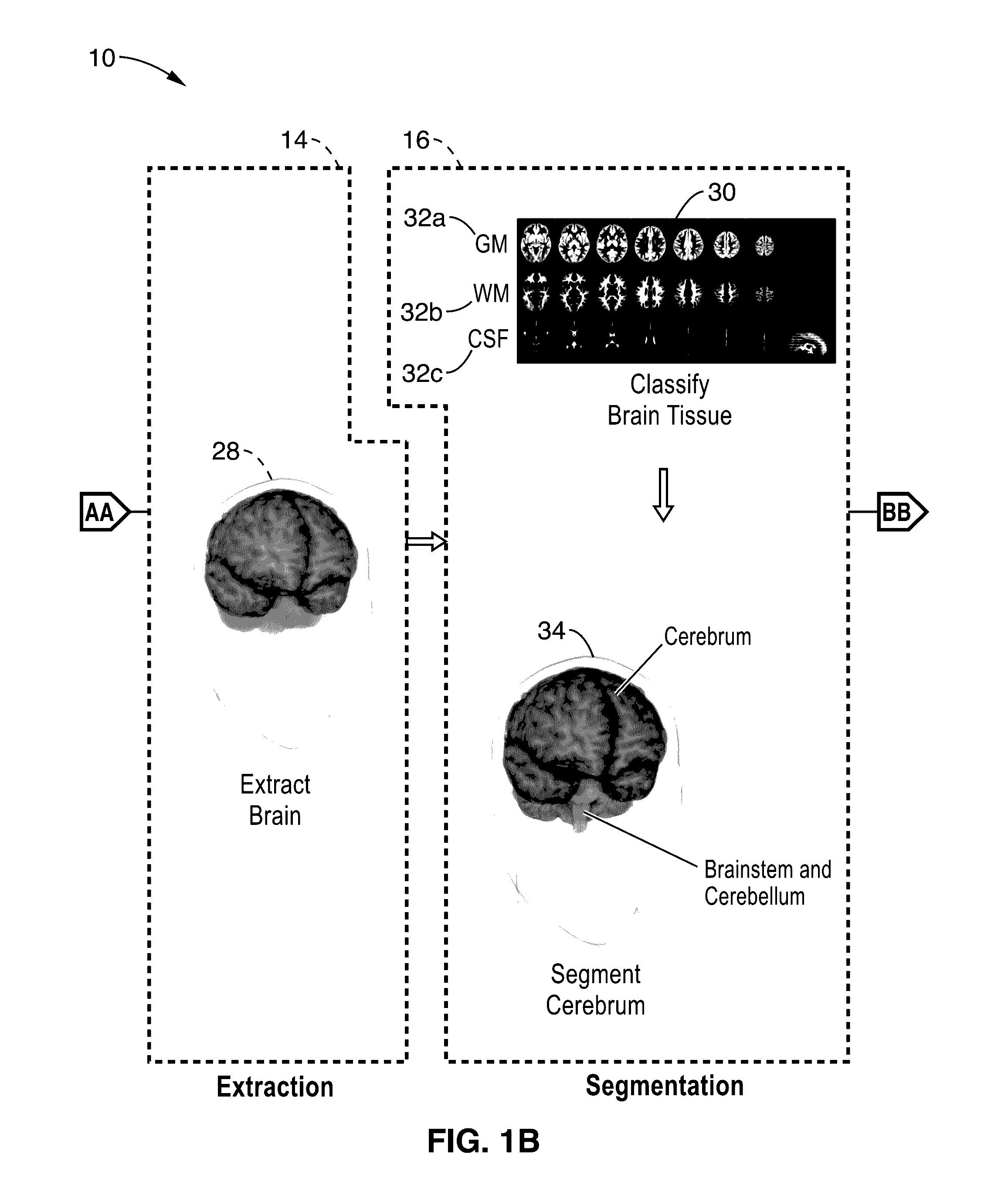
[0026]This disclosure addresses the problem of segmentation and reconstruction of cortical surfaces, including reconstructing both inner-surfaces and outer-surfaces of the cortex. The outer-surface is the pial surface, hereafter referred to Gray Matter (GM) surface. The inner-surface is gray-white boundary, hereafter referred to White Matter (WM) surface. The reconstruction of the cortical surfaces is a complex procedure which is broken into a number of subtasks.

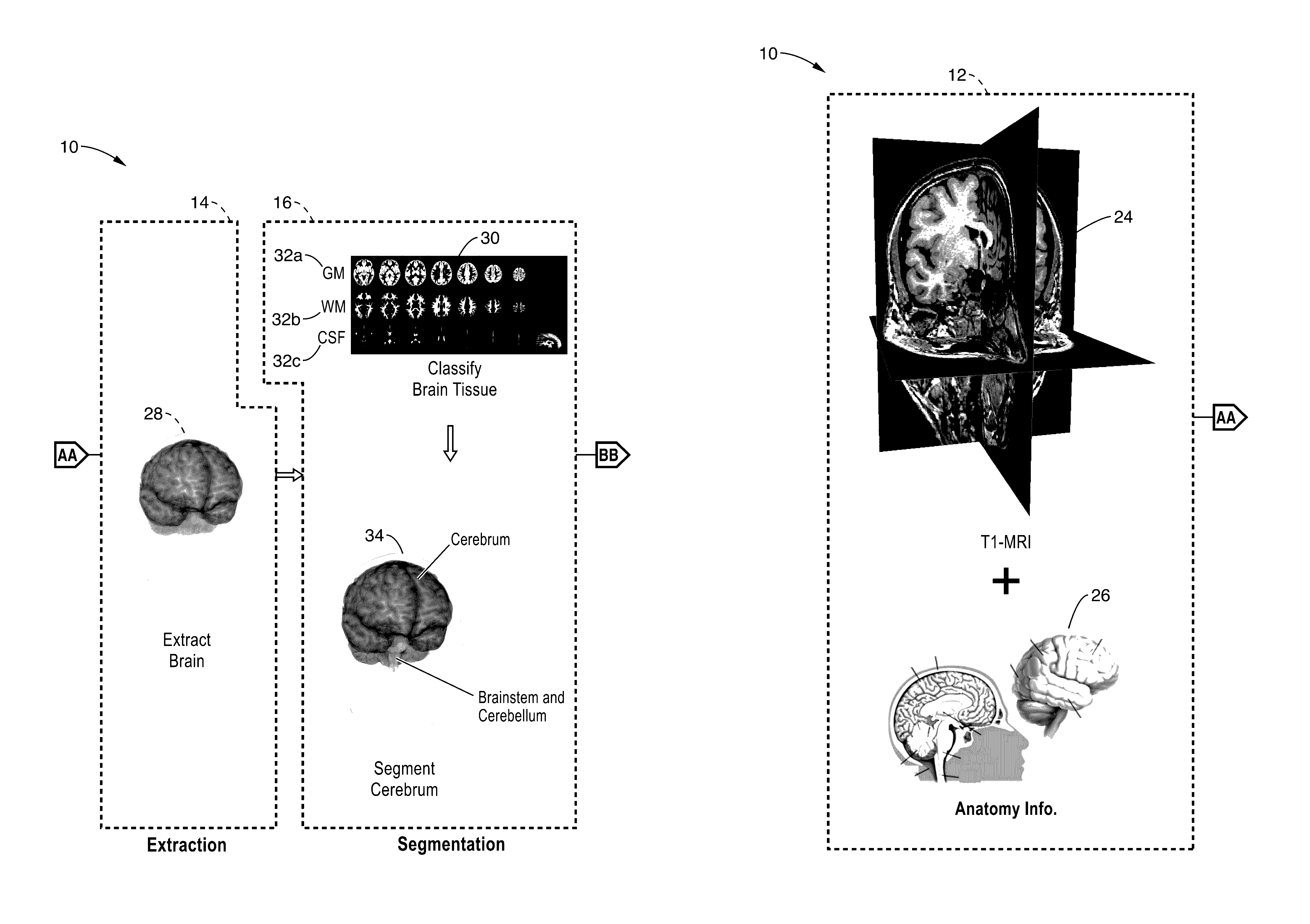
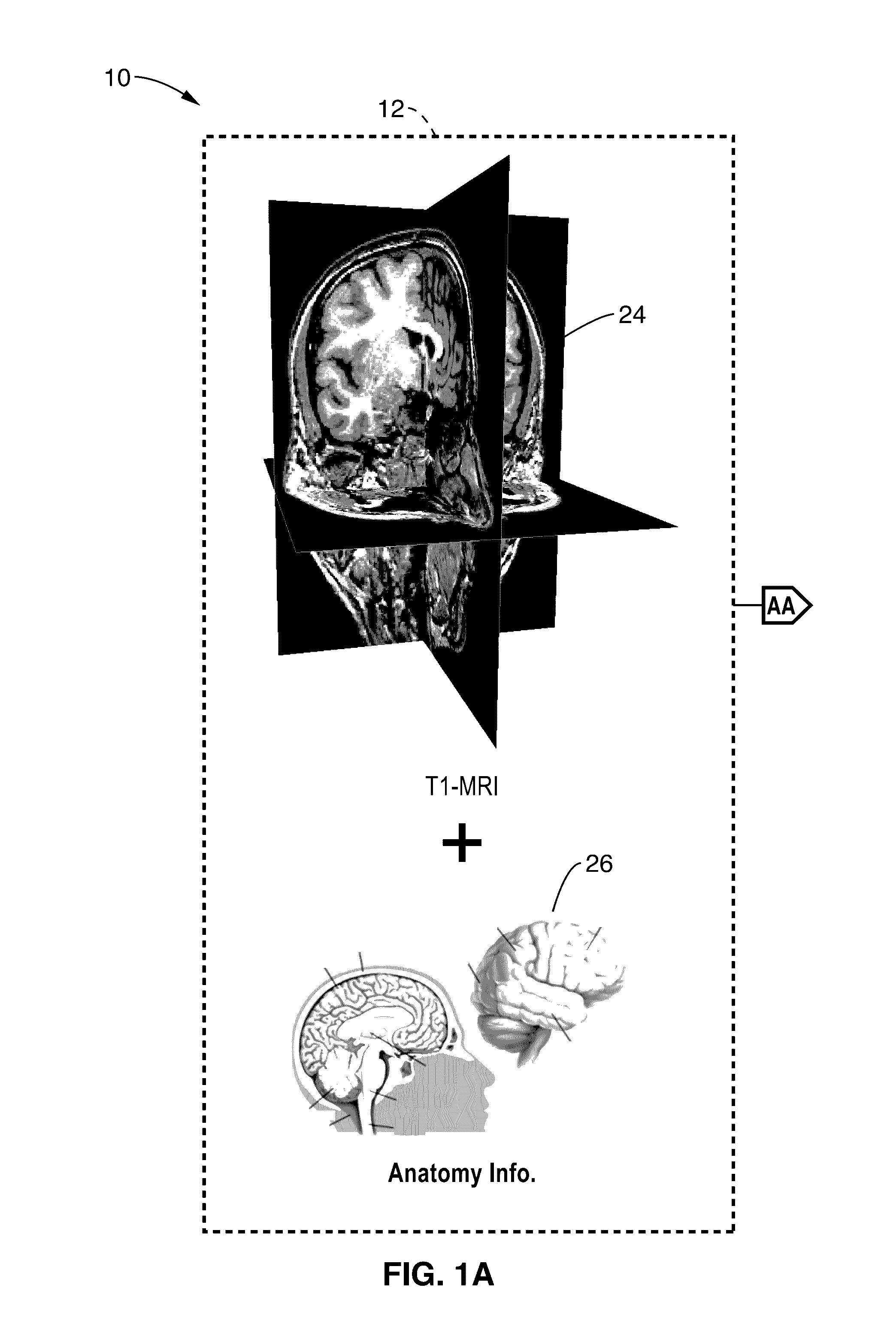
[0027]FIG. 1A through FIG. 1D illustrate an example embodiment 10 of automatic 3D segmentation and cortical surfaces reconstruction process from T1-MRI data. The process is shown in principle steps of receiving input information 12, extraction 14, segmentation 16, anatomical refinement 18, reconstruction 20, and output 22.

[0028]In FIG. 1A raw brain MRI data 24 is received from an MRI machine, with additional anatomical information 26 received, such as from a database. Extraction processing 14 commences in FIG. 1B to remove n...


PUM


Abstract

An apparatus and method for performing automatic 3D image segmentation and reconstruction of organ structures, which is particularly well-suited for use on cortical surfaces is presented. A brain extraction process removes non-brain image elements, then classifies brain tissue as to type in preparation for a cerebrum segmentation process that determines which portions of the image information belong to specific physiological structures. Ventricle filling is performed on the image data based on information from a ventricle extraction process. A reconstruction process follows in which specific surfaces, such as white matter (WM) and grey matter (GM), are reconstructed.

Description

CROSS-REFERENCE TO RELATED APPLICATIONS[0001]Not ApplicableSTATEMENT REGARDING FEDERALLY SPONSORED RESEARCH OR DEVELOPMENT[0002]Not ApplicableINCORPORATION-BY-REFERENCE OF COMPUTER PROGRAM APPENDIX[0003]Not ApplicableNOTICE OF MATERIAL SUBJECT TO COPYRIGHT PROTECTION[0004]A portion of the material in this patent document is subject to copyright protection under the copyright laws of the United States and of other countries. The owner of the copyright rights has no objection to the facsimile reproduction by anyone of the patent document or the patent disclosure, as it appears in the United States Patent and Trademark Office publicly available file or records, but otherwise reserves all copyright rights whatsoever. The copyright owner does not hereby waive any of its rights to have this patent document maintained in secrecy, including without limitation its rights pursuant to 37 C.F.R. §1.14.BACKGROUND[0005]1. Technological Field[0006]This disclosure pertains generally to medical imag...

Claims


Application Information

Patent Timeline
IPC IPC(8): G06T7/00G06K9/62G06K9/46G06T15/00G06T11/00
CPCG06T7/0012G06T15/00G06T11/003G06K9/4604G06T2207/10088G06T7/0079G06T2207/10004G06T2207/10081G06K9/6267G06T2207/30016G06T7/68G06T19/20G06T2219/028G06T7/11G06T17/205G06T7/12G06T2207/20092G06V10/44G06F18/24G06T7/10
Inventor LIU, MING-CHANGSONG, BI
Owner SONY CORP
Why Eureka
Try Eureka