
Systems and methods for deep-learning-based segmentation of composite images

Active Publication Date: 2022-07-12
EXINI DIAGNOSTICS
View PDF103 Cites 10 Cited by
  • Summary
  • Abstract
  • Description
  • Claims
  • Application Information

AI Technical Summary

Benefits of technology

This method significantly improves segmentation accuracy and hotspot detection, providing more reliable risk assessments and disease localization, especially in challenging regions like the bladder and prostate, by integrating anatomical and functional imaging data.

Problems solved by technology

The process may also involve repeating the imaging study due to inconclusive results, or ordering further tests based on initial results.

Method used


Image

  • Systems and methods for deep-learning-based segmentation of composite images
  • Systems and methods for deep-learning-based segmentation of composite images
  • Systems and methods for deep-learning-based segmentation of composite images

Examples


Embodiment Construction

[0072]It is contemplated that systems, devices, methods, and processes of the claimed invention encompass variations and adaptations developed using information from the embodiments described herein. Adaptation and / or modification of the systems, devices, methods, and processes described herein may be performed by those of ordinary skill in the relevant art.

[0073]Throughout the description, where articles, devices, and systems are described as having, including, or comprising specific components, or where processes and methods are described as having, including, or comprising specific steps, it is contemplated that, additionally, there are articles, devices, and systems of the present invention that consist essentially of, or consist of, the recited components, and that there are processes and methods according to the present invention that consist essentially of, or consist of, the recited processing steps.

[0074]It should be understood that the order of steps or order for performin...


PUM


Abstract

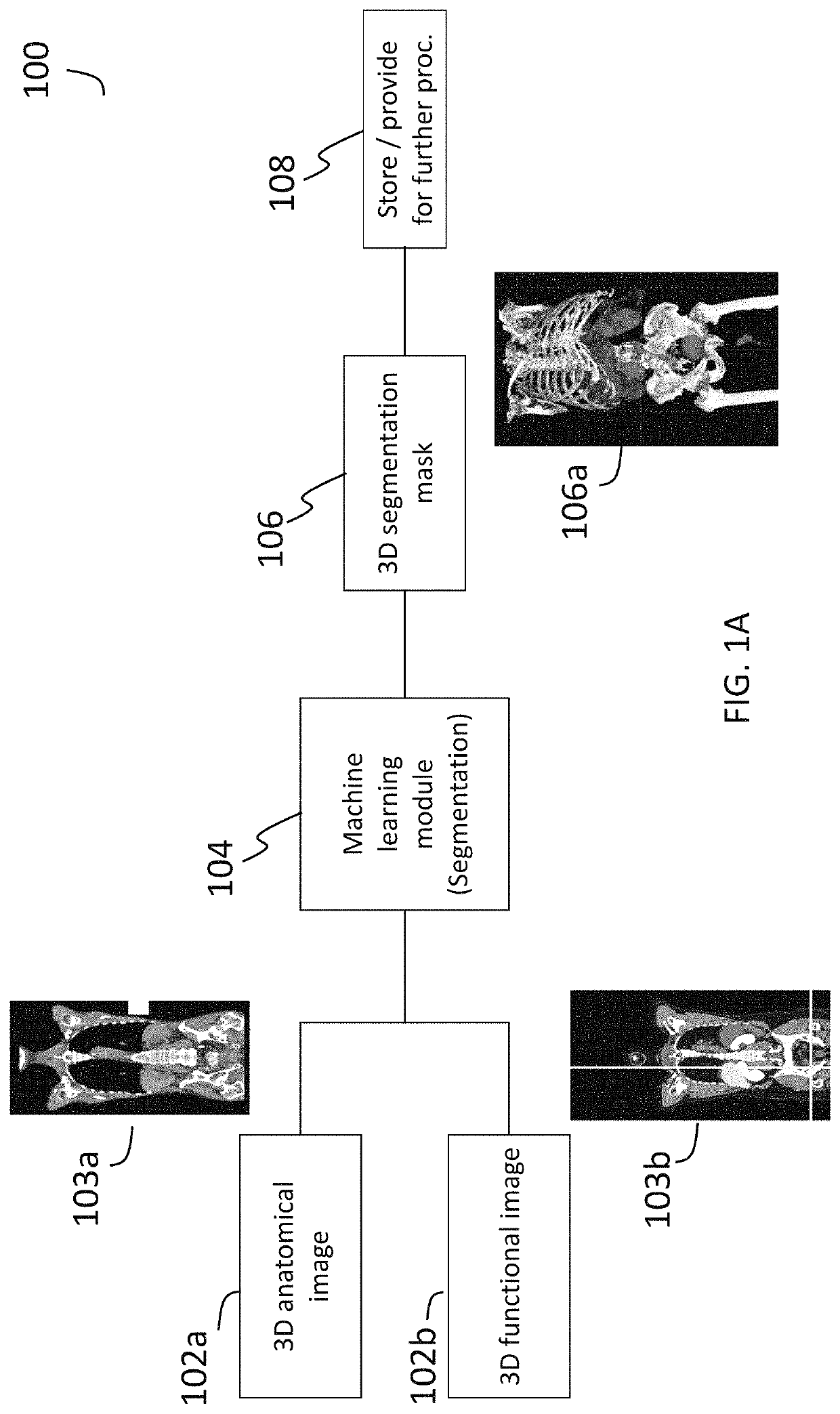
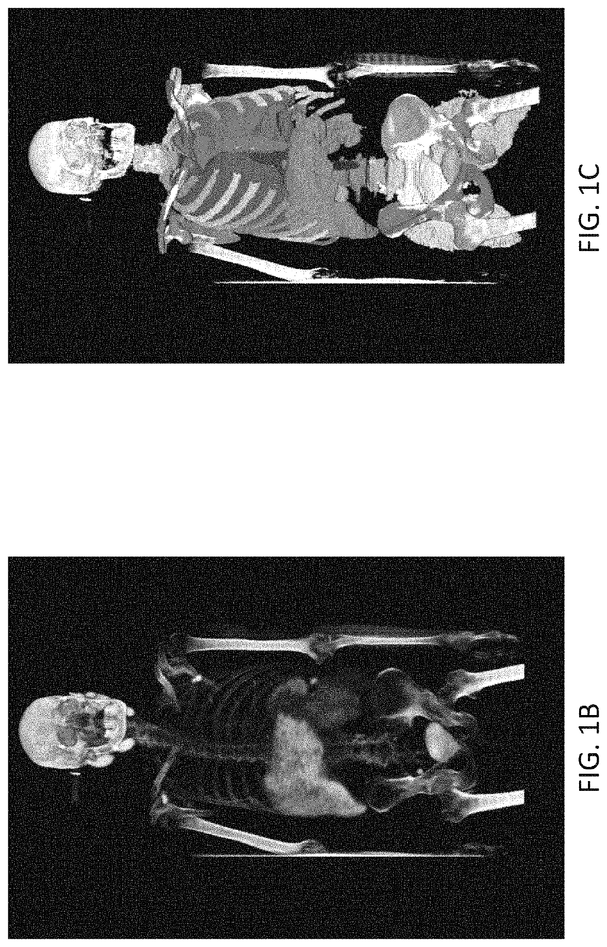
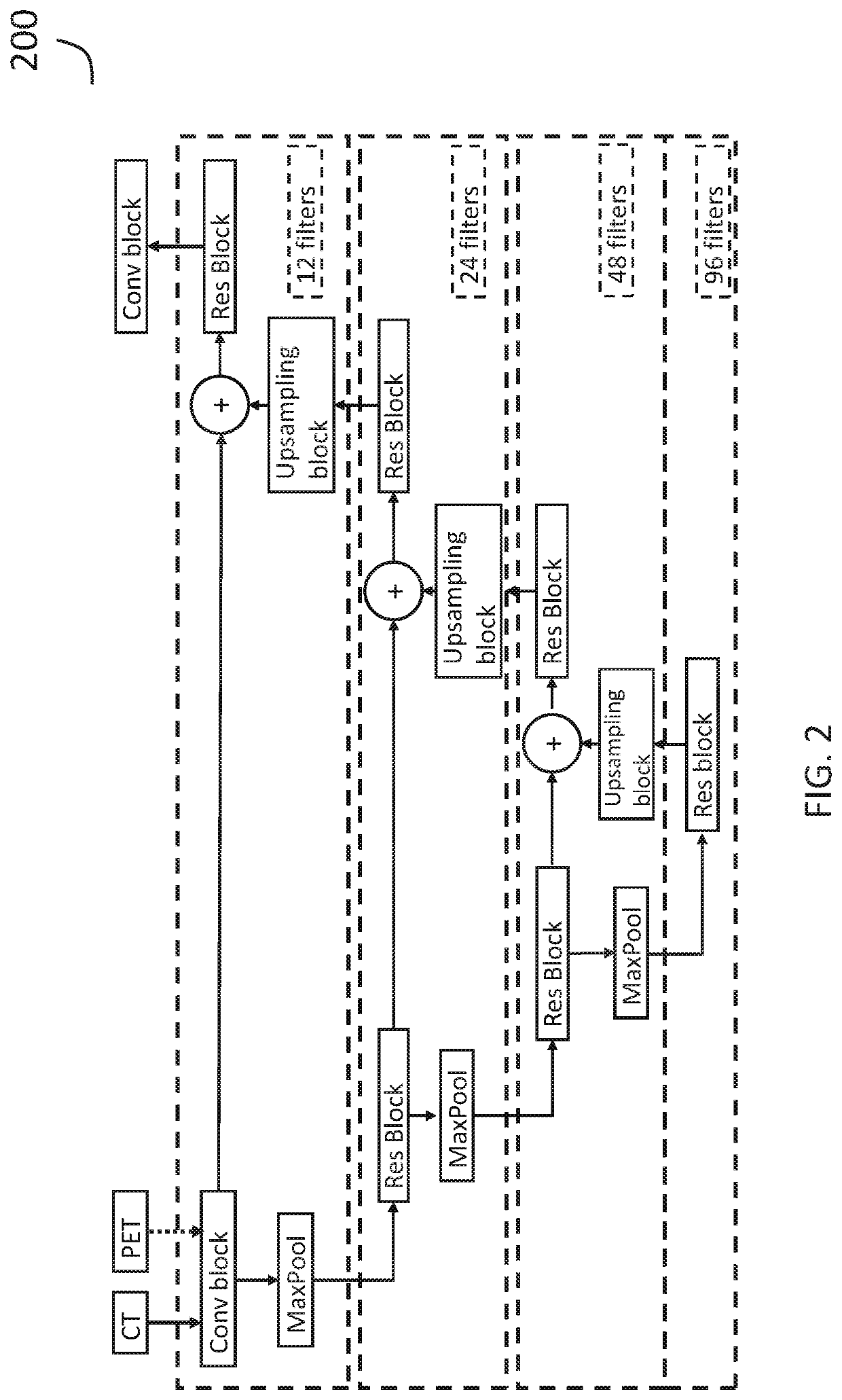
Presented herein are systems and methods that provide for improved 3D segmentation of nuclear medicine images using an artificial intelligence-based deep learning approach. For example, in certain embodiments, the machine learning module receives both an anatomical image (e.g., a CT image) and a functional image (e.g., a PET or SPECT image) as input, and generates, as output, a segmentation mask that identifies one or more particular target tissue regions of interest. The two images are interpreted by the machine learning module as separate channels representative of the same volume. Following segmentation, additional analysis can be performed (e.g., hotspot detection / risk assessment within the identified region of interest).

Description

CROSS REFERENCE TO RELATED APPLICATIONS[0001]This application is a continuation-in-part of U.S. Non-Provisional application Ser. No. 17 / 008,404, filed Aug. 31, 2020, which claims priority to and benefit of U.S. Provisional Application No. 63 / 014,637, filed Apr. 23, 2020, the content of each of which are hereby incorporated by reference in their entirety.TECHNICAL FIELD[0002]This invention relates generally to systems and methods for creation, analysis, and / or presentation of medical image data. More particularly, in certain embodiments, the invention relates to systems and methods for identifying 3D volumes of interest in medical images.BACKGROUND[0003]Nuclear medicine imaging involves the use of radiolabeled compounds, referred to as radiopharmaceuticals. Radiopharmaceuticals are administered to patients and accumulate in various regions in the body in manner that depends on, and is therefore indicative of, biophysical and / or biochemical properties of tissue therein, such as those ...

Claims


Application Information

Patent Timeline
Patent Type & Authority Patents(United States)
IPC IPC(8): G16H30/40G06T7/00G06T7/187G06N20/00G16H30/20G06N3/08G06T15/08
CPCG16H30/20G06N3/08G06N20/00G06T7/0012G06T7/187G06T15/08G16H30/40G06T2207/10081G06T2207/10108G06T17/00G06T2210/41G16H50/20G16H50/30G06T7/11G06T2207/10104G06T2207/20084G06T2207/30081G06T2207/30084G06T2207/30056G06T2207/30048G06T2207/30101G06N3/045
Inventor JOHNSSON, KERSTIN ELSA MARIABRYNOLFSSON, JOHAN MARTINSAHLSTEDT, HANNICKA MARIA ELEONORA
Owner EXINI DIAGNOSTICS
Learn More