
Automatic segmentation method for breast MRI focus based on Inter-frame correlation

An automatic segmentation and correlation technology, applied in image analysis, image data processing, instruments, etc., can solve the problems of low efficiency, noise sensitivity, uneven gray level of MRI image area, etc., to reduce manual interaction and achieve high accuracy. Effect

Inactive Publication Date: 2017-02-22
TIANJIN UNIV
View PDF4 Cites 28 Cited by
  • Summary
  • Abstract
  • Description
  • Claims
  • Application Information

AI Technical Summary

Problems solved by technology

However, various segmentation methods have their limitations. For example, the threshold method and region growth method are inefficient and sensitive to noise. Algorithms such as FCM and MRF are too dependent on data and cannot guarantee that the segmentation results are anatomically correct.
The latest research proposes a segmentation method combining two algorithms, such as a segmentation algorithm combining SLIC superpixels and level sets [11] (SLIC+ and DRLSE), this method is used for tumor segmentation of mammography images, and achieved good segmentation results. However, due to the uneven gray level of the MRI image, the experimental results of this algorithm on MRI images are not ideal
First of all, the simple linear iterative clustering algorithm (SLIC) used in this algorithm produces a large difference in the shape of superpixels, which will affect the subsequent segmentation
The fine segmentation step uses the distance regularized level set method (DRLSE), but this method needs to manually determine the sign of the constant evolution rate according to the position of the initial curve, which requires manual intervention and cannot achieve adaptive segmentation
In addition, this method only considers two-dimensional segmentation, and does not make full use of the three-dimensional image space information provided by breast MRI scanning.
On the MRI image, especially when the tumor on the start and end frames is small and the gray level is close to the surrounding tissue, the experimental results of this method are not ideal, and even the lesion cannot be segmented
Some literature incorporating inter-frame correlation [12-13] , although inter-frame information is combined, it requires manual annotation by physicians, which is not suitable for processing large amounts of data

Method used


Image

  • Automatic segmentation method for breast MRI focus based on Inter-frame correlation
  • Automatic segmentation method for breast MRI focus based on Inter-frame correlation
  • Automatic segmentation method for breast MRI focus based on Inter-frame correlation

Examples


Embodiment Construction

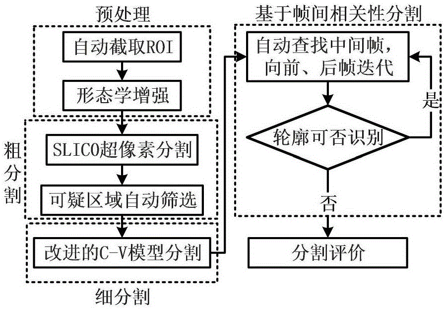
[0029] refer to figure 1 As shown, the following execution steps are included: first read the MRI image 10; then perform preprocessing 20 on the obtained image; then coarsely segment the preprocessed image 30 to determine the initial outline of the lesion; finely segment the coarsely segmented image 40, refine the edge of the lesion; finally adopt the segmentation 50 based on inter-frame correlation to improve the segmentation accuracy.

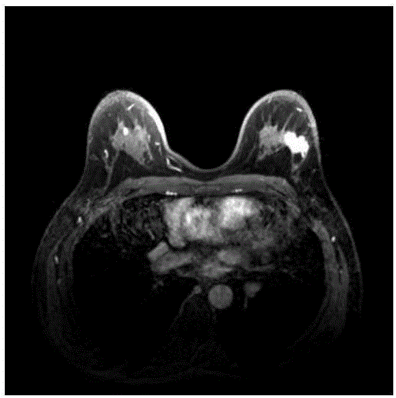
[0030] In the above steps, the breast MRI image 10 is read, and the obtained image is as follows figure 2 shown. The acquisition device of the above images is a Philips Intera Achieva 1.5T magnetic resonance scanner. The axial rapid volume acquisition dynamic enhanced fat suppression sequence (dyn_eTHRIVE) scan was used, and the relevant imaging parameters were: repetition time TR=4.4ms, echo time TE=2.2ms, flip angle FA=12°. The layer thickness is 2mm, the FOV is 100×100cm, the gray scale of the image is 12 bits, the matrix size is 352×3...


PUM


Abstract

The present invention relates to an automatic segmentation method for a breast MRI focus based on inter-frame correlation. The method comprises: reading an MRI image; preprocessing the image, and performing coarse segmentation on the preprocessed image to determine an initial contour of a focus; and by adopting an improved C- V level set model method, performing fine segmentation on a coarsely segmented image that is obtained in the prior step, further refining the tumor contour on the basis of a coarsely segmented contour, and optimizing an obtained finely segmented result by combining inter-frame correlation. The method provided by the present invention has higher accuracy and can better segment the focus.

Description

technical field [0001] The invention relates to the technical field of medical image segmentation, in particular to a breast magnetic resonance image segmentation method Background technique [0002] Breast cancer is one of the most common malignant tumors in women, which seriously threatens women's physical and mental health and life. Early detection, diagnosis and treatment of breast cancer can help improve the annual survival rate and quality of life of patients. Magnetic resonance imaging (MRI) has recognized high sensitivity (more than 90%), and has strong resolution energy for various soft tissue structures. It provides multiple sequences and images in multiple directions, and provides tissue structures and lesions for clarifying the etiology. The morphology, mass distribution and other richer image information. Breast MRI has a great auxiliary effect on the early diagnosis of breast cancer, the observation during the treatment period, the formulation of a reasonable...

Claims


Application Information

Patent Timeline
Patent Type & Authority Applications(China)
IPC IPC(8): G06T7/12G06T5/00
CPCG06T2207/30068G06T2207/20036G06T2207/10088G06T5/77
Inventor 褚晶辉王星宇吕卫
Owner TIANJIN UNIV
Learn More