Ultrasonic diagnosis apparatus, breast imaging system, and breast imaging method

a technology of ultrasound diagnosis and ultrasound image, which is applied in the field of ultrasound diagnosis apparatus, breast imaging system, and ultrasound imaging method, can solve the problems of long operation time to acquire ultrasonic image, pain in subjects to be examined, and difficulty in accurately positioning mammographic images with ultrasonic images, so as to shorten the time required to acquire ultrasonic images, reduce pain accompanying compression, and accurately position the effect of imaging

Inactive Publication Date: 2008-10-09
TOSHIBA MEDICAL SYST CORP
View PDF9 Cites 24 Cited by
  • Summary
  • Abstract
  • Description
  • Claims
  • Application Information

AI Technical Summary

Benefits of technology

[0010]The present invention has been made in consideration of the above situation, and has as its object to provide an ultrasonic diagnosis apparatus, breast imaging system, and breast imaging method which can accurately positionally associate mammographic images with ultrasonic images while shortening the time required to acquire ultrasonic images and reducing pain accompanying compression as compared with the prior art.

Problems solved by technology

However, the following problems arise in integrated breast imaging of conventional X-ray imaging and ultrasonic imaging.
For this reason, the shapes of breasts change at the time of image acquisition, and hence it is difficult to accurately positionally associate mammographic images with ultrasonic images.
This operation requires long time to acquire an ultrasonic image.
In addition, in some cases, compressing breasts with compression paddles having a plurality of holes causes pain in subjects to be examined.
This imposes heavy burden on the subjects.
In addition, compressing a breast with compression paddles having a plurality of holes may lead to a reduction in compression strength.

Method used


Image

  • Ultrasonic diagnosis apparatus, breast imaging system, and breast imaging method
  • Ultrasonic diagnosis apparatus, breast imaging system, and breast imaging method
  • Ultrasonic diagnosis apparatus, breast imaging system, and breast imaging method

Examples


first embodiment

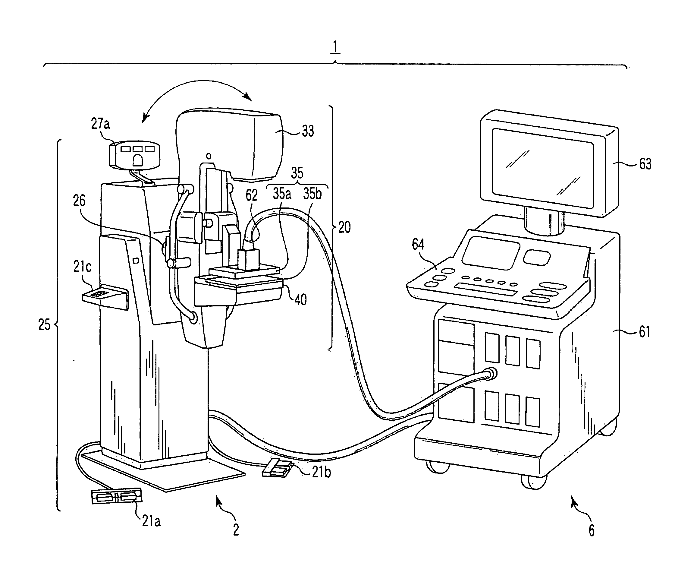
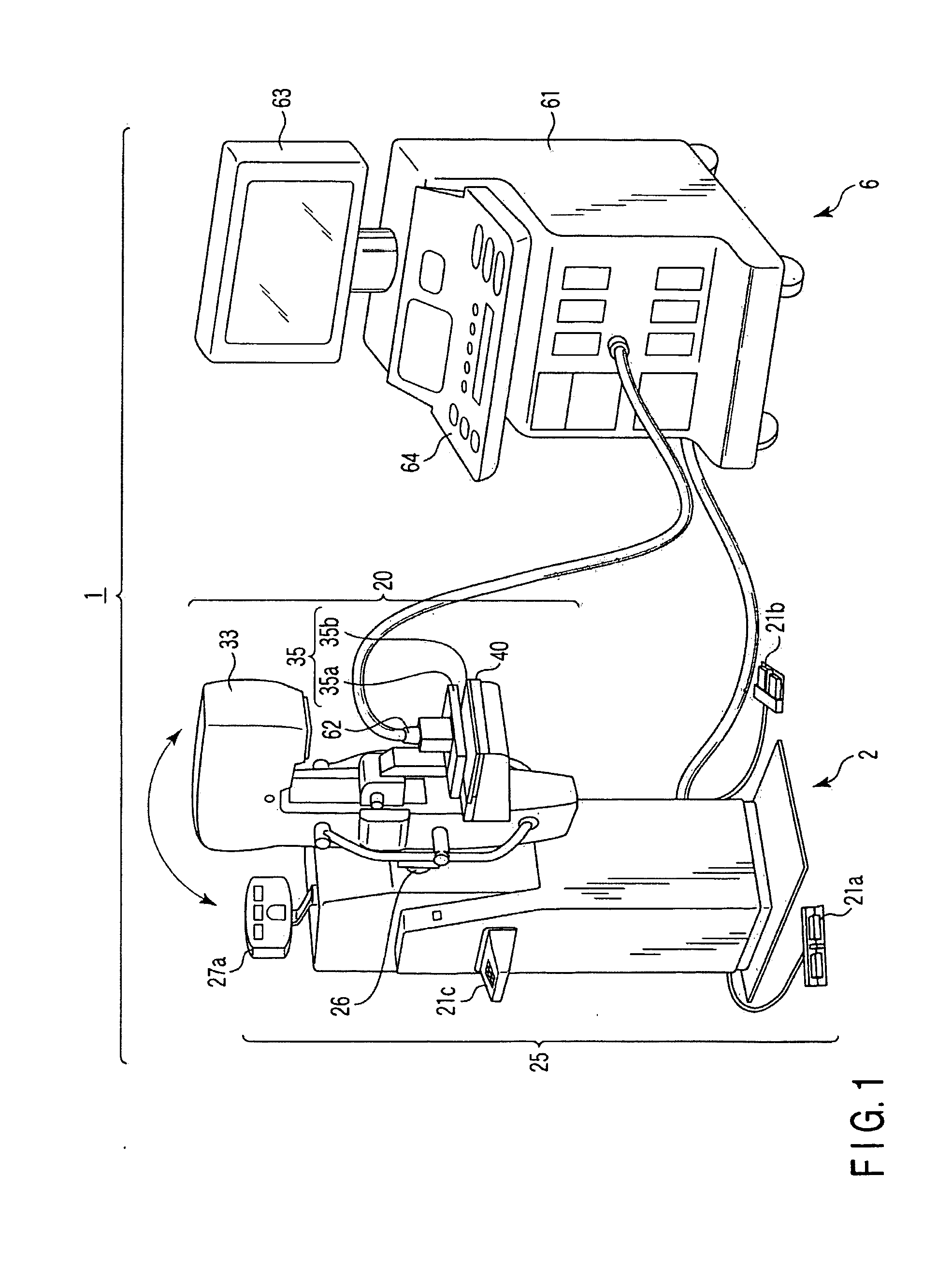
[0027]FIG. 1 is a perspective view showing the outer appearance of a breast imaging system 1 according to this embodiment. As shown in FIG. 1, the breast imaging system 1 comprises a mammographic diagnosis apparatus 2 and an ultrasonic diagnosis apparatus 6. The arrangement of each apparatus will be described below.

(Mammographic Diagnosis Apparatus)

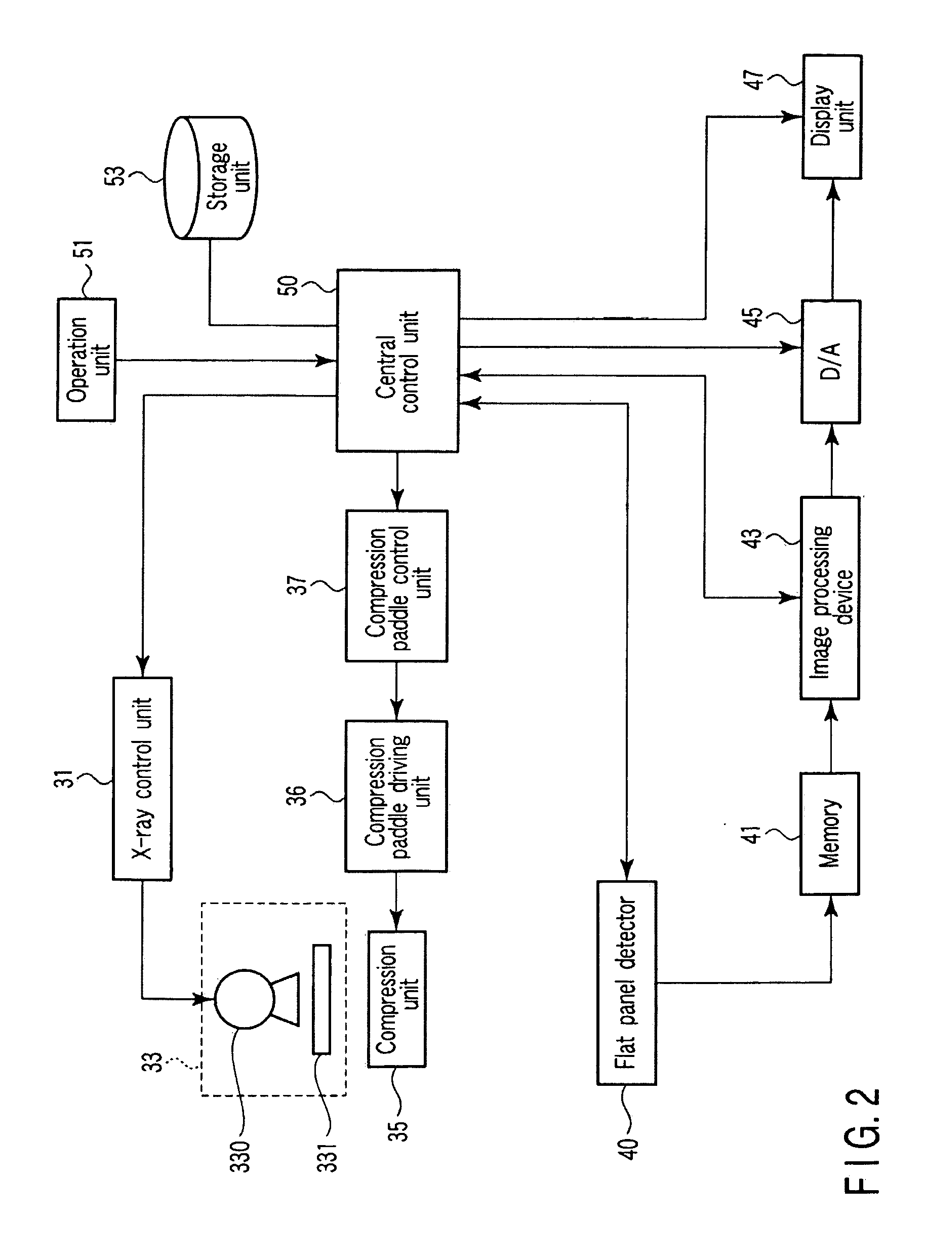
[0028]As shown in FIG. 1, the mammographic diagnosis apparatus 2 comprises an arm portion 20 and a support portion 25. The arm portion 20 is provided with an X-ray source device 33 and flat panel detector 40 which are placed to face each other, a compression unit 35 comprising upper and lower paddles 35a and 35b, and the like. The support portion 25 is provided with a display panel 27a, foot pedals 21a and 21b, a touch panel 21c, and the like. The arm portion 20 can move vertically with respect to the support portion 25 and rotate about a shaft 26. When the arm portion 20 is kept in a predetermined posture and a breast is compressed by th...

second embodiment

[0081]The second embodiment of the present invention will be described next. This embodiment exemplifies a breast imaging system 1 which implements a compressed breast image reconstruction function using a two-dimensional ultrasonic probe.

[0082]When a two-dimensional array probe having a two-dimensional array of ultrasonic transducers is to be used, volume data can be acquired by three-dimensional scanning without moving the probe along an opening portion 350. If an ultrasonic probe 62, therefore, is a two-dimensional array probe, a compression unit 35 has an opening portion 351 having a shape conforming to the two-dimensional array probe as shown in FIG. 10.

[0083]Note that an image combining unit 77 can generate an arbitrary compressed breast image by performing compressed breast image reconstruction processing for each arbitrary slice constituting acquired volume data. In addition, performing compressed breast image reconstruction processing for each slice constituting the acquire...

third embodiment

[0086]The third embodiment of the present invention will be described next. This embodiment exemplifies a breast imaging system 1 which implements a compressed breast image reconstruction function using a one-dimensional array ultrasonic probe.

[0087]When a one-dimensional array probe having a one-dimensional array of ultrasonic transducers is to be used, an ultrasonic probe 62 is placed in, for example, an opening portion 351 shown in FIG. 10, and ultrasonic transmission / reception is performed such that an ultrasonic scanning plane becomes a fan-like shape (e.g., a trapezoidal shape).

[0088]This arrangement can acquire an ultrasonic image of a desired slice of a breast without moving the probe 62 along the opening portion 350. This can further reduce pain in a breast at the time of image diagnosis.


PUM


Abstract

A compression paddle of a mammographic diagnosis apparatus is provided with an opening portion which allows an ultrasonic probe to be fitted therein and slide. This apparatus performs ultrasonic scanning on a breast compressed / fixed to the compression paddle, and reconstructs a compressed breast image by using the obtained ultrasonic image as a virtual image, thereby acquiring an ultrasonic image and volume data of the compressed / fixed breast.

Description

CROSS-REFERENCE TO RELATED APPLICATIONS[0001]This application is based upon and claims the benefit of priority from prior Japanese Patent Application No. 2007-099691, filed Apr. 5, 2007, the entire contents of which are incorporated herein by reference.BACKGROUND OF THE INVENTION[0002]1. Field of the Invention[0003]The present invention relates to a system used for combined breast imaging of X-ray imaging and ultrasonic imaging, an ultrasonic diagnosis apparatus used for the integrated imaging, and a breast imaging system.[0004]2. Description of the Related Art[0005]An ultrasonic diagnosis technique can display, in real time, how a heart beats or a fetus moves, with simple operation of bringing an ultrasonic probe into contact with the body surface. In addition, this technique offers a high level of safety, and hence can be repeatedly used for examination. Furthermore, the system size is smaller than those of other diagnosis apparatuses such as X-ray, CT, and MRI apparatuses. Theref...

Claims


Application Information

Patent Timeline
Patent Type & Authority Applications(United States)
IPC IPC(8): A61B6/00A61B8/14
CPCA61B6/0414A61B6/502A61B6/5247A61B8/483A61B8/4416A61B6/4417A61B8/0825A61B8/403A61B8/4405A61B8/5207
Inventor OKAMURA, YOKOKAMIYAMA, NAOHISASUGIYAMA, ATSUKOYAMAGATA, HITOSHINISHIKI, MASAYUKIKURATOMI, NAOKOSEO, YASUTSUGU
Owner TOSHIBA MEDICAL SYST CORP
Why Eureka
Try Eureka