
Sigmoidoscope With Integral Obturator

a sigmoidoscope and integral technology, applied in the field of medical instruments, can solve the problems of inadvertent contact with non-disposable parts, remained in relation to the obturator, and potential cross-infection of the reusable components of the sigmoidoscope, and achieve the effect of effectively insulating the reusable light sour

Inactive Publication Date: 2008-11-06
DALTRAY
View PDF11 Cites 36 Cited by
  • Summary
  • Abstract
  • Description
  • Claims
  • Application Information

AI Technical Summary

Benefits of technology

[0020]In the preferred embodiment, the engagement formations include a spiral drive slot formed in the retractable tubular member, and at least one complementary drive boss extending radially outwardly from the inner tubular member for engagement with the drive slot, such that relative rotation between the inner and outer tubular members progressively effects a corresponding axial displacement of the retractable tubular member between the retracted and the extended positions. Preferably, the outer tubular member includes at least one complementary locating formation to prevent relative rotation between the retractable tubular member and the outer tubular member while the inner and outer members rotate relative to one another.
[0025]The forward end of the obturation tube is preferably configured such that in the extended configuration, the domed head provides a substantially closed, rounded tip protruding smoothly and generally continuously beyond the forward end of the speculum, thereby to provide an integral obturator adapted to facilitate initial insertion of the speculum, without requiring subsequent withdrawal and disposal of a discrete obturating element during the examination procedure.
[0033]Preferably, the instrument further includes a gas-tight observation window disposed to prevent passage of the insufflation medium from the lumen of the speculum to the connection means for the light source. Preferably also, the instrument includes contamination prevention means, effectively insulating the reusable light source from exposure to any contaminated insufflation medium.

Problems solved by technology

The fibre-optic head together with the hinged window mounted thereto and the fibre-optic illumination means are relatively expensive components and are therefore not disposed of, but rather are retained for use with another speculum.
This practice gives rise to the potential for cross-infection from the reusable components of the sigmoidoscope, most notably the insufflation bulb and the fibre optic head.
However, a problem remained in relation to the obturator, which has not hitherto been addressed by the applicant's earlier invention, or the prior art.
However, a fundamental problem with known sigmoidoscope designs is that as the obturator is withdrawn, it can and in practice almost inevitably does inadvertently come into contact with non-disposable parts of the sigmoidoscope, or with disposable parts that subsequently contact non-disposable parts.
This creates significant potential for cross contamination.
A further problem is that when inserting the instrument or subsequently during the examination procedure, faecal matter may partially or completely occlude the open end of the speculum and hence obstruct the view.
This is inconvenient, time-consuming and gives rise to further potential for contamination of the instrument.
While addressing the problem of the potential for initial contamination from the obturator, this arrangement is not necessarily effective in clearing the insertion end of the speculum should it become occluded by faecal matter during a sigmoidoscopy procedure, and also presented other problems.
In the insertion configuration, longitudinal gaps or slots remain between the petals, bounded by the relatively sharp edges of the petals, giving rise to the possibility of pinching of the tissue of the patient.
This in turn gives rise to the possibility of discomfort or even perforation of the bowel, with potentially serious consequences.
Although these problems have been described in the context of sigmoidoscopy, it will be appreciated that similar difficulties may arise in analogous medical procedures.

Method used


Image

  • Sigmoidoscope With Integral Obturator
  • Sigmoidoscope With Integral Obturator
  • Sigmoidoscope With Integral Obturator

Examples


Embodiment Construction

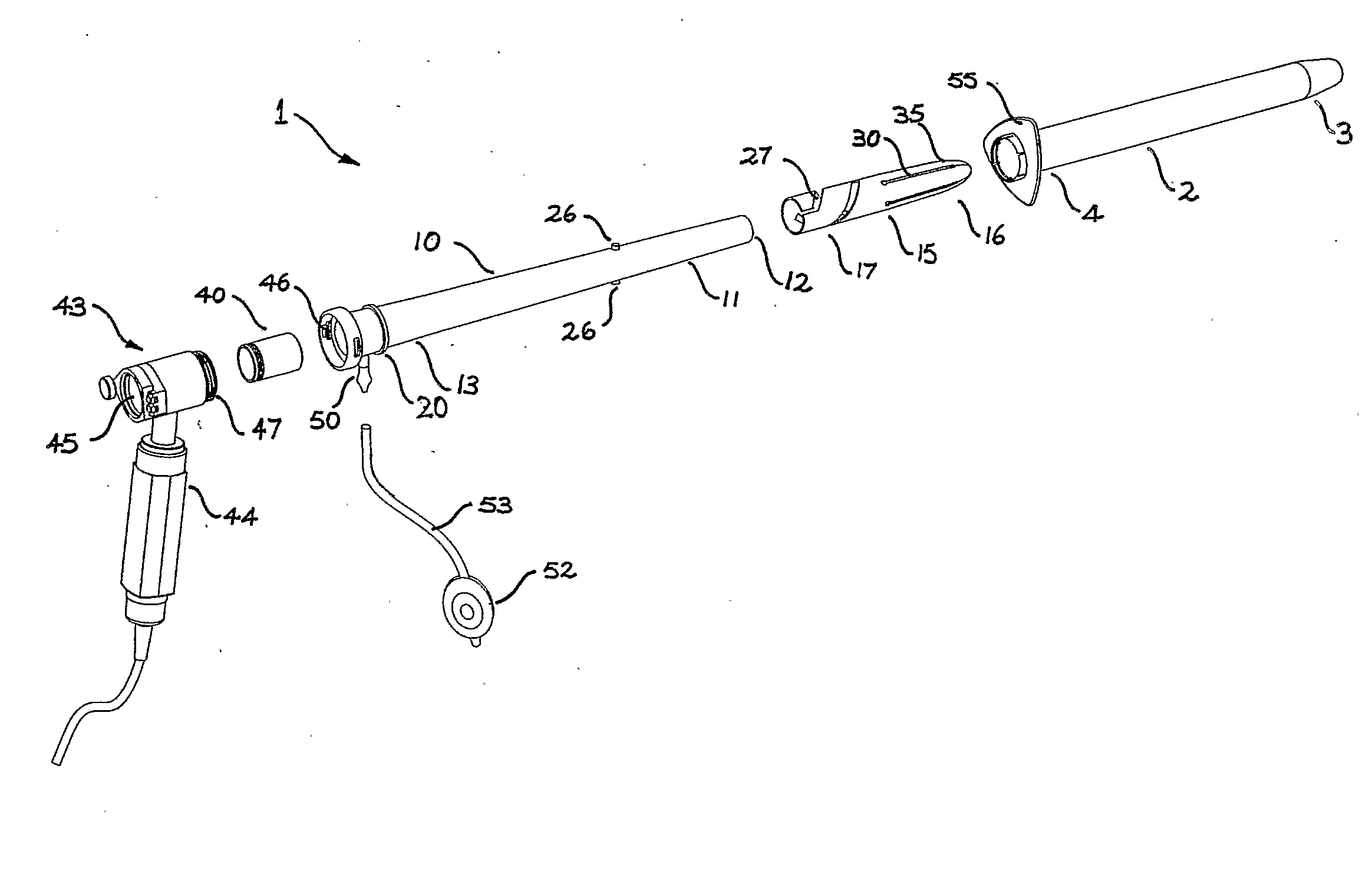
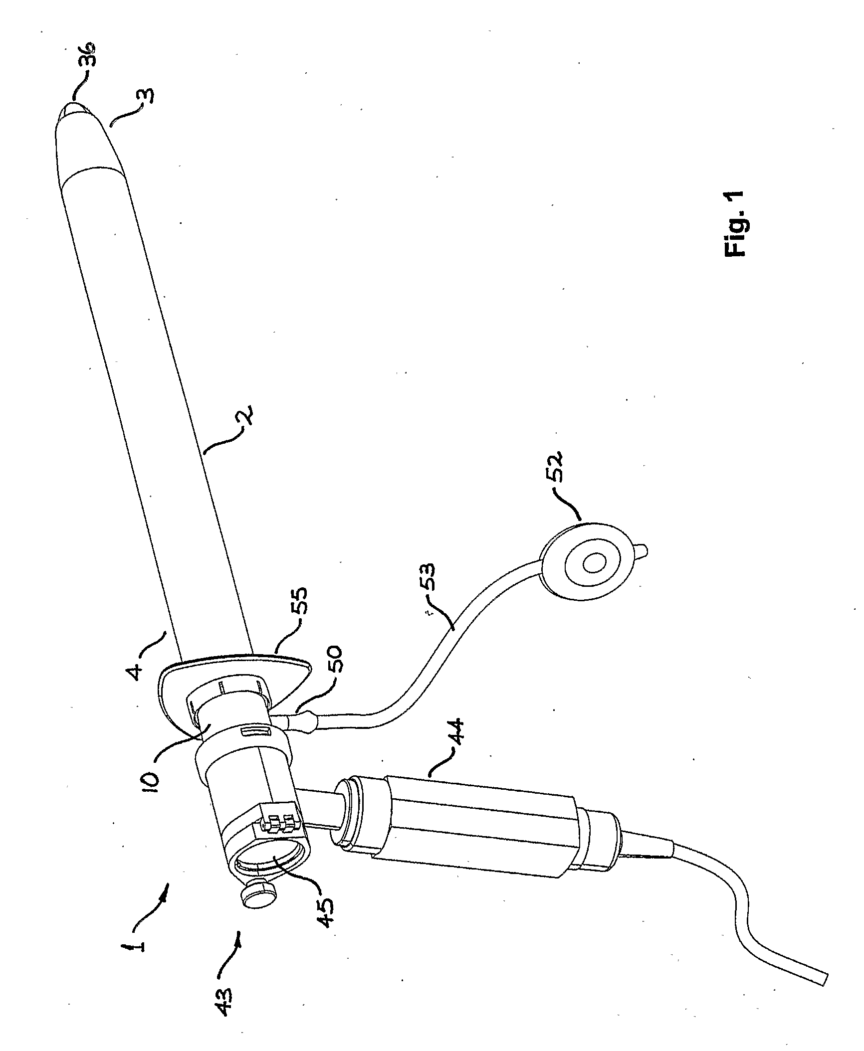
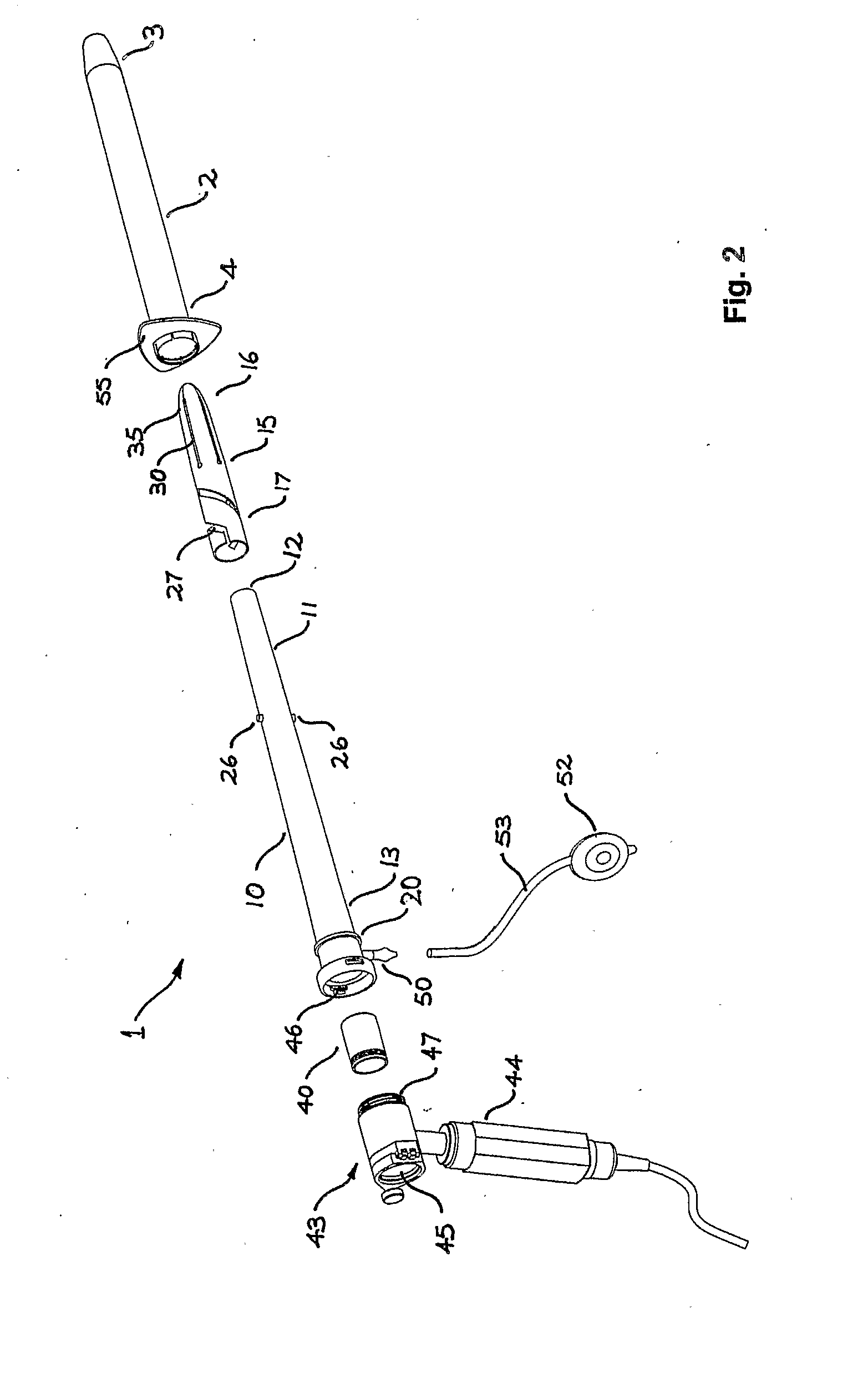
[0050]Referring to the drawings, the invention provides a medical instrument adapted for use as a sigmoidoscope 1. The sigmoidoscope includes an outer tubular member in the form of a speculum 2, having a forward insertion end 3 and a rearward observation end 4. An inner tubular member in the form of tapered inner guide tube 10 incorporates a forward end 11 including an outer guide rim 12 and a rearward end 13. The guide tube is disposed substantially within the speculum 2, with the forward end 11 recessed well behind the forward end 3 of the speculum.

[0051]The sigmoidoscope 1 further includes a retractable tubular member in the form of obturation tube 15, having a forward obturation end 16 and a rearward driven end 17. The obturation tube 15 is movable between a retracted configuration in which the forward end 16 is positioned substantially within the speculum as shown in FIG. 11, and an extended configuration in which the forward end protrudes longitudinally beyond the forward end ...


PUM


Abstract

A medical instrument used as a sigmoidoscope (1) for internal examination of the rectum and sigmoid colon of a patient. The instrument includes an outer tubular member in the form of a speculum (2) having a forward insertion end (3) and a rearward observation end (4). An inner tapered guide tube (10) is disposed at least partially within the speculum (2). A retractable obturation tube (15) is disposed between the speculum (2) and the inner guide tube (10) for relative movement between a retracted position substantially within the speculum (2) and an extended position in which the obturation tube (15) protrudes longitudinally beyond the forward end (3) of the speculum (2).

Description

FIELD OF THE INVENTION[0001]This invention relates to medical instruments, and in particular to an instrument adapted for insertion into bodily cavities. The invention has been developed primarily as a sigmoidoscope for use in examination of the bowel cavity of a patient and for sigmoidoscopy generally. It will be appreciated, however, that the invention is not limited to this particular field of use.BACKGROUND OF THE INVENTION[0002]The following description of the prior art is provided to place the invention in an appropriate technical context, and to enable the advantages flowing from it to be fully appreciated. References to the prior art should not, however, be interpreted as indicating that such art was well known, or formed part of the common general knowledge, in the relevant field at the priority date.[0003]It is frequently necessary for specialised medical practitioners to be required to inspect the colon or rectum of a patient. This procedure is commonly conducted with a r...

Claims


Application Information

Patent Timeline
IPC IPC(8): A61B1/31A61B1/32A61B17/02A61B17/34A61B19/00
CPCA61B1/31A61B1/32A61B17/0218A61B17/3439A61B19/38A61B90/40
Inventor LUBOWSKI, DAVIDMARSH, PETER CRISPIN
Owner DALTRAY
Learn More