Apparatus and method for endothelial keratoplasty donor tissue transport and delivery

a technology of endothelial keratoplasty and donor tissue, applied in the field of medical instruments, can solve the problems of poor wound strength, fast failure rate of cornea transplantation, and retained surface sutures, and achieve the effect of improving the manipulation of donor tissue and minimizing endothelial layer damag

Inactive Publication Date: 2008-11-13
ENSION +1
View PDF13 Cites 42 Cited by
  • Summary
  • Abstract
  • Description
  • Claims
  • Application Information

AI Technical Summary

Benefits of technology

[0025]It is an object of the present invention to provide an apparatus for endothelial keratoplasty donor tissue transport and delivery and method of using same that minimizes endothelial layer damage of the donor tissue 30 and improves manipulation of the donor tissue 30 for proper positioning.

Problems solved by technology

This PK procedure has been shown to yield healthy donor tissue with good endothelial layer 14 function, but this procedure has the inherent problems of unpredictable surface topography, retained surface sutures, and poor wound strength.
The process of inserting the donor corneal button 30 may be very traumatic to endothelial cells and might possibly cause faster failure rates for the cornea transplant.
Therefore, any increase in cell loss incurred in the surgical procedure reduces the success of the procedure.
The outcome of this condition results in loss of the eye in the majority of the cases.
Sutures are well known to induce infections as a result of bacterial organisms tracking through the suture tract to inside the cornea and inducing serious vision threatening infections.
Thus the corneal sutures are prone to breakage after any amount of trauma to the eye.
DSAEK can result in an increased risk of endothelial cell damage.
In addition, as a result of using stromal tissue in the transplantation process, haze at the junction between the donor and recipient corneal stroma can occasionally reduce maximum visual acuity attained.
This system does not cure the deficiencies of the prior art.

Method used


Image

  • Apparatus and method for endothelial keratoplasty donor tissue transport and delivery
  • Apparatus and method for endothelial keratoplasty donor tissue transport and delivery
  • Apparatus and method for endothelial keratoplasty donor tissue transport and delivery

Examples


Embodiment Construction

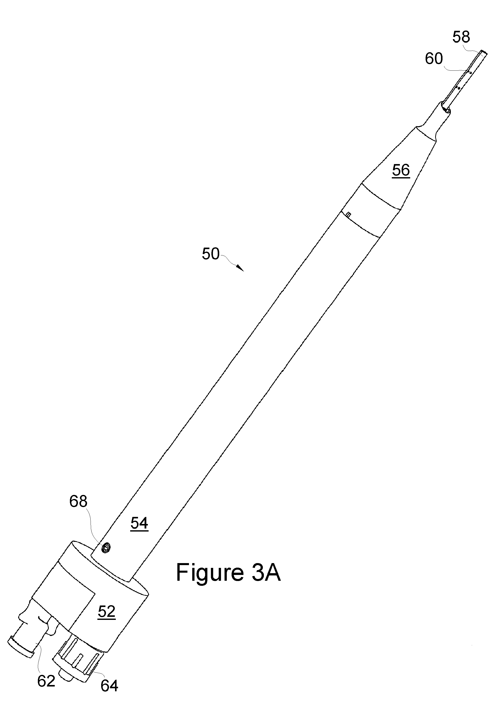
[0057]FIGS. 3a-c are perspective views of an apparatus or instrument 50 for endothelial keratoplasty donor tissue transport and delivery in accordance with one embodiment of the present invention. As will be evident in this application the apparatus 50 is applicable for current DLEK, DSEK and DSAEK procedures, and will likely be useful for future modifications of these techniques. Consequently the apparatus 50 is not intended to be limited to these procedures as will be evident to those of ordinary skill in the art.

[0058]The apparatus 50 includes an elongated actuator 52 or apparatus body upon which a device shaft or sleeve 54 is slidably received as described below. A replaceable insertion tip 56 is provided at a distal end of the shaft 54 with the insertion tip 56 having a reduced distal end. The insertion tip 56 is movable relative to the actuator 52 through the movement of the sleeve 54.

[0059]An arcuate shaped tissue holder 58 is attached to the actuator 52, whereby the tip 56 i...


PUM


Abstract

A method for endothelial keratoplasty donor tissue transport and delivery to a recipient's eye, such as in DLEK, DSEK and DSAEK procedures, comprises the steps of: attaching endothelial keratoplasty donor tissue to a tissue holder, wherein the attachment between the tissue holder and the donor tissue is not on the endothelial layer of the endothelial keratoplasty donor tissue; placing the tissue holder and the attached donor tissue within a surrounding insertion tip; transporting the insertion tip to a position adjacent an access opening in the recipient's eye; inserting the tissue holder with the attached donor tissue through the opening and extending the tissue holder from within the insertion tip to position the donor tissue within the recipient's eye; and separating the attached donor tissue from the tissue holder with the tissue holder within the recipient's eye. An associated apparatus is disclosed.

Description

RELATED APPLICATIONS[0001]The Present application claims the benefit of U.S. Provisional Patent Application Ser. No. 60 / 917,627 entitled “Apparatus and Method for Endothelial Keratoplasty Donor Tissue Transport and Delivery” filed May 11, 2007 and U.S. Provisional Patent Application Ser. No. 60 / 917,359 entitled “Instrument for Descemet Stripping with Automated Endothelial Keratoplasty” filed May 11, 2007.BACKGROUND OF THE INVENTION[0002]1. Field of the Invention[0003]The present invention relates to medical instruments, and more particularly to such instruments for inserting tissue into the eye, such as an apparatus for endothelial keratoplasty donor tissue transport and delivery and method of using same.[0004]2. Background Information[0005]The cornea 10 is the optical window to the eye 12 and must remain clear to achieve excellent vision. FIG. 1 illustrates the elements of the eye 12 and cornea 10 for the understanding of the present invention. The most internal layer of the cornea...

Claims


Application Information

Patent Timeline
Patent Type & Authority Applications(United States)
IPC IPC(8): A61F9/007
CPCA61F2/14A61F2/142A61F9/0017A61F9/007
Inventor MILLER, JASONSPEAKMAN, JEFFREY W.AL-GHOUL, AHMEDDHALIWAL, DEEPINDER
Owner ENSION
Why Eureka
Try Eureka