Non-Rotating Transaxial Radionuclide Imaging

a radionuclide and transaxial technology, applied in the field of radionuclide imaging, can solve the problem that the gantry is a relatively expensive subsystem of the diagnostic tool, and achieve the effect of reducing cost and efficient manufacturing

Active Publication Date: 2009-01-01
SIEMENS MEDICAL SOLUTIONS USA INC
View PDF1 Cites 63 Cited by
  • Summary
  • Abstract
  • Description
  • Claims
  • Application Information

AI Technical Summary

Benefits of technology

[0009]An advantage of the present invention is a collimator for use in medical imaging, particularly SPECT imaging, wherein the collimator is structured so that it is not necessary to rotate the radiation detectors relative to the patient.
[0010]Another advantage of the present invention is a nuclear medical imaging device that can be manufactured efficiently and at a significantly reduced cost.
[0011]A further advantage of the present invention is a radionuclide imaging method comprising linearly displacing a patient along a longitudinal axis without relative rotation between the radiation detectors and patient.
[0012]Additional advantages and other features of the present invention will be set forth in the description which follows and in part will be apparent to those having ordinary skill in the art upon examination of the following or may be learned from the practice of the present invention. The advantages of the present invention may be realized and obtained as particularly pointed out in the appended claims.
[0013]According to the present invention, the foregoing and other advantages are achieved in part by a collimator comprising a plurality of adjacently stacked collimating segments sharing a common central axis, wherein, adjacent collimating segments are annularly displaced from one another about the common central axis.
[0014]Another advantage of the present invention is a radionuclide imaging method comprising: positioning a patient having a radiating mass within a single photon emission computed tomography (SPECT) system and moving the patient along a longitudinal axis, the SPECT system comprising: a polygonal-shaped radiation detector; and a collimator on at least two sides of the polygonal-shaped detector, wherein the collimator comprises a plurality of adjacently stacked collimating segments sharing a common central axis, each segment having a plurality of apertures extending therethrough, wherein the adjacent collimating segments are angularly displaced from one another about the common central axis; wherein each aperture forms a passageway for radiation rays emanating from the radiating mass in a direction substantially aligned with a longitudinal axis of the respected passageway so that aligned radiation rays strike the detector; and motive means for effective longitudinal relative motion between the detector and the patient for taking multi-angular SPECT radiation sampling of the radiating mass in the patient utilizing the passageways, without relative rotation between the patient and detector.

Problems solved by technology

A drawback associated with this requirement, however, is that such gantries are relatively expensive subsystems of the diagnostic tool, because they must be capable of providing rapid rotation of large, heavy camera heads through very precise orbits about the patient.

Method used


Image

  • Non-Rotating Transaxial Radionuclide Imaging
  • Non-Rotating Transaxial Radionuclide Imaging
  • Non-Rotating Transaxial Radionuclide Imaging

Examples


Embodiment Construction

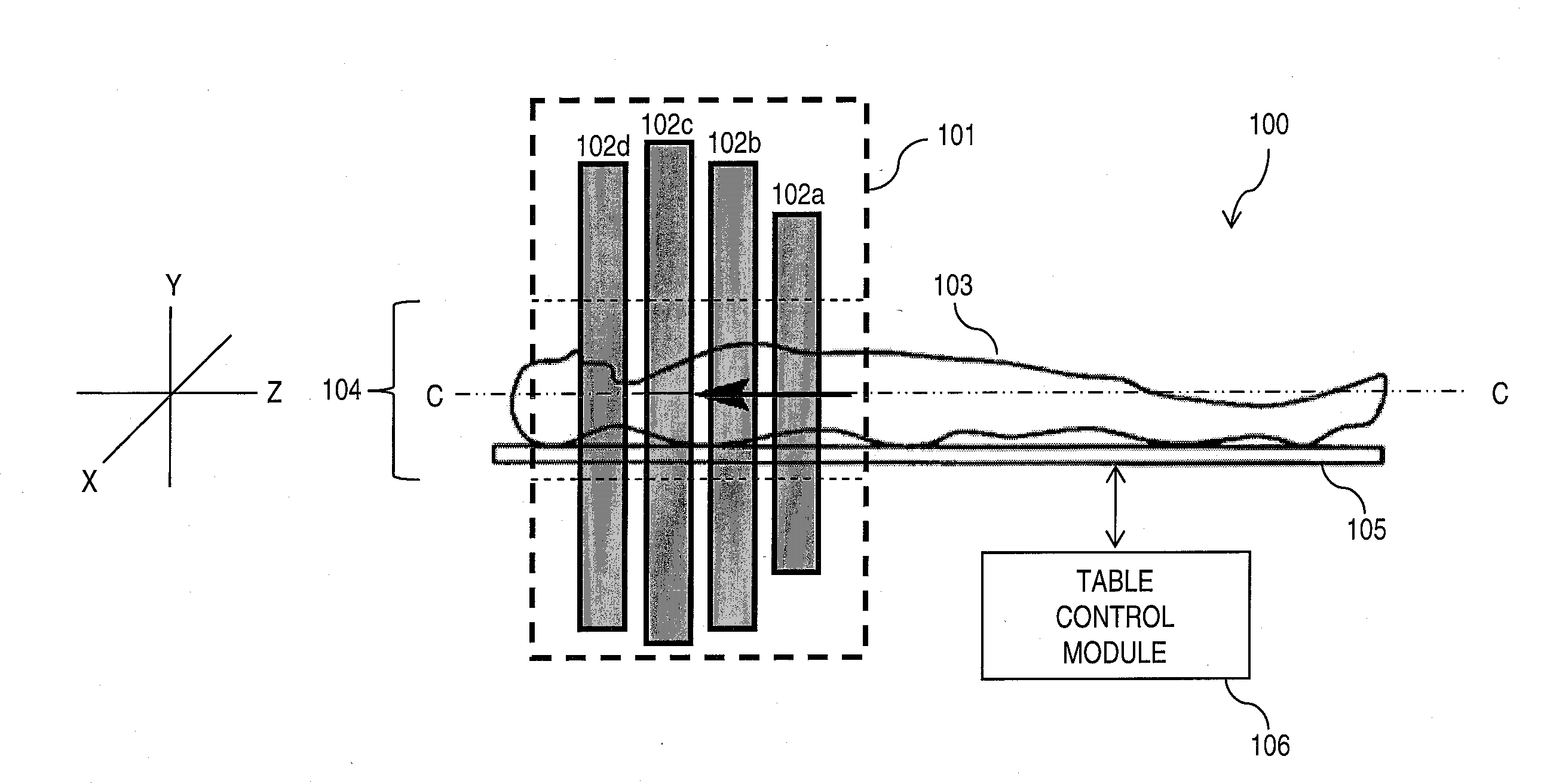
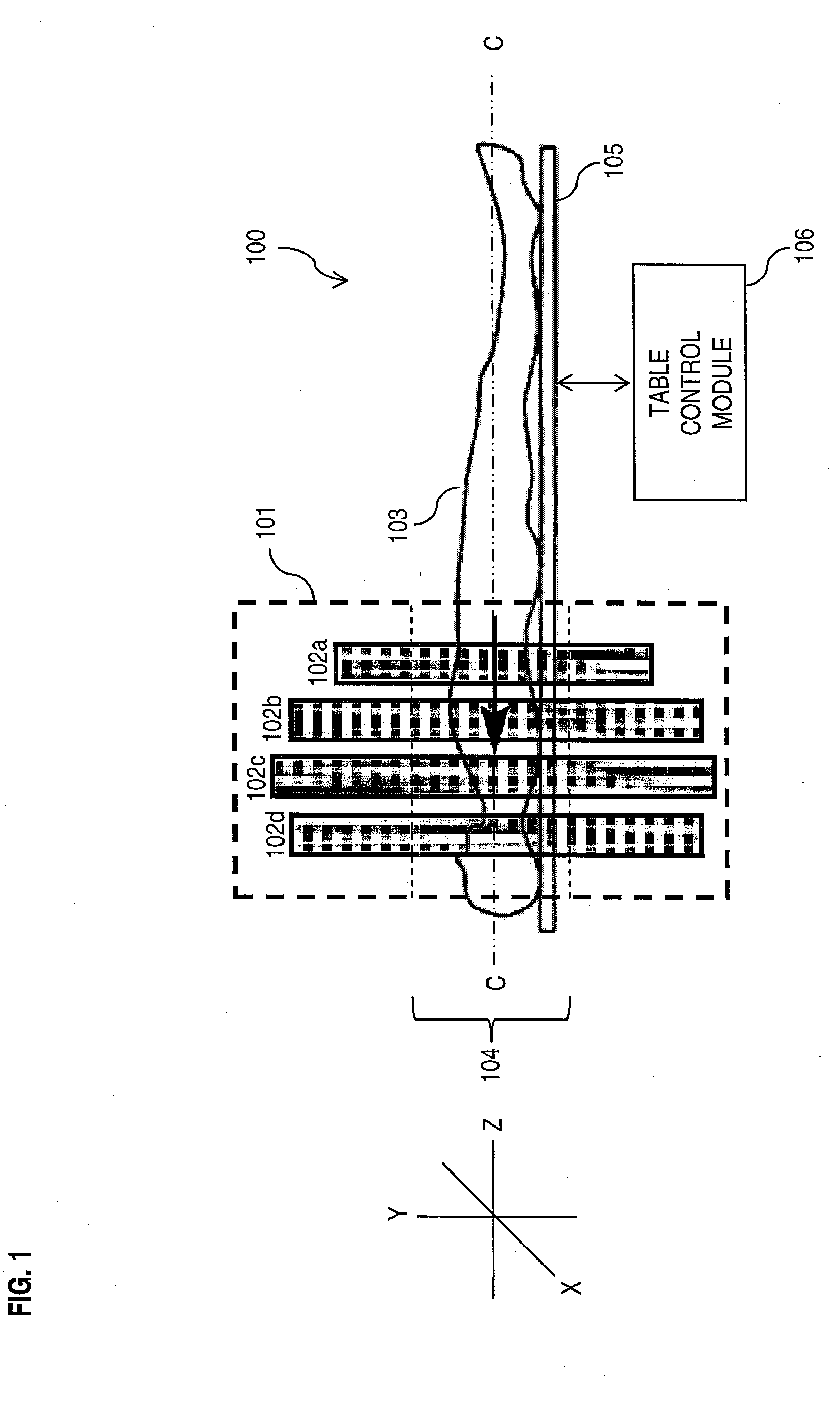
[0026]The present invention addresses the need for nuclear medical imaging devices that can be manufactured efficiently and at reduced cost. Embodiments of the present invention achieve that objective by providing collimators that can be used in various types of nuclear medical imaging devices, such as those employed in SPECT scanning, without the necessity of causing relative rotation between the detectors and patient. In this way, the use of expensive gantry systems to support and rotate heavy detectors, such as gamma cameras, around the patient is avoided. Thus, in accordance with embodiments of the present invention, effective and accurate imaging is performed simply by causing linear movement of the patient along a longitudinal axis, without causing relative rotation between the detectors and patient.

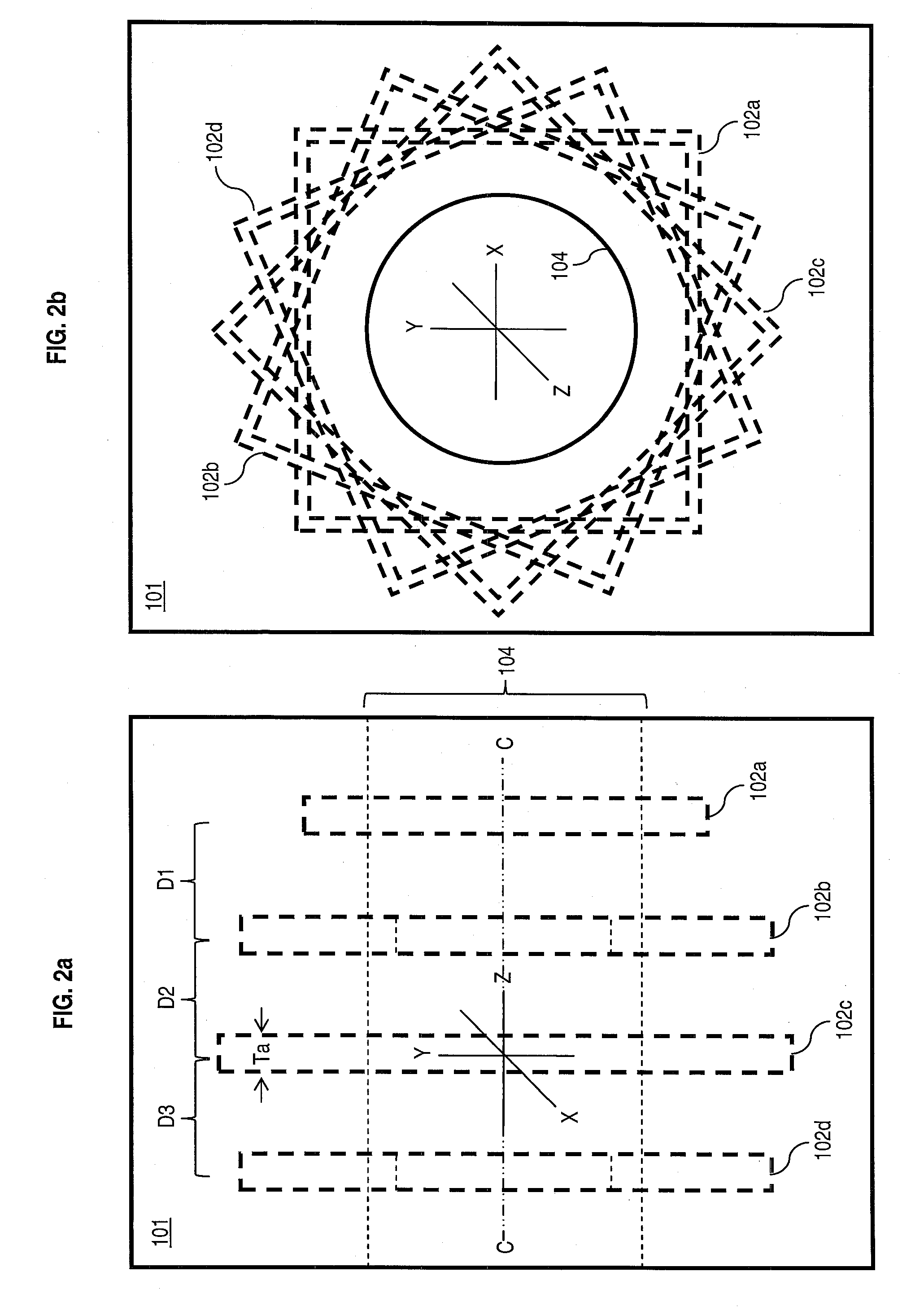
[0027]In accordance with embodiments of the present invention, a collimator is provided comprising a plurality of stacked parallel collimating segments, each containing a plurality...


PUM


Abstract

Transaxial radionuclide imaging is implemented without relative rotation between detectors and a patient by employing a collimator comprising segments sharing a common central axis, each segment having a plurality of apertures extending therethrough, wherein the segments are angularly displaced from one another about the common central axis. Embodiments include SPECT systems comprising a polygonal detector having a collimator on at least two sides thereof. Embodiments further include collimators comprising six segments, each offset by an angle of 7 to 9°.

Description

FIELD OF THE INVENTION[0001]The present invention relates to radionuclide imaging. The present invention is particularly applicable to mechanisms for directionally filtering incident gamma quanta upon detectors (or cameras) within diagnostic imaging apparatuses, such as those used in single photon emission computed tomography (SPECT) systems.BACKGROUND OF THE INVENTION[0002]Medical radionuclide imaging, commonly referred to as nuclear medicine, is a significant diagnostic tool that involves the use of ionizing radiation to obtain accurate, three-dimensional (3D) maps of an in vivo patient. Typically, one or more biologically appropriate radiopharmaceuticals are administered to a patient, as by ingestion, inhalation, or injection. Tracer amounts of these radioactive substances emanate gamma quanta while localizing at specific organs, bones, or tissues of interest (hereinafter collectively referred to as the “study area”) within the patient's body. One or more radiation detectors are ...

Claims


Application Information

Patent Timeline
Patent Type & Authority Applications(United States)
IPC IPC(8): G01T1/166
CPCA61B6/037G21K1/025G01T1/1648B33Y80/00
Inventor HAWMAN, ERIC GRANT
Owner SIEMENS MEDICAL SOLUTIONS USA INC
Why Eureka
Try Eureka