Feature Dependent Extended Depth of Focusing on Semi-Transparent Biological Specimens

a semi-transparent, biological technology, applied in image enhancement, image analysis, instruments, etc., can solve the problems of microscopists constantly manually manually, objects that appear outside the narrow depth of field or focal plane become blurred and out of focus, and suffer from limited depth of field in high-resolution microscopy

Active Publication Date: 2009-02-19
CDX MEDICAL IP INC
View PDF5 Cites 35 Cited by
  • Summary
  • Abstract
  • Description
  • Claims
  • Application Information

AI Technical Summary

Benefits of technology

[0016]Objects of no diagnostic importance are removed by performing a morphological erosion on all sharp appearing objects. In a preferred embodiment, an erosion algorithm is implemented which erodes pixels from each side of an image. Consequently, thin cell walls that appear sharp and in focus, but confer no diagnostically important information, will be eliminated or significantly diminished The larger image of the cell's nucleus on the other hand, will survive the erosion process, thereby preserving the diagnostic objects.

Problems solved by technology

High-resolution microscopy suffers from limited depth of field, which prevents thick or uneven specimen preparations from being imaged entirely in focus.
Objects that appear outside the narrow depth of field or focal plane become quickly blurred and out of focus, forcing the microscopist to constantly manually focus back and forth.
This not only limits the productivity of the microscopist but also increases the likelihood he or she will miss a subtle feature that may appear only in a narrow focal plane.
Moreover, this limited depth of field becomes worse as the magnification increases because it is directly dependent on the numerical aperture of the microscope objective, which increases with the magnification.
As such, when analyzing a specimen that has dimensionality, often there will be areas of the specimen that are out of focus.
As a result, the computer may classify cells found in these out of focus areas as abnormal because they may appear to the computer as exhibiting features that it is trained to classify as abnormal.
A single high-resolution image cannot be in-focus everywhere.
However, the EDF algorithms of the prior art—which are ideal for creating a composite image of the top surface of a three-dimensional object—do not work well for biological specimens, which present an additional complexity of having multiple semi-transparent objects or cells stacked on each other.
These methods are of no utility when attempting to construct a composite image of a semi-transparent biological sample for diagnostic purposes, whereby it is critical to have all the pixels come from a single object or cell and to avoid pixel contamination.
Additionally, for diagnostic purposes, a pathologist prefers to see a composite image that only includes cell nuclei This presents a further limitation of standard EDF algorithms; the nucleus' sharpness might be less than the cell boundary's sharpness, causing the composite image to only display the cell boundary and not the diagnostic important cell nucleus.
A pathologist needs to see the nucleus, but prior art EDF algorithms, which select in-focus images based solely on the sharpness of the image, cannot distinguish between different objects, both of which appear sharp and in-focus.
This image will be sharp and in-focus everywhere, but will not convey any diagnostically important information to an examining pathologist or to a specialized computer system that may be analyzing the slide.
In summary, the semi-transparent quality of biological specimens presents a problem with applying prior art EDF algorithms to thick cellular specimens.
Additionally due to the disaggregated nature of the cellular samples obtained by a brush biopsy, a slide prepared in connection with a brush biopsy will inevitably contain cells that overlap and overlay each other.
Furthermore, there are objects of no diagnostic significance, such as cell walls or artifacts that should be deemphasized or eliminated from a composite image.

Method used


Image

  • Feature Dependent Extended Depth of Focusing on Semi-Transparent Biological Specimens
  • Feature Dependent Extended Depth of Focusing on Semi-Transparent Biological Specimens
  • Feature Dependent Extended Depth of Focusing on Semi-Transparent Biological Specimens

Examples


Embodiment Construction

[0024]Embodiments of the present invention will now be described with reference to the above-identified figures of the Drawings. However, the Drawings and the description herein of the invention are not intended to limit the scope of the invention. It will be understood that various modifications of the present description of the invention are possible without departing from the spirit of the invention. Also, features described herein may be omitted, additional features may be included, and / or features described herein may be combined in a manner different from the specific combinations recited herein, all without departing from the spirit of the invention

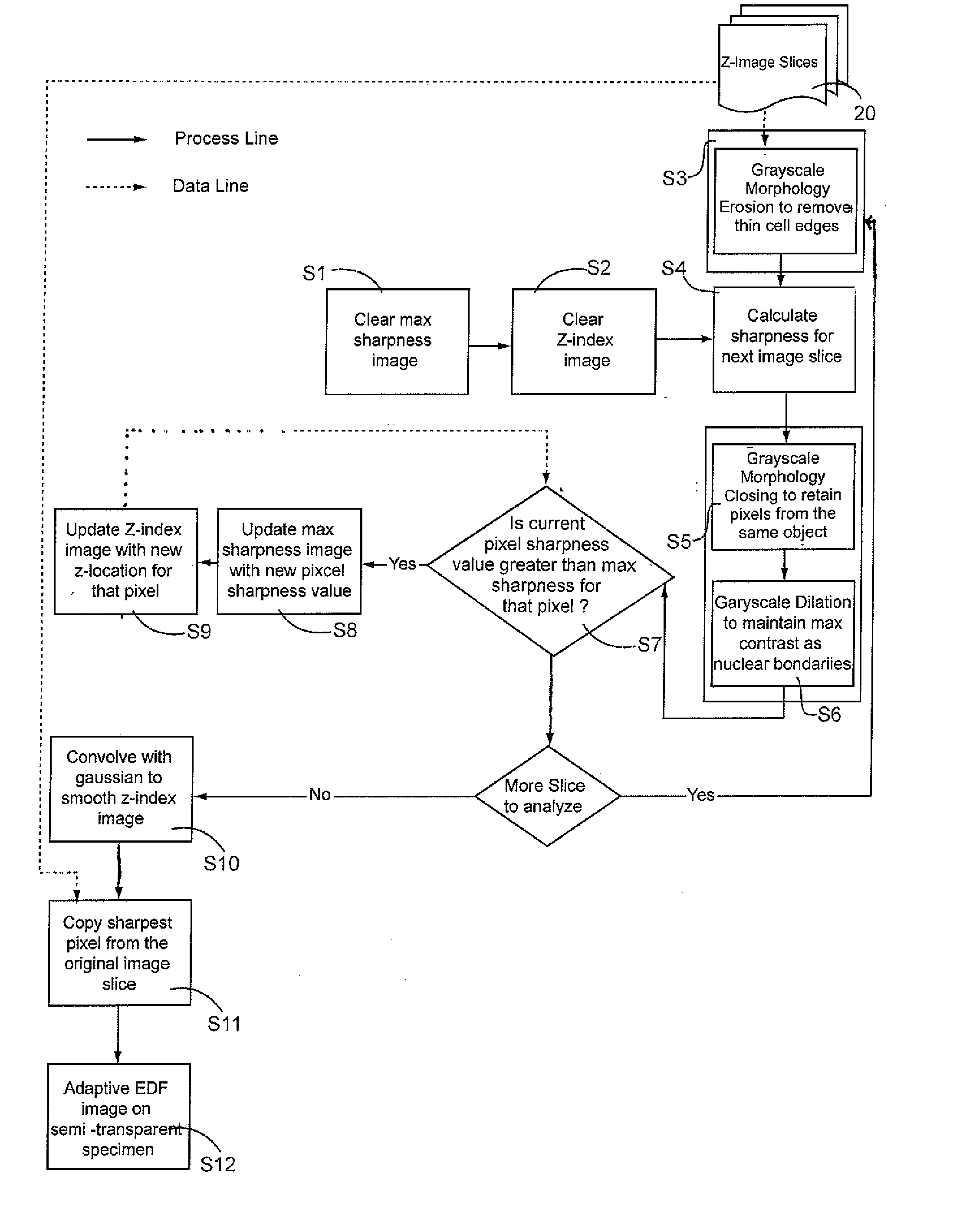
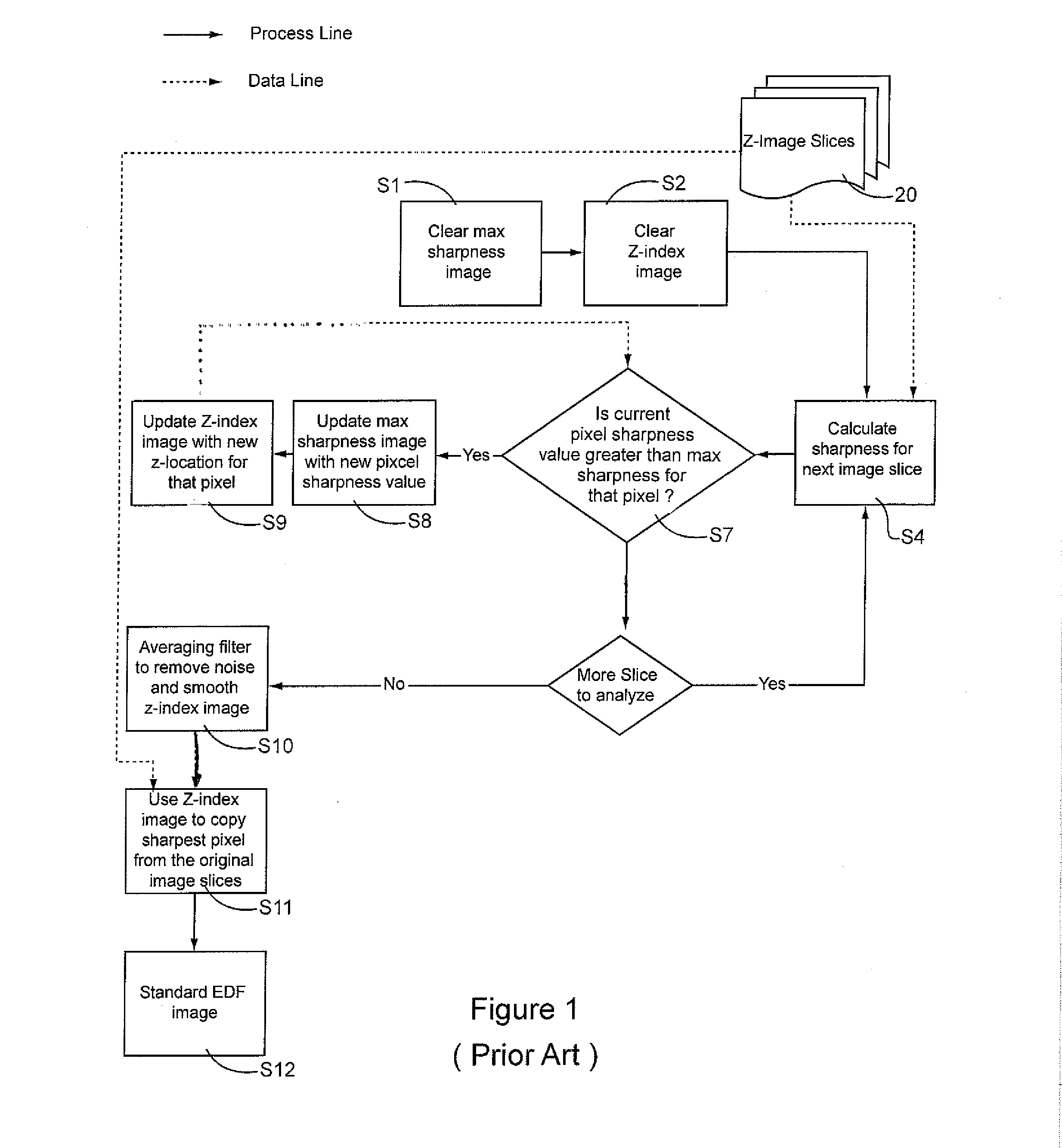
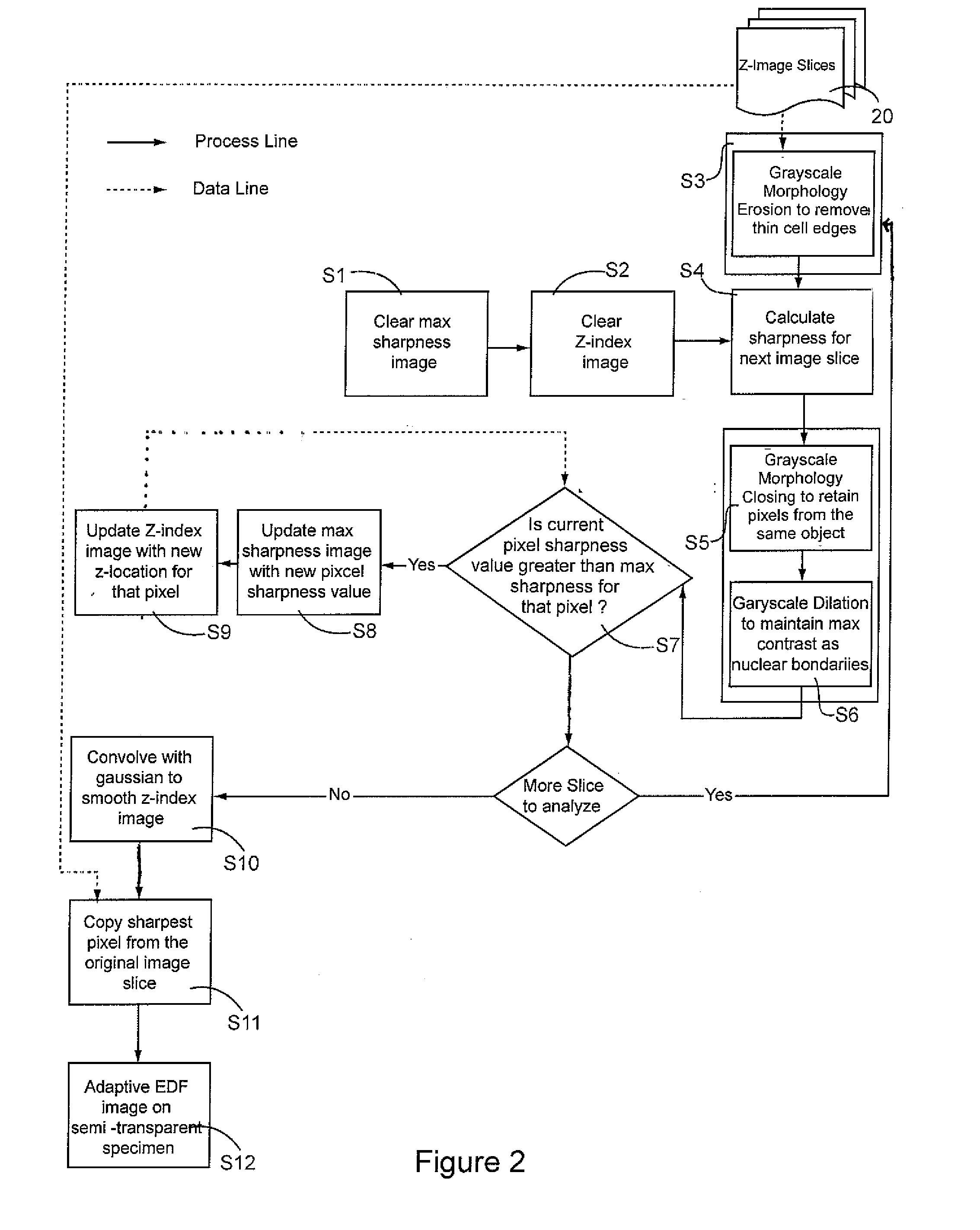
[0025]Limited depth-of-focus is a deficiency in microscope imaging of thick, semi-transparent biological samples that is effectively be rectified using EDF processing techniques that is the subject of the current invention.

[0026]Typically, in the preparation of a cellular specimen for pathology, a clinician will transfer and affix ...


PUM


Abstract

A method and system for constructing a digital image of a three-dimensional biological specimen that displays diagnostically important information—substantially to the exclusion of unimportant information. The system de-enhances features in a cellular specimen which are not diagnostically important and enhances those which are The system selects the sharpest pixel for each pixel location from among a stack of image slices and copies them into a composite image

Description

RELATED APPLICATIONS[0001]This application claims the benefit of provisional application 60 / 956,504 filed on Aug. 17, 2007.FIELD OF THE INVENTION[0002]The present invention relates to the field of digital processing of microscope images, specifically to an extended depth-of-focus (EDF) of thick or three-dimensional semi-transparent biological specimens.BACKGROUND OF THE INVENTION[0003]High-resolution microscopy suffers from limited depth of field, which prevents thick or uneven specimen preparations from being imaged entirely in focus. Objects that appear outside the narrow depth of field or focal plane become quickly blurred and out of focus, forcing the microscopist to constantly manually focus back and forth. This not only limits the productivity of the microscopist but also increases the likelihood he or she will miss a subtle feature that may appear only in a narrow focal plane.[0004]Moreover, this limited depth of field becomes worse as the magnification increases because it i...

Claims


Application Information

Patent Timeline
Patent Type & Authority Applications(United States)
IPC IPC(8): G06K9/36
CPCG06T5/30G06T2207/30024G06T2207/10056G06T5/50
Inventor RUTENBERG, MARKSCOTT, RICHARDTJON, ROBERTFRIST, STEPHEN
Owner CDX MEDICAL IP INC
Why Eureka
Try Eureka