Tumor vasculature markers and methods of use thereof
- Summary
- Abstract
- Description
- Claims
- Application Information
AI Technical Summary
Benefits of technology
Problems solved by technology
Method used
Image
Examples
example 1
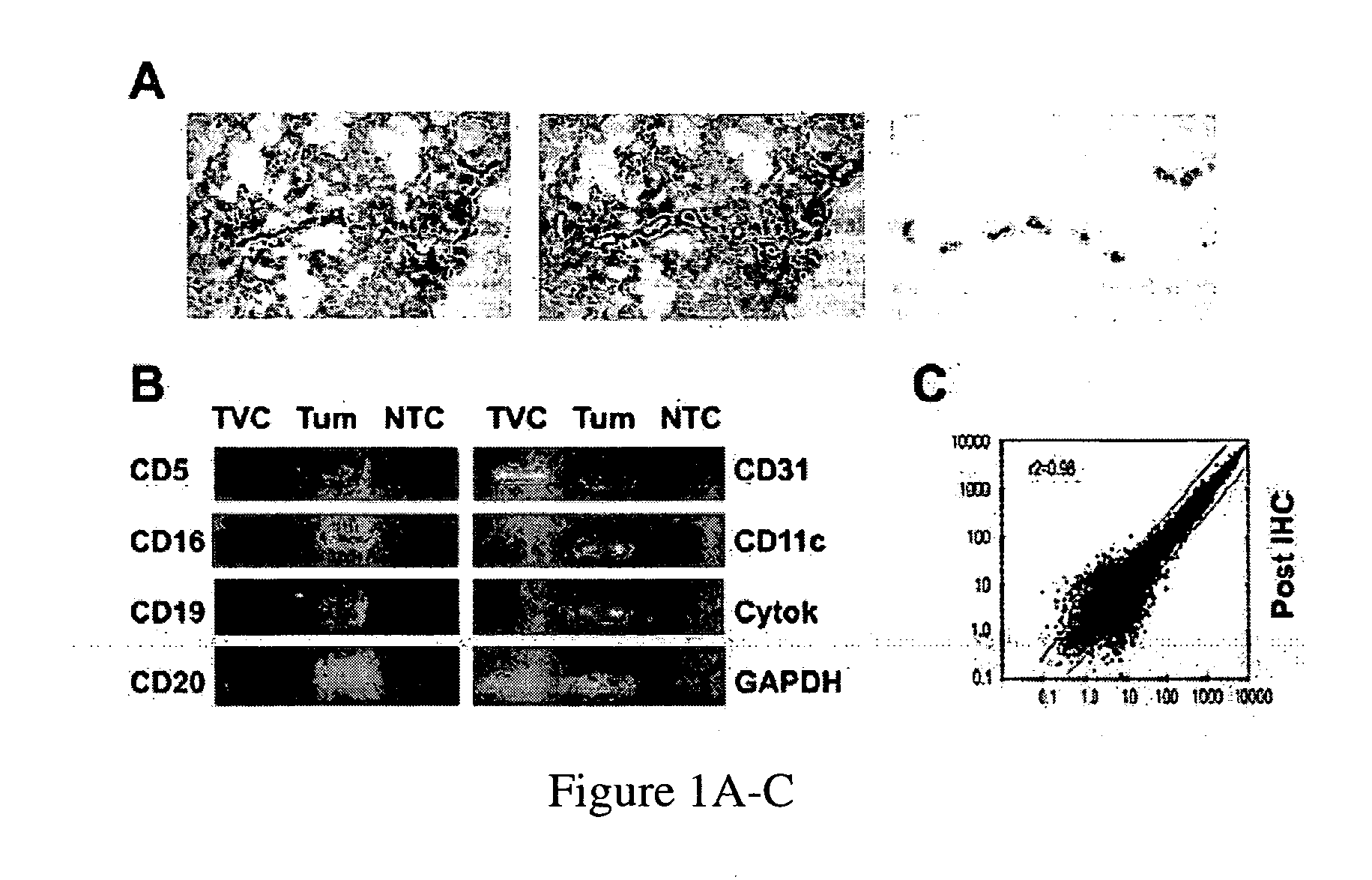
CD31-Based Immuno-LCM Successfully Captures Tumor Vascular Cells, Including Myeloid Cells
Materials and Experimental Methods
Tissues
[0345]Stage-III epithelial ovarian cancer and ductal breast cancer specimens were collected at the University of Turin, Italy, following informed consent, from previously untreated patients. Additional ovarian cancer specimens, and normal ovaries were collected at the University of Pennsylvania Medical Center after obtaining written informed consent under Institutional Review Board (IRB)-approved protocols. Malignant mesothelioma (n=3), non-small cell lung carcinoma (n=3) (provided by Dr. Steven M. Albelda) and malignant melanoma (n=3) (provided by Dr. David Elder) were collected after obtaining written informed consent under IRB-approved protocols. A panel of normal human tissues (FIG. 3B) was provided by the Cooperative Human Tissue Network. All specimens were processed in compliance with HIPAA requirements.
Reagents
[0346]Antibody against human CD31 (BD ...
example 2
mRNA Profile of Immuno-LCM Procured Tumor Vascular Cells
[0372]The mRNA profiles of micro-dissected TVC from 2 tumor samples were analyzed in parallel to 3 micro-dissected normal ovary vascular cells and to cultured human umbilical vein endothelial cells (HUVEC) using Affymetrix-U133 arrays. 13 / 13 known pan-endothelial markers in were detected in the TVC arrays. Similarly, 14 / 15 known tumor endothelial-specific markers (those listed below, Tem-4, and Tem-9) were exclusively expressed or markedly overexpressed (p<0.001) in TVC arrays (Table 4). These findings indicate that the protocol exhibits a sensitivity of greater than 90%.
TABLE 4Expression of pan-endothelial and tumor endothelial markers inHUVEC; endothelial cells from normal ovary isolated throughimmuno-LCM (Normal); and tumor endothelial cells from ovariancancer specimens isolated through immuno-LCM (Tumor1 andTumor2).HUVECNormalTumor 1Tumor 2Pan Endothelial MarkersAngiomodulin++++Hevin++++Connective tissue growth++++factorCol...
example 3
Further Validation of TVM
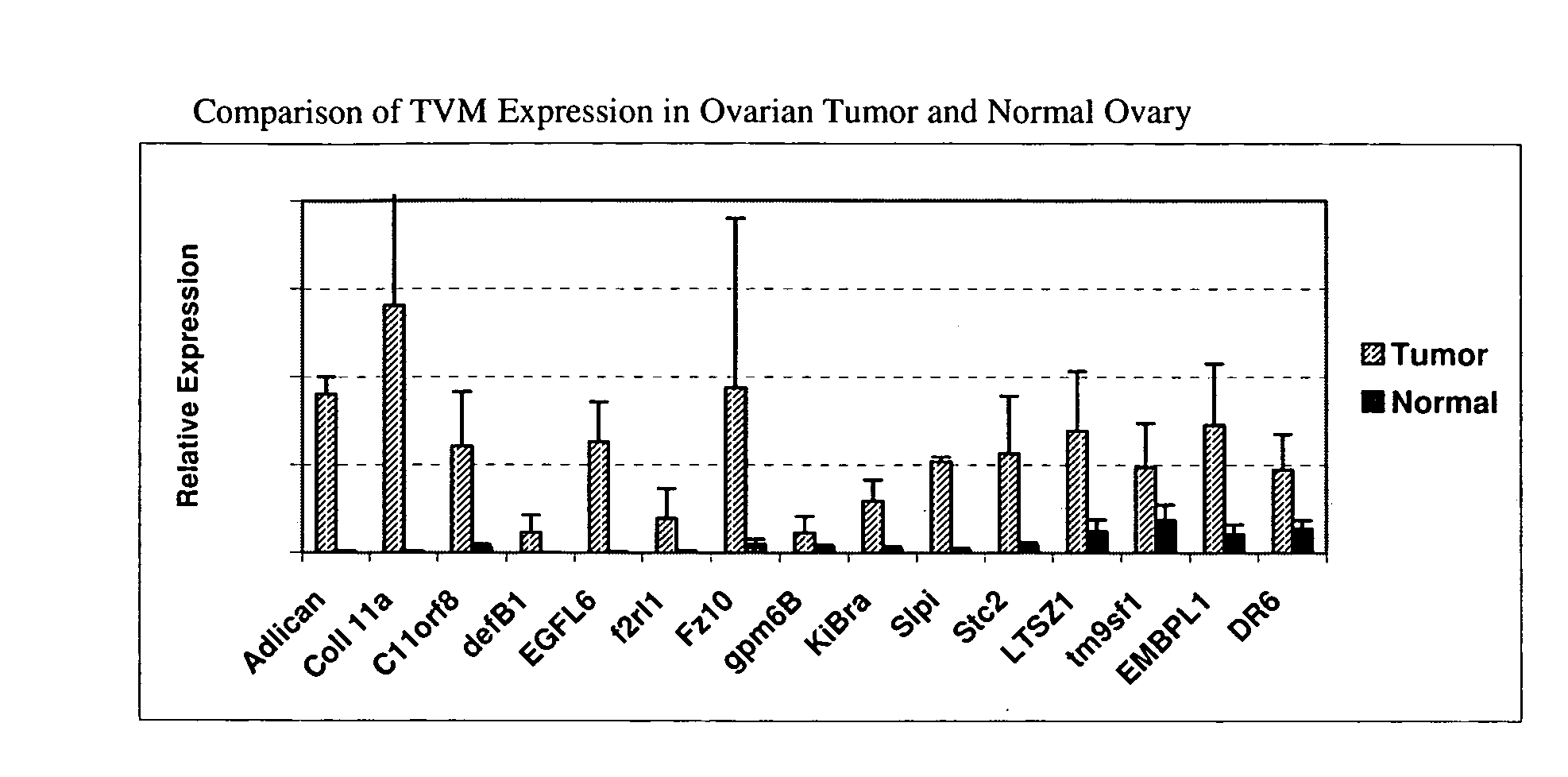
[0375]To test the specificity of identified genes to tumors relative to normal tissues, previously uninvestigated genes were selected from the above candidates for further validation. As positive controls for our assays, known TVM were included. First, 12 selected TVM were tested for enrichment of expression in ovarian tumors versus normal ovarian tissue, by analyzing their whole tissue expression by qRT-PCR in an independent set of stage-III ovarian cancers (n=20) and normal ovaries (n=5). All 12 TVM tested were upregulated in cancer tissue versus normal ovaries (p<0.05 for all). Many TVM were expressed at or below the lowest limits of detection in normal ovaries (FIG. 3A). TVM expression in normal tissues was also examined by analyzing pre-existing SAGE data (Boon K et al, An anatomy of normal and malignant gene expression. Proc Natl Acad Sci USA 2002 Aug. 20; 99(17):11287-92.) (FIG. 3E). Genes exhibiting no or minimal in silico expression in normal tissue...
PUM
| Property | Measurement | Unit |
|---|---|---|
| Temperature | aaaaa | aaaaa |
| Volume | aaaaa | aaaaa |
| Fraction | aaaaa | aaaaa |
Abstract
Description
Claims
Application Information
 - R&D
- Intellectual Property
- Life Sciences
- Materials
- Tech Scout
- Unparalleled Data Quality
- Higher Quality Content
- 60% Fewer Hallucinations
Browse by: Latest US Patents, China's latest patents, Technical Efficacy Thesaurus, Application Domain, Technology Topic, Popular Technical Reports.
© 2025 PatSnap. All rights reserved.Legal|Privacy policy|Modern Slavery Act Transparency Statement|Sitemap|About US| Contact US: help@patsnap.com


