Method and system for evaluating image segmentation based on visibility

a technology of image segmentation and visibility, applied in image enhancement, instruments, applications, etc., can solve the problems of blood filled structures that cannot be differentiated from surrounding tissue using conventional radiology, information that is difficult to decipher, and no quantitative measurement of visibility that can be used to evaluate segmentation techniques

Active Publication Date: 2009-03-26
SIEMENS HEALTHCARE GMBH
View PDF13 Cites 14 Cited by
  • Summary
  • Abstract
  • Description
  • Claims
  • Application Information

AI Technical Summary

Problems solved by technology

Since blood has the same radiodensity as the surrounding tissues, these blood filled structures cannot be differentiated from the surrounding tissue using conventional radiology.
Such fluoroscopic image sequences contain useful information that can be difficult to decipher due to the collapsing of 3-dimensional information into the 2-dimensional images.
Although human perception can be used to see that the visibility of the segmented vessels has increased, there is no quantitative measurement of visibility that can be used to evaluate segmentation techniques.

Method used


Image

  • Method and system for evaluating image segmentation based on visibility
  • Method and system for evaluating image segmentation based on visibility
  • Method and system for evaluating image segmentation based on visibility

Examples


Embodiment Construction

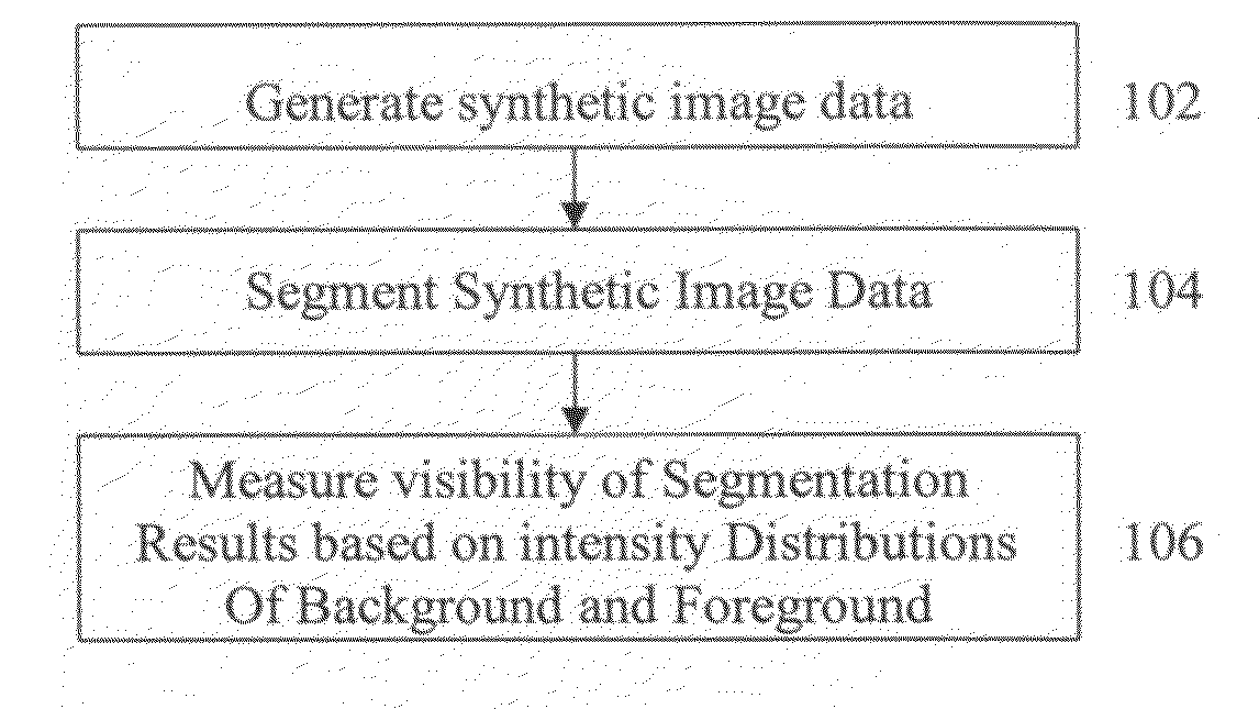
[0026]The present invention is directed to a method for evaluating image segmentation based on visibility. Although embodiments of the present invention described herein are directed toward evaluating segmentation of fluoroscopic images, the present invention is not limited thereto, and may be similarly applied to segmentation of other types of images, such as computed tomography (CT), magnetic resonance (MR), and ultrasound images. Embodiments of the present invention are described herein to give a visual understanding of segmentation evaluation method. A digital image is often composed of digital representations of one or more objects (or shapes). The digital representation of an object is often described herein in terms of identifying and manipulating the objects. Such manipulations are virtual manipulations accomplished in the memory or other circuitry / hardware of a computer system. Accordingly, is to be understood that embodiments of the present invention may be performed withi...


PUM


Abstract

A method and system for evaluating image segmentation is disclosed. In order to quantitatively evaluate an image segmentation technique, synthetic image data is generated and the synthetic image data is segmented to extract an object using the segmentation technique. This segmentation results in a foreground containing the extracted object and a background. The visibility of the extracted object is quantitatively measured based on the intensity distributions of the segmented foreground and background. The visibility is quantitatively measured by calculating the Jeffries-Matusita distance between the foreground and background intensity distributions. This method can be used to evaluate segmentation of vessels in fluoroscopic image sequences by coronary digital subtraction angiography (DSA).

Description

[0001]This application claims the benefit of U.S. Provisional Application No. 60 / 974,099, filed Sep. 21, 2007, the disclosure of which is herein incorporated by reference.BACKGROUND OF THE INVENTION[0002]The present invention relates to object segmentation in medical images, and more particularly to evaluating image segmentation based on visibility.[0003]Angiography is a medical imaging technique in which X-ray images are used to visualize internal blood filled structures, such as arteries, veins, and the heart chambers. Since blood has the same radiodensity as the surrounding tissues, these blood filled structures cannot be differentiated from the surrounding tissue using conventional radiology. Thus, in angiography, a contrast agent is added to the blood, usually via a catheter, to make the blood vessels visible via X-ray. In many angiography procedures, X-ray images are taken over a period of time, which results in a sequence of fluoroscopic images, which show the motion of the b...

Claims


Application Information

Patent Timeline
Patent Type & Authority Applications(United States)
IPC IPC(8): A61B5/00
CPCG06T7/0081G06T7/0097G06T7/2033G06T2207/30101G06T2207/10121G06T2207/30048G06T7/60G06T7/11G06T7/174G06T7/246
Inventor ZHANG, WEIBARBU, ADRIANPRUMMER, SIMONEOSTERMEIER, MARTINCOMANICIU, DORIN
Owner SIEMENS HEALTHCARE GMBH
Why Eureka
Try Eureka