Slit collimator scatter correction

a collimator and scatter correction technology, applied in the field of medical imaging, can solve the problems of slow healing and poor healing, physical abnormalities, and changes in postur

Active Publication Date: 2009-04-09
GENERAL ELECTRIC CO
View PDF7 Cites 18 Cited by
  • Summary
  • Abstract
  • Description
  • Claims
  • Application Information

AI Technical Summary

Problems solved by technology

Osteoporosis predisposes a person to fractures, which are often slow to heal and heal poorly.
Unchecked osteoporosis can lead to changes in posture, physical abnormality (particularly a condition known colloquially as “dowager's hump”), and decreased mobility.
Scatter can produce a significant level of error in the quantitative values obtained during DXA.
However, these techniques attenuate the X-rays and do not completely remove the effect of scatter.

Method used


Image

  • Slit collimator scatter correction
  • Slit collimator scatter correction
  • Slit collimator scatter correction

Examples


Embodiment Construction

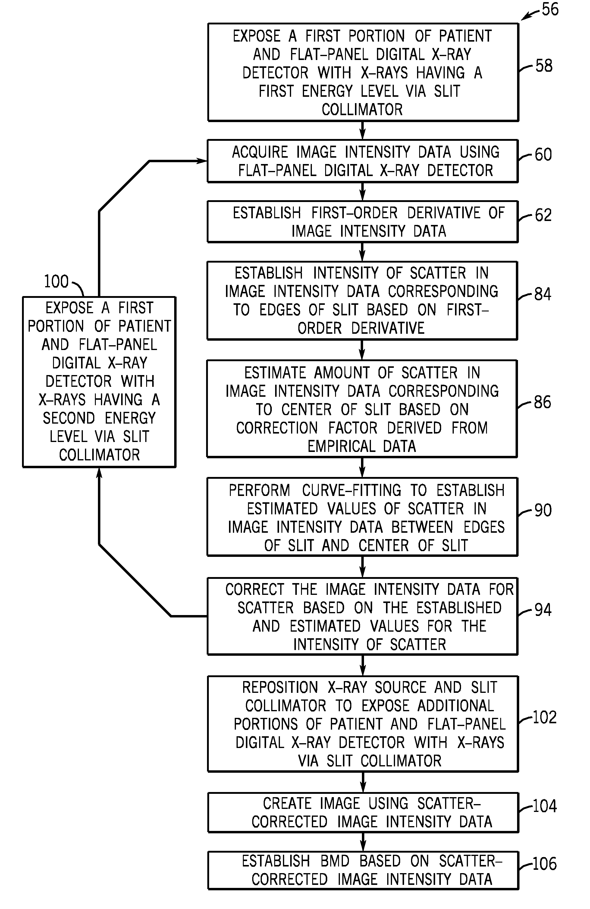
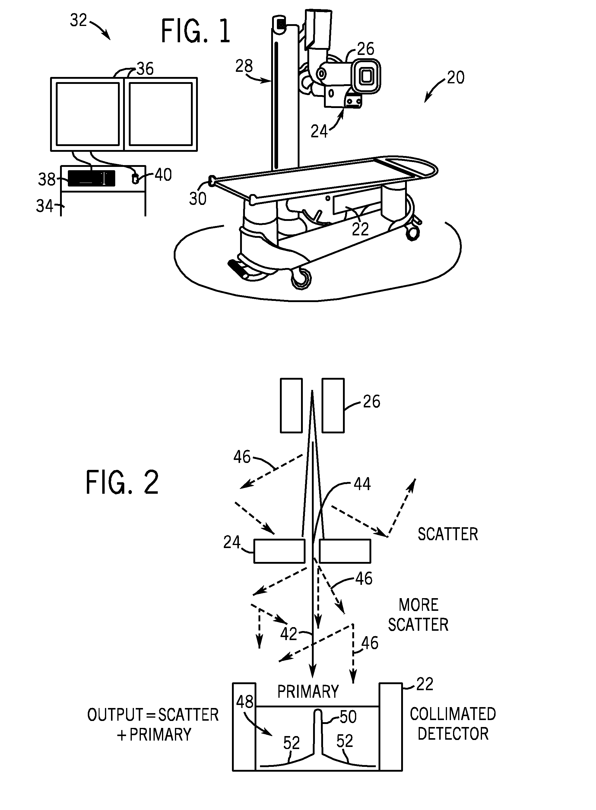
[0024]Referring now to FIG. 1, the present invention will be described as it might be applied in conjunction with an exemplary imaging system, in this case a dual-energy X-ray imaging system, as represented generally by reference numeral 20. In this embodiment, the dual-energy X-ray imaging system 20 is operable to perform dual-energy X-ray absorpiometry (DXA). In general, however, it should be borne in mind that the present techniques may be used with any suitable imaging modality. In particular, this technique is applicable for any imaging system using a large, flat-panel digital detector.

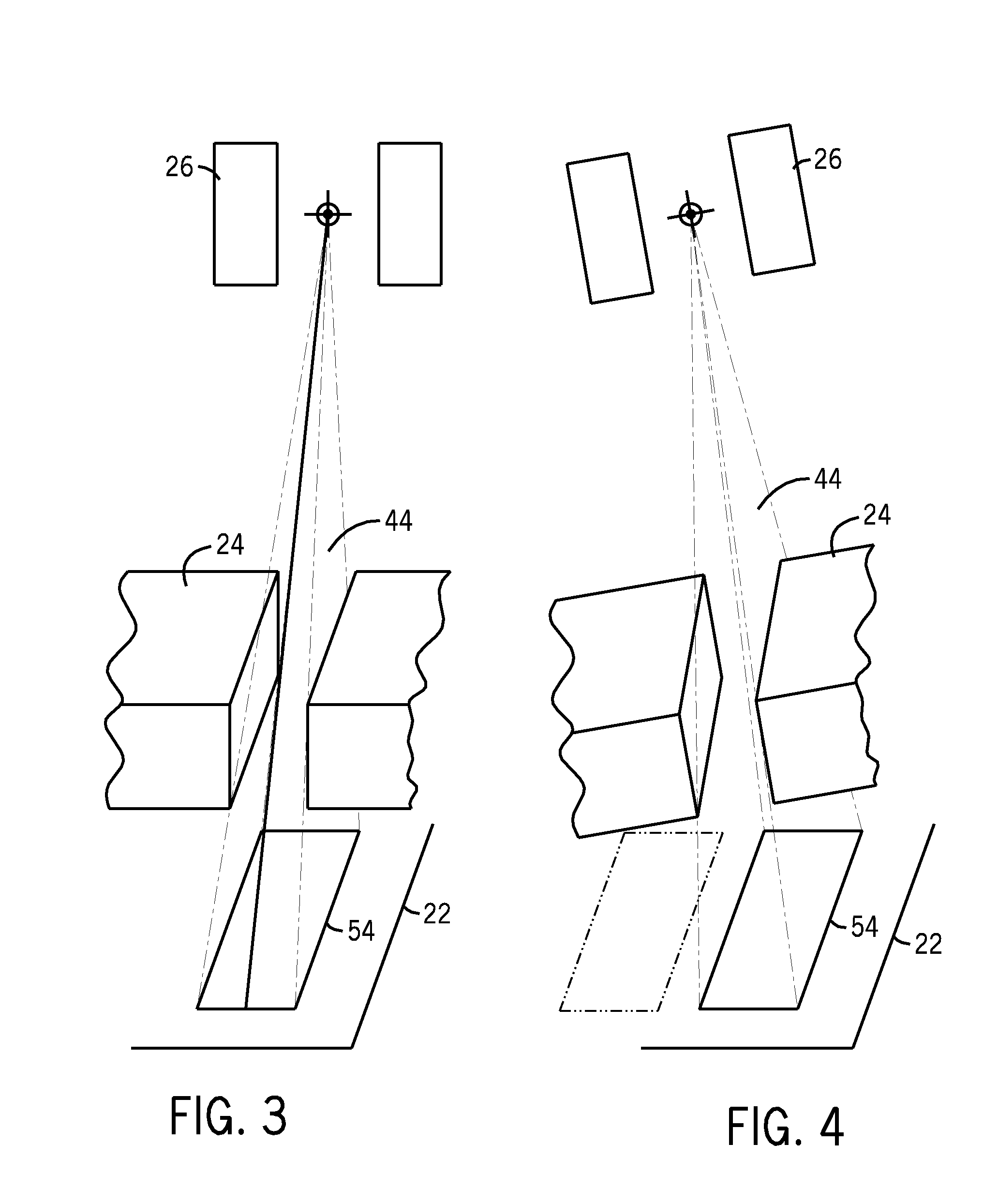
[0025]In the illustrated embodiment, the system 20 has a large, flat-panel digital X-ray detector 22, and a slit collimator 24 disposed over an X-ray source 26. The dual-energy X-ray imaging system 20 is capable of producing images of bone and soft tissue by taking images using X-rays of different energy. In particular, the system 20 is capable of producing an image of a region of interest using ...


PUM


Abstract

A technique is presented for establishing a patient's BMD using a dual-energy X-ray imaging system. In the technique, the dual-energy X-ray imaging system utilizes a slit collimator to expose a series of portions of a region of interest within a patient with X-rays of two different energies. A flat-panel digital X-ray detector detects the X-rays passing through the patient's region of interest and produces data representative of the intensity of the X-rays reaching the detector. The image intensity data is corrected for scatter based on identifying the regions of the image intensity data that are produced from scatter only, and not primary X-rays. A first-order derivative of the image intensity data is used to identify these regions. A value for the intensity of the scatter at the boundary of the scatter-only region is established. The value for the intensity of the scatter at the boundary of the scatter-only region is used to estimate scatter intensity in the region of the image intensity data that is produced from primary X-rays, as well as scatter. To correct the image intensity data for scatter, the known and estimated scatter intensities are subtracted from the image intensity data.

Description

BACKGROUND[0001]The invention relates generally to medical imaging. In particular, the invention relates to digital X-ray medical imaging systems having a flat-panel digital X-ray detector.[0002]The bone mineral density (BMD) of a bone reflects the strength of the bone as represented by calcium content. It is defined as the integral mass of bone mineral per unit of projected area in grams per square centimeter. BMD is a useful tool for the diagnosis and treatment of several diseases and conditions, one of which is osteoporosis.[0003]Osteoporosis is a disease of bone in which the BMD is reduced due to depletion of calcium and bone protein. Osteoporosis predisposes a person to fractures, which are often slow to heal and heal poorly. It is more common in older adults, particularly post-menopausal women; in patients on steroids; and in those who take steroidal drugs. Unchecked osteoporosis can lead to changes in posture, physical abnormality (particularly a condition known colloquially ...

Claims


Application Information

Patent Timeline
Patent Type & Authority Applications(United States)
IPC IPC(8): G06K9/00
CPCA61B6/06A61B6/482A61B6/583G01N2223/401G01N23/20083
Inventor SABOL, JOHN MICHAELJABRI, KADRI NIZARBODEN, ADAM ELLISYUE, MEGHAN LYNN
Owner GENERAL ELECTRIC CO
Why Eureka
Try Eureka