Method for segmentation of lesions

a technology for segmentation and lesions, applied in the field of digital imaging, can solve the problems of large inter-observer variation of nodule size estimation, difficult task of segmenting lesions from normal anatomy, etc., and achieve the effect of segmentation

Active Publication Date: 2009-05-21
PHILIPS HEALTHCARE INFORMATICS INC
View PDF3 Cites 81 Cited by
  • Summary
  • Abstract
  • Description
  • Claims
  • Application Information

AI Technical Summary

Benefits of technology

[0009]It is an object of the present invention to effectively segment an anatomical structure, such as a pulmonary lesion, from the background tissue in a volumetric medical image.

Problems solved by technology

This is a tedious task due to the complex shape of nodules and that the nodule spans multiple slices.
As a result boundary outlines are subject to individual radiologist interpretation and can lead to large inter-observer variation of the nodule's size estimate.
Thus, segmenting the lesion from normal anatomy is a difficult task as the image differences between the lesion and normal anatomy often are not discernable in terms of voxel intensity values, e.g., Hounsfield units HU.
Too high a threshold leads to an under segmentation of the nodule and an underestimation of the nodule's volume.
As a result it is harder to segment the nodule from the normal anatomy and consequently more sophisticated segmentation algorithms are required.
One problem with known volumetric segmentation methods is the tendency to include part of the normal anatomy with the detected nodule, because of an inability to distinguish between the two.
As mentioned before using a low enough Hounsfield threshold to capture the non-solid component of a nodule exacerbates this problem.
Another problem with known volumetric segmentation methods stems from the use of Hounsfield thresholds to distinguish between target structures such as, for example, nodules or lesions, and anatomical structures such as, for example, local pulmonary vasculature or the pleural surface adjoining the thoracic wall.
Segmenting at such a relatively high threshold, however, may not allow a specialist to determine the full extent of the target structure.
Similarly, although a relatively low Hounsfield threshold can be used to determine a greater extent of the target structure, segmenting at such a relatively low threshold may not allow a specialist to distinguish between the target structure and the surrounding anatomical structure.

Method used


Image

  • Method for segmentation of lesions
  • Method for segmentation of lesions
  • Method for segmentation of lesions

Examples


Embodiment Construction

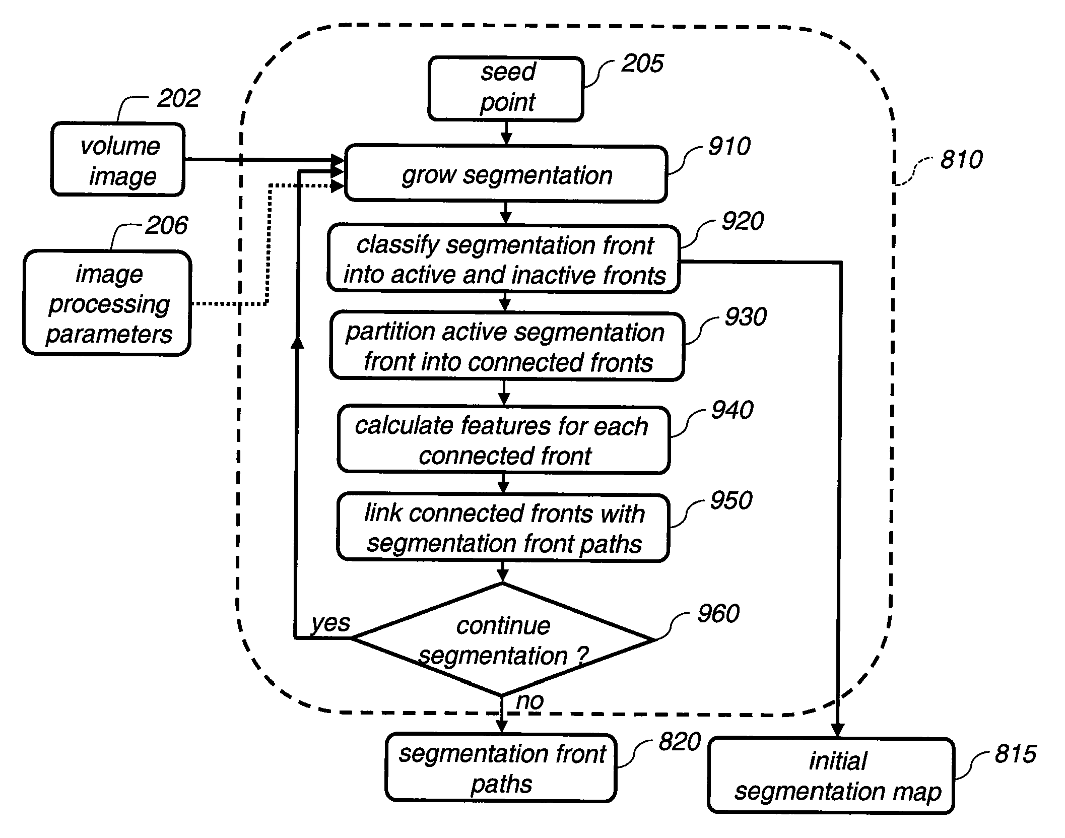
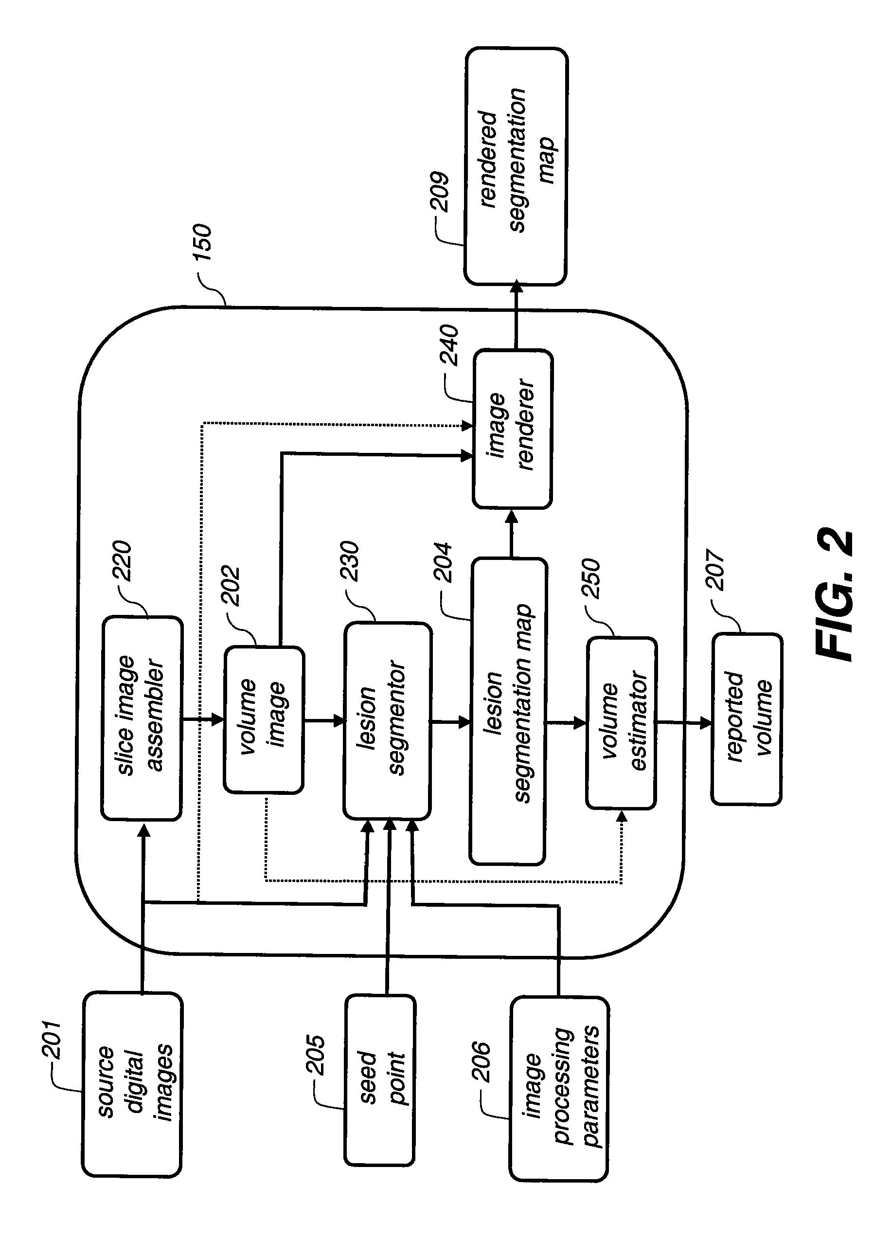
[0028]The current invention will be elucidated in the context of segmenting a pulmonary lesion, in particular for the cases where the pulmonary lesion is attached to normal anatomy such as the local pulmonary vasculature and the pleural surface. The current invention can be applied to segmenting any anatomical structure that is attached to other anatomical structures where the image differences between the anatomical structures are not readily discernable in terms of voxel intensity values.

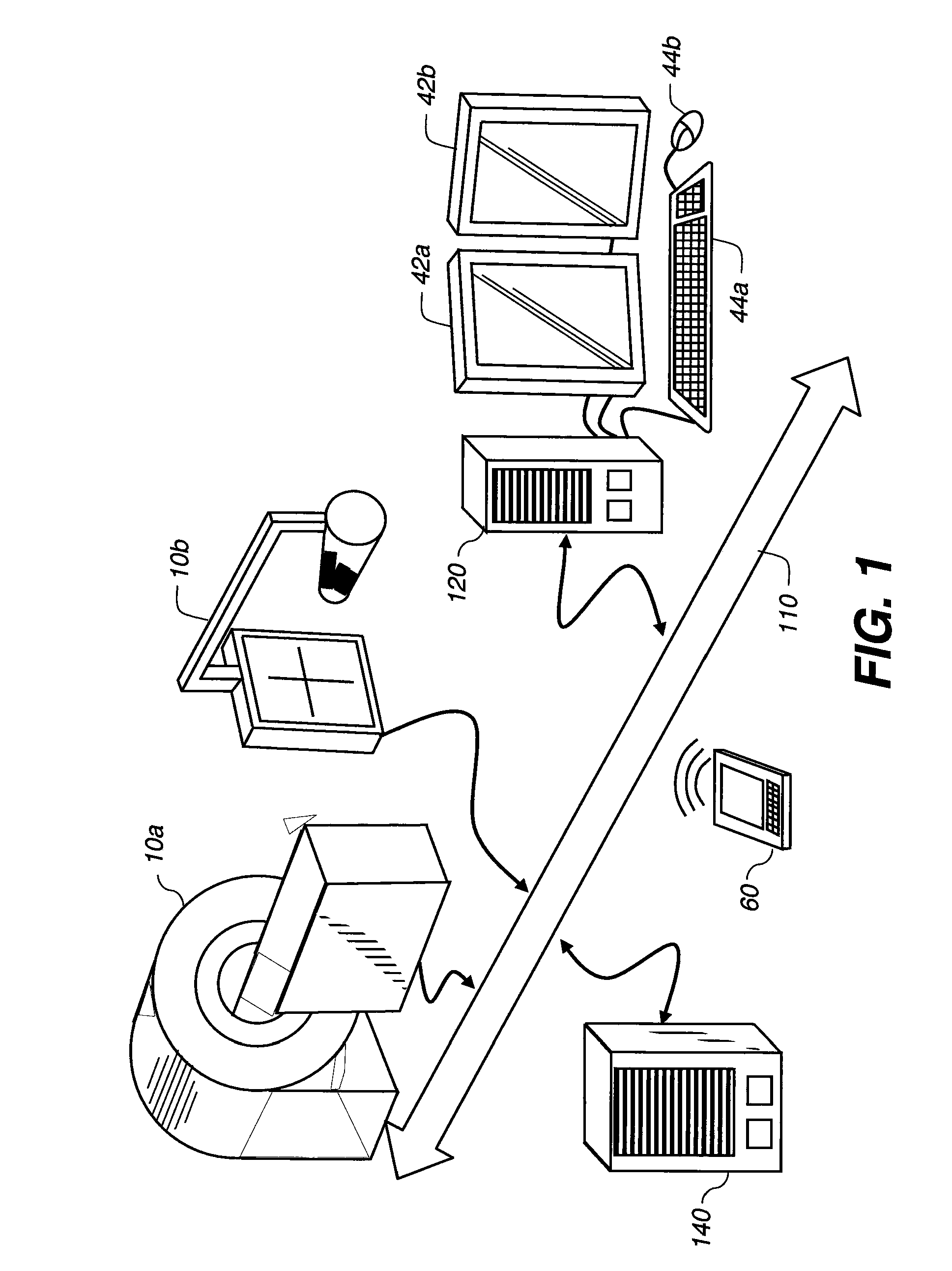
[0029]Many medical imaging applications are implemented via a picture archiving and communications systems (PACS). These systems provide a means for displaying digital images acquired by a wide variety of medical imaging modalities such as, but not limited to, projection radiography (x-ray images), computed tomography (CT images), ultrasound (US images), and magnetic resonance (MR images). Each of the above mentioned medical imaging modalities contain a slightly different set of diagnostic informa...


PUM


Abstract

A method of image segmentation includes receiving a set of voxels, segmenting the set of voxels into a foreground group and a background group, and classifying voxels of the foreground group as either lesion voxels or normal anatomy voxels. The method also includes blocking the normal anatomy voxels and performing a second segmentation on voxels of the background group and the lesion voxels, the second segmentation forming a stage two foreground group comprising the lesion voxels and a portion of the voxels of the background group. The method further includes classifying voxels of the stage two foreground group as either stage two lesion voxels or stage two normal anatomy voxels.

Description

CROSS REFERENCE TO RELATED APPLICATIONS[0001]Reference is made to commonly-assigned copending U.S. patent application Ser. No. 11 / 567,857, filed Dec. 7, 2006 entitled ANALYZING LESIONS IN A MEDICAL DIGITAL IMAGE, by Simon et al., the disclosure of which is incorporated herein.FIELD OF THE INVENTION[0002]The present invention relates to the field of digital imaging, and more particularly to processing a digital image to segment an object in a 3-dimensional data set.BACKGROUND OF THE INVENTION[0003]Lung nodule size and growth rates are strong predictors of malignancy (and are used to distinguish benign from malignant). The determination of nodule size involves the manual outlining of a nodule's boundary. This is a tedious task due to the complex shape of nodules and that the nodule spans multiple slices. As a result boundary outlines are subject to individual radiologist interpretation and can lead to large inter-observer variation of the nodule's size estimate.[0004]A method of autom...

Claims


Application Information

Patent Timeline
Patent Type & Authority Applications(United States)
IPC IPC(8): G06K9/34G06K9/00
CPCG06T7/0012G06T7/0081G06T2207/30101G06T2207/30096G06T2207/10072G06T7/11
Inventor SIMON, RICHARD A.GINDELE, EDWARD B.RAY, LAWRENCE A.
Owner PHILIPS HEALTHCARE INFORMATICS INC
Why Eureka
Try Eureka