Precise endoscopic planning and visualization

a planning and visualization technology, applied in the field of precise endoscopic planning and visualization, can solve the problems of difficult bronchoscopy, high cost, and high difficulty of bronchoscopy, and achieve the effect of ensuring the safety of the patien

Inactive Publication Date: 2009-06-18
PENN STATE RES FOUND
View PDF62 Cites 101 Cited by
  • Summary
  • Abstract
  • Description
  • Claims
  • Application Information

AI Technical Summary

Benefits of technology

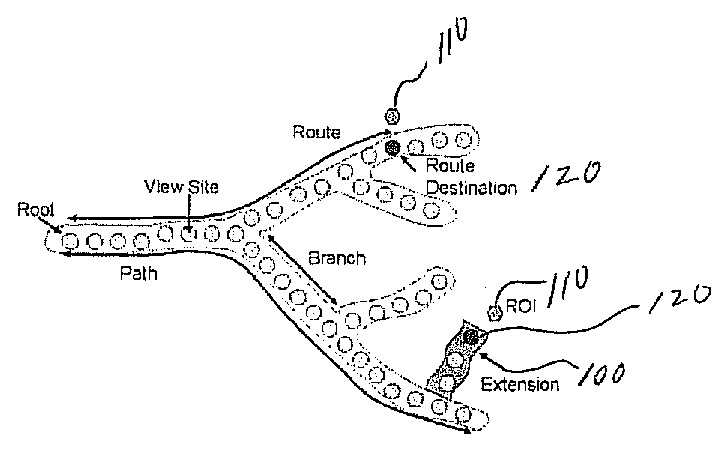
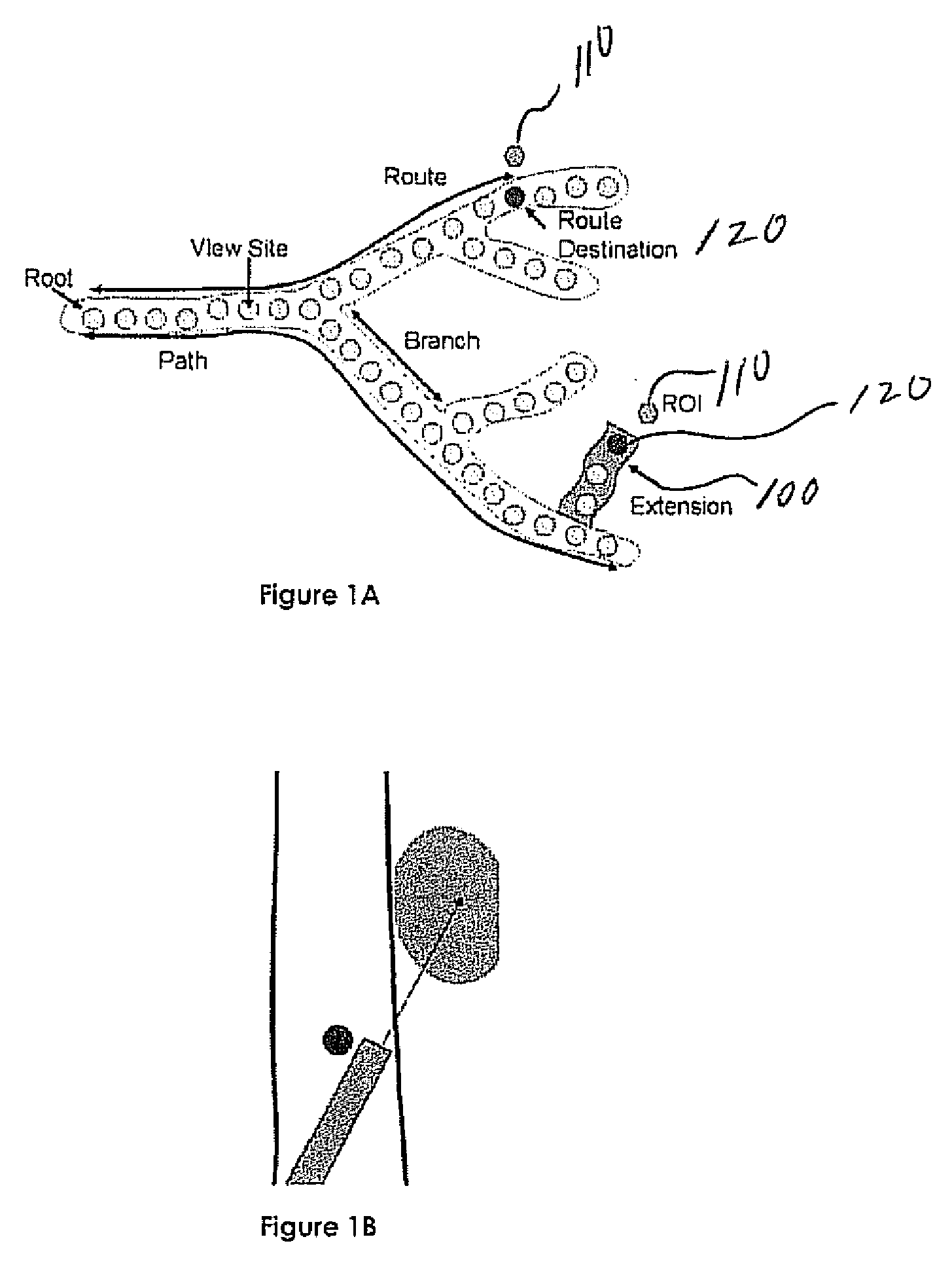
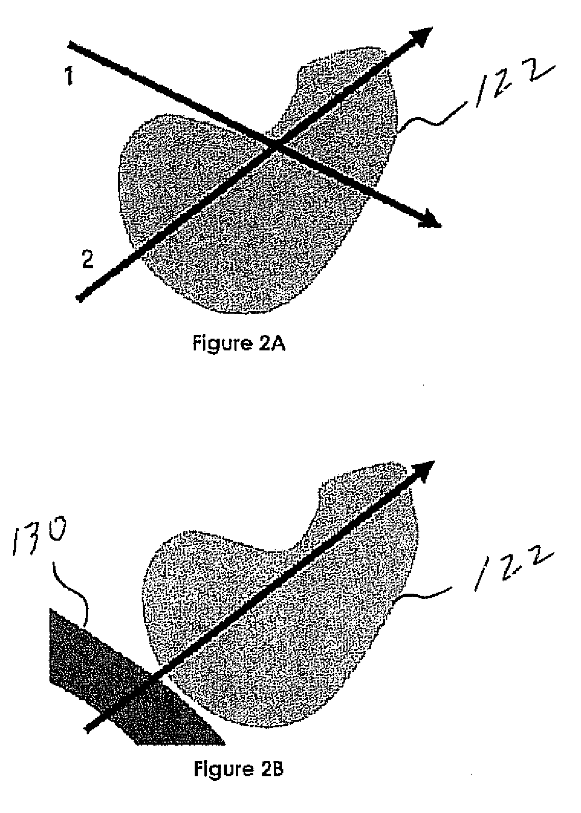
[0008]In determining a first pose, optimum pose, or a best pose(s), the physician is directed to bronchoscopic configurations that maximize the core sample of the ROI, namely, the size or depth of the core sample. Using a patient-specific model of the anatomy derived from a 3D image such as, for example, a 3D MDCT image, bronchoscopic poses are chosen to be realizable given the physical characteristics of the bronchoscope and the relative geometry of the patient's airways and the ROI. To help ensure the safety of the patient, in one embodiment, calculations account for obstacles such as the aorta and pulmonary arteries, precluding the puncture of these sensitive blood vessels. In another embodiment, a real-time visualization system conveys the calculated pose orientation and the quality of any arbitrary bronchoscopic pose orientation. In this system, a suggested pose orientation is represented as an arrow within a virtual bronchoscopic (VB) rendering of the patient's airway tree. The location and orientation of the arrow indicates the suggested pose orientation to which the physician should align during the procedure. In another embodiment, the visualization system shows the ROI, which is differentially colored to indicate the depth of sample of the ROI at a given VB camera location. The ROI, which may be located outside the airway wall, is blended into the scene at varying color intensities, with brighter intensities indicating a larger depth of sample. The physician can freely maneuver within the VB world, with the visual representation of the quality of a bronchoscopic sample at the current VB camera location updating in real time.

Problems solved by technology

However, physicians often get disoriented in the complex branching airway tree.
Furthermore, ROIs are often located beyond the airway walls and therefore outside the view of the bronchoscopic camera.
For these reasons, bronchoscopies are difficult, error-prone procedures [5, 3, 22, 11, 25, 21].

Method used


Image

  • Precise endoscopic planning and visualization
  • Precise endoscopic planning and visualization
  • Precise endoscopic planning and visualization

Examples


example

[0129]We have conducted an MDCT image-analysis study to determine the efficacy and performance of a pose-orientation selection strategy in accordance with the present invention. In this study, we examined high-resolution MDCT chest scans of 11 patients with 20 diagnostic ROIs in the central chest region, defined at the direction of a physician, that were identified for potential follow-on transbronchial needle aspiration (TBNA) procedures. The purpose of the study was to verify the route-planning methods give appropriate routes to ROIs in a clinically-reasonable timeframe while accommodating realistic procedural, anatomical, and physical constraints.

[0130]Table I, below, summarizes the cases examined in this study. For most patients, ROIs were located along the trachea or in a subcarinal position at Mountain stations M4 (lower paratracheal) and M7 (inferior mediastinal), which are typically accessible for TBNA with modern videobronchoscopes [26]. Patients 20349.3.29, 20349.3.37, and...


PUM


Abstract

Endoscopic poses are used to indicate the exact location and direction in which a physician must orient the endoscope to sample a region of interest (ROI) in an airway tree or other luminal structure. Using a patient-specific model of the anatomy derived from a 3D MDCT image, poses are chosen to be realizable given the physical characteristics of the endoscope and the relative geometry of the patient's airways and the ROI. To help ensure the safety of the patient, the calculations also account for obstacles such as the aorta and pulmonary arteries, precluding the puncture of these sensitive blood vessels. A real-time visualization system conveys the calculated pose orientation and the quality of any arbitrary bronchoscopic pose orientation. A suggested pose orientation is represented as an icon within a virtual rendering of the patient's airway tree or other structure. The location and orientation of the icon indicates the suggested pose orientation to which the physician should align during the procedure.

Description

REFERENCE TO RELATED APPLICATIONS[0001]This application is a continuation-in-part of U.S. patent application Ser. No. 12 / 018,953, filed Jan. 24, 2008, which claims priority from U.S. Provisional Patent Application Ser. No. 60 / 887,472, filed Jan. 31, 2007, the entire content of both of which is incorporated herein by reference.GOVERNMENT SPONSORSHIP[0002]This work was partially supported by Grant Nos. CA074325 and CA091534 from the National Cancer Institute of the NIH NIBIB Grant No. EB000305. The U.S. Government may have rights in this invention.FIELD OF THE INVENTION[0003]This invention relates generally to endoscopic planning and visualization and, in particular, to methods and apparatus facilitating the precise determination of an optimal endoscopic configuration for sampling a region of interest (ROI).BACKGROUND OF THE INVENTION[0004]Co-pending U.S. patent application Ser. No. 12 / 018,953, entitled “Methods and Apparatus for 3D Route Planning Through Hollow Organs” describes auto...

Claims


Application Information

Patent Timeline
Patent Type & Authority Applications(United States)
IPC IPC(8): A61B1/012
CPCG06T19/003A61B2019/505G06T2210/41G06T2207/30061A61B2019/507A61B2034/105A61B2034/107
Inventor HIGGINS, WILLIAM E.GIBBS, JASON D.
Owner PENN STATE RES FOUND
Why Eureka
Try Eureka