Fluoroscopy operator protection device

a fluoroscopic operator and protection device technology, applied in the direction of x-ray tubes, patient positioning for diagnostics, applications, etc., can solve the problems of many life-saving interventional procedures that cannot be carried out, exposure of patient and attendant medical personnel to potentially harmful x-ray radiation, and significant doses of x-ray radiation for medical personnel involved in fluoroscopic imaging

Active Publication Date: 2009-09-17
RADIACTION
View PDF52 Cites 48 Cited by
  • Summary
  • Abstract
  • Description
  • Claims
  • Application Information

AI Technical Summary

Benefits of technology

[0010]In another embodiment, the radiation protection device has a first conical or cylindrical radiation shield that extends from the X-ray source to the patient or to the procedure table and a second conical or cylindrical radiation shield that extends from the patient to the image intensifier. The first radiation shield and the second radiation shield have length or height adjustments to fit the device to the patient and to accommodate motion of the C-arm relative to the patient. A soft, flexible material surrounds the openings of the first radiation shield and the second radiation shield to comfortably fit them to the contours of the patient's body. Optiona

Problems solved by technology

Many life saving interventional procedures would not be possible without the use of fluoroscopy.
However, fluoroscopic imaging exposes the patient and attendant medical personnel to potentially harmful X-ray radiation.
However, medical personnel involved in fluoroscopic imaging are exposed to significant doses of X-ray radiation on a daily basis.
While scattered X-rays are much lower intensity than the direct X-ray beam, it is much more likely for the operator to be exposed to scattered X-rays and the damagi

Method used


Image

  • Fluoroscopy operator protection device
  • Fluoroscopy operator protection device
  • Fluoroscopy operator protection device

Examples


Embodiment Construction

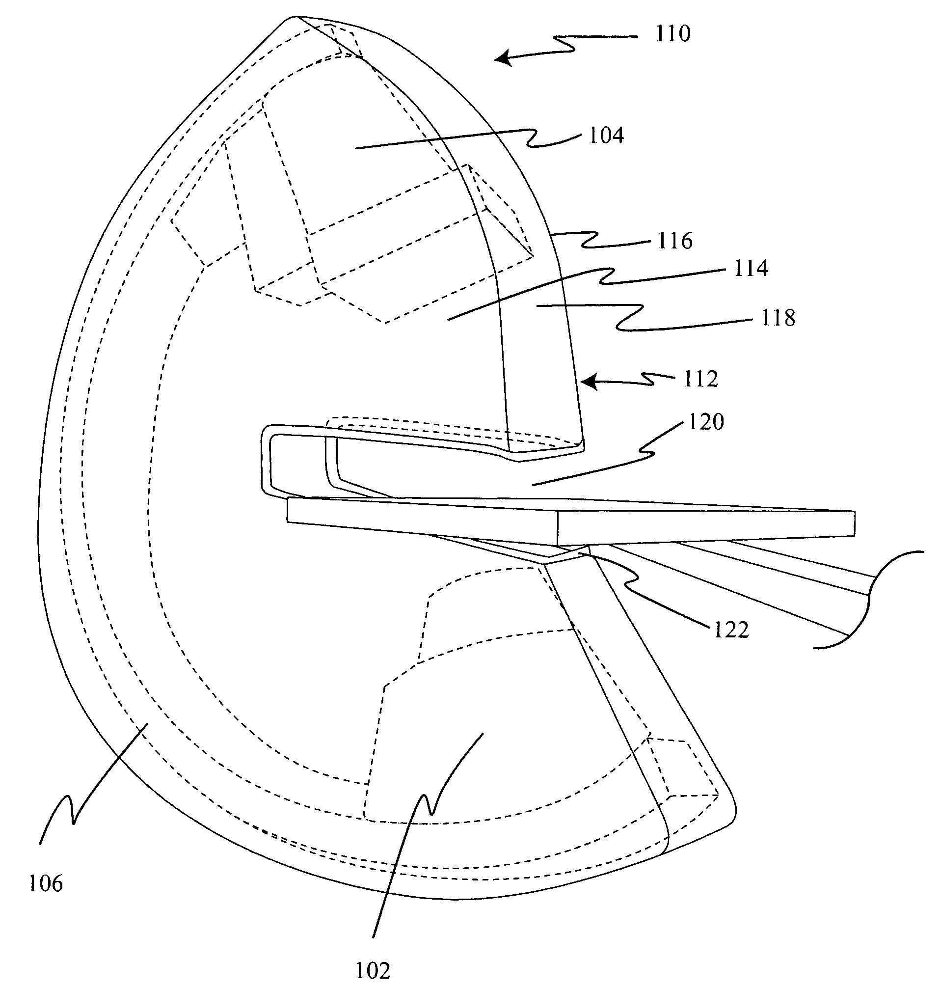
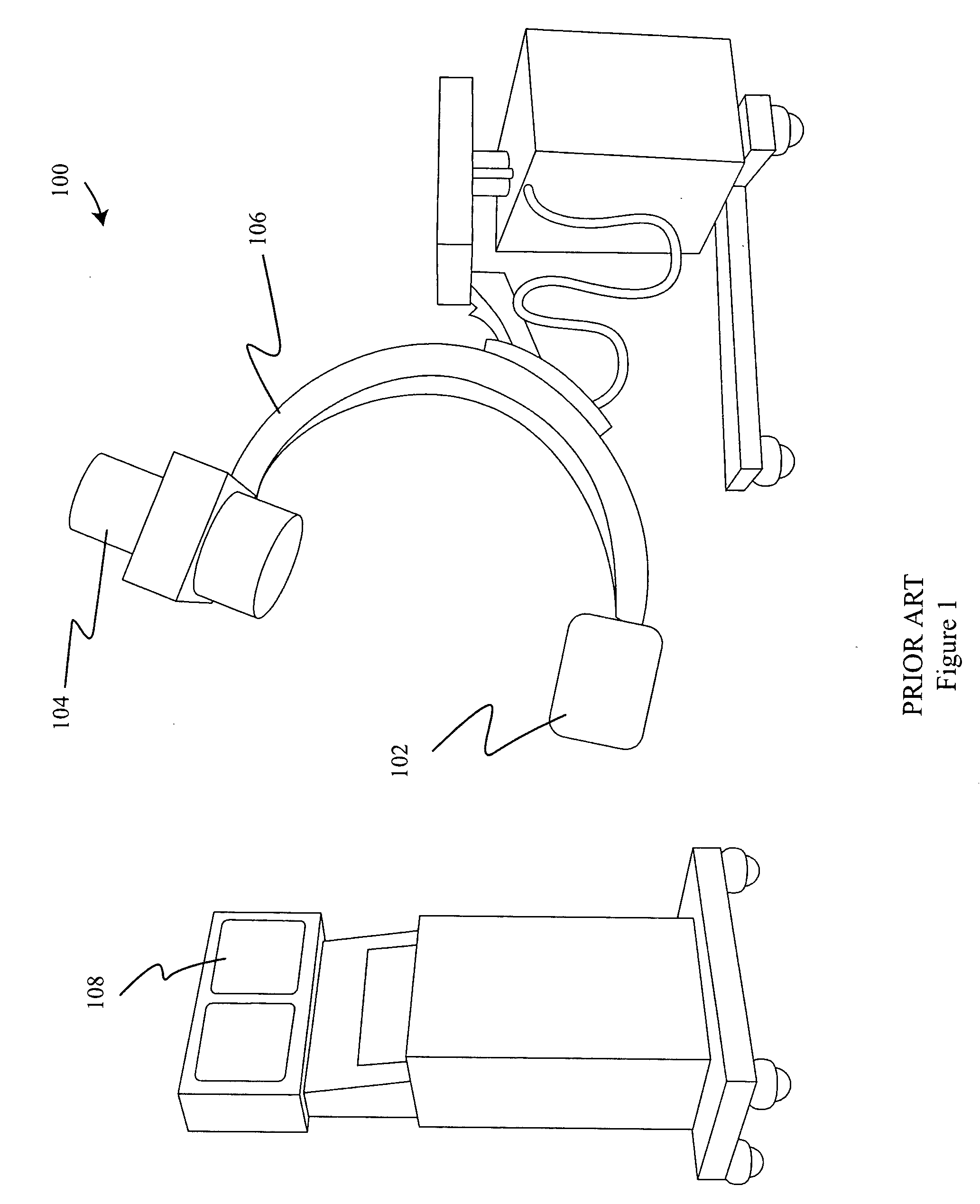
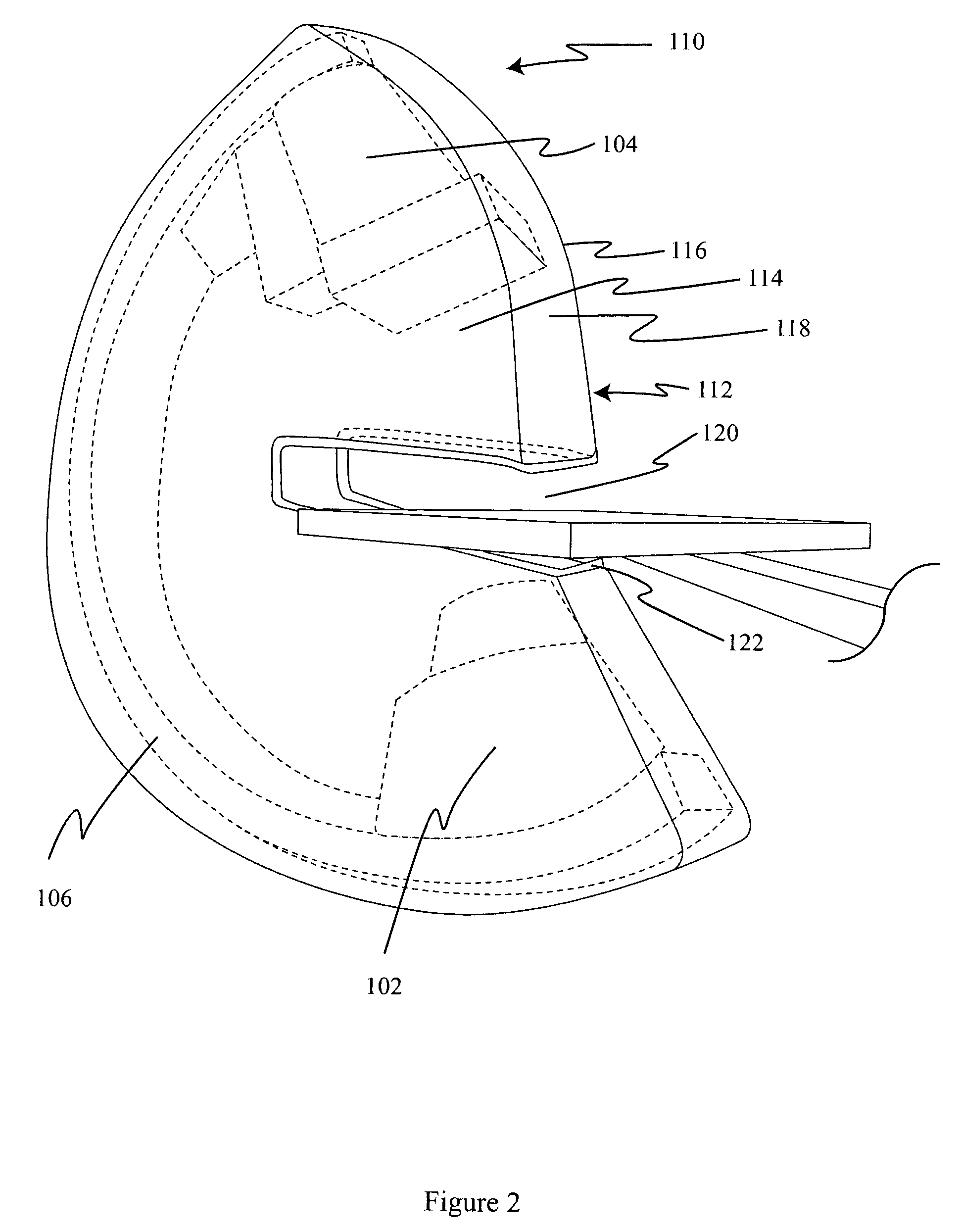
[0039]FIG. 1 illustrates a prior art C-arm fluoroscopy system 100. The fluoroscope 100 includes an X-ray source 102 and an image intensifier 104 mounted on opposite ends of a C-arm 106. The C-arm 106 may be mounted on a mobile base with wheels, as shown, or it may be mounted via a support arm to the floor or the ceiling of the fluoroscopy suite. In use, the X-ray source 102 and the image intensifier 104 are placed on opposite sides of the portion of the patient's body to be imaged. The X-ray source 102 directs an X-ray beam through the patient's body toward the image intensifier 104, which captures the X-ray image and displays it on a monitor 108 in real time. Often, the X-ray source 102 is positioned below the patient and the image intensifier 104 is positioned above as shown, however for some applications these positions may be reversed or the C-arm 106 may be positioned horizontally or at an oblique angle. The system may also include electronic memory for storing and replaying fl...


PUM


Abstract

A radiation protection device attaches to the C-arm of a fluoroscope and shields and collimates the X-ray beam between the X-ray source and the patient and between the patient and the image intensifier. One embodiment has a radiation shield of X-ray opaque material that surrounds the C-arm of the fluoroscopy system, the X-ray source and the image intensifier. A padded slot fits around the patient's body. Another embodiment has conical or cylindrical radiation shields that extend between the X-ray source and the patient and between the patient and the image intensifier. The radiation shields have length adjustments and padded ends to fit the device to the patient. The radiation protection device may be motorized to advance and withdraw the radiation shields. A blanket-like radiation shield covers the patient in the area surrounding where the X-ray beam enters the body.

Description

CROSS REFERENCE TO OTHER APPLICATIONS[0001]The present application is a continuation-in-part of PCT International application PCT / US2007 / 023892, filed on Nov. 7, 2007, which claims the benefit of U.S. Provisional Application 60 / 858,058, filed on Nov. 11, 2006 and U.S. Provisional Application 60 / 923,481, filed on Apr. 13, 2007. The disclosures of these and all patents and patent applications referred to herein are hereby incorporated by reference.FIELD OF THE INVENTION[0002]The present invention relates to radiation protection devices to protect fluoroscopy operators and other medical personnel from radiation exposure during fluoroscopic imaging procedures.BACKGROUND OF THE INVENTION[0003]Fluoroscopy, a real-time X-ray imaging technique, has long been important as a medical diagnostic tool, particularly in disciplines where still X-rays do not provide sufficient diagnostic information of the movement and function of living, moving organs. Fluoroscopy is widely used in cardiology, ele...

Claims


Application Information

Patent Timeline
IPC IPC(8): H01J35/16A61B6/04
CPCA61B6/107A61B6/589A61B6/4423A61B6/4441A61B6/487
Inventor BELSON, AMIRLEARY, JAMES J.
Owner RADIACTION
Why Eureka
Try Eureka