System and method for analyzing medical images

a medical image and system analysis technology, applied in the field of medical image processing and analysis, can solve the problems of low efficiency of the process, inability to obtain much data using these techniques, and inability to reliably detect pregnancies at risk of loss,

Inactive Publication Date: 2009-10-08
GOYAIKE A C I Y F
View PDF4 Cites 17 Cited by
  • Summary
  • Abstract
  • Description
  • Claims
  • Application Information

AI Technical Summary

Benefits of technology

[0004]Accordingly, a system and method are provided for analyzing and processing medical images of animal patients during pregnancy. The system and method enable the monitoring of pregnancies throughout gestation for possible abnormalities.

Problems solved by technology

However, these processes have shown low efficiency, mainly due to a high mortality rate of embryos and fetuses during pregnancy.
However, in veterinary medicine, not much data is available using these techniques, and reliable ultrasound markers of pregnancies at risk of loss have not previously been realized.
Even if parameters could be determined which indicated at risk pregnancy using medical imaging techniques, using these reproductive technologies in livestock animals research would result in voluminous amounts of data.

Method used


Image

  • System and method for analyzing medical images
  • System and method for analyzing medical images
  • System and method for analyzing medical images

Examples


Embodiment Construction

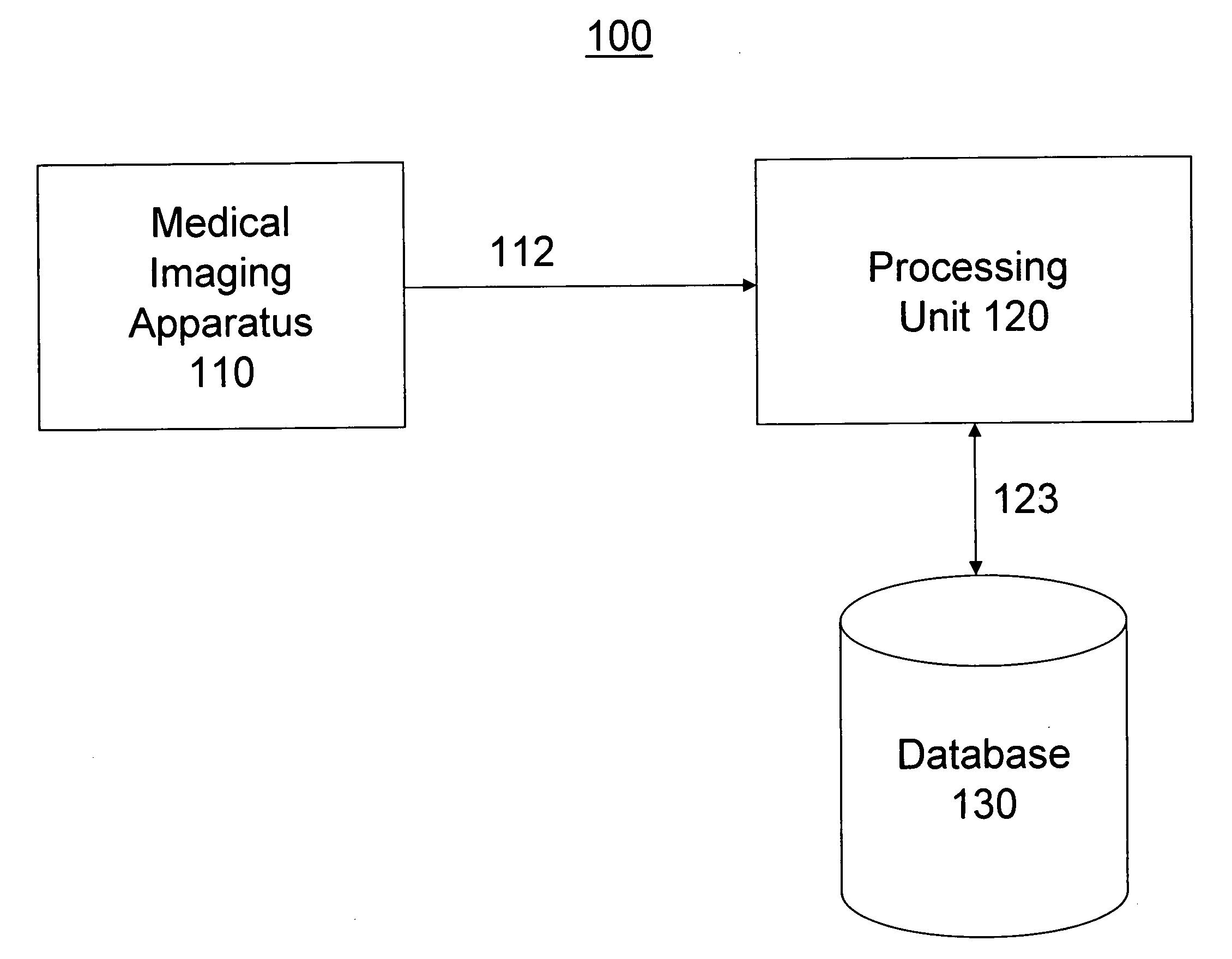
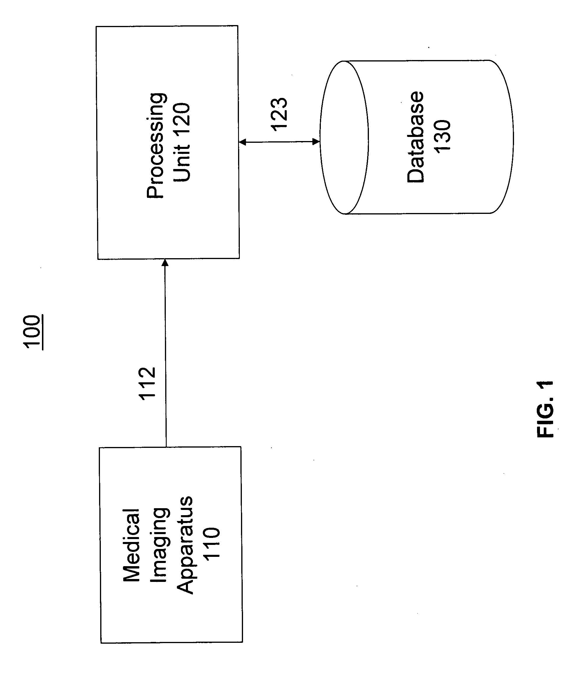
[0012]FIG. 1 depicts an overall configuration of a system 100 for analyzing medical images. System 100 may include one or more medical imaging apparatus 110, a processing unit 120, and a database 130. Medical imaging apparatus 110 may be communicatively coupled to processing unit 120 via communication link 112. Likewise, processing unit 120 may be communicatively coupled to database 130 via communication link 123. While depicted as separate entities, processing unit 120 and database 130 may both be components of a single computer workstation, such as, for example, a server.

[0013]Medical imaging apparatus 110 may include apparatus such as echo-doppler and / or ultrasound imaging machines. These apparatus are known to capture digital images in human patients which contain information that may be used to diagnose patient conditions and to monitor pregnancies. The inventors have determined several factors that may be used to monitor pregnancies in animal patients. Parameters may include, ...


PUM


Abstract

A system and method are provided for analyzing medical images and monitoring pregnancies for a plurality of animal patients. Medical images obtained from medical imaging devices are scanned, and extracted data are stored in a database. A color histogram is generating characterizing the image based on placental blood flow.

Description

FIELD OF THE INVENTION[0001]This application relates generally to systems and methods for analyzing medical images, and more specifically to systems and methods for processing and analyzing medical images to detect pregnancy abnormalities.BACKGROUND[0002]Biotechnological techniques, such as nuclear transfer and cloning, have become popular areas of research and development. However, these processes have shown low efficiency, mainly due to a high mortality rate of embryos and fetuses during pregnancy. To date, the causes of the mortalities have not yet been completely diagnosed until after they have occurred, or after signs of abortion present. Medical imaging technology, such as ultrasound and Doppler have been used to monitor human patients during pregnancy. However, in veterinary medicine, not much data is available using these techniques, and reliable ultrasound markers of pregnancies at risk of loss have not previously been realized.[0003]Even if parameters could be determined w...

Claims


Application Information

Patent Timeline
Patent Type & Authority Applications(United States)
IPC IPC(8): G06K9/00
CPCG06F19/3487G06F19/321G16H15/00G16H30/20
Inventor PANARACE, MARTIN
Owner GOYAIKE A C I Y F
Why Eureka
Try Eureka