Extended electron tomography

an electron microscope and electron beam technology, applied in material analysis using wave/particle radiation, instruments, nuclear engineering, etc., can solve the problem that the low-dose electron beam may be below the damaging level of the molecule conformation, and achieve the effect of improving the resolution of the three-dimensional structur

Active Publication Date: 2009-11-19
OKINAWA INST OF SCI & TECH PROMOTION CORP
View PDF3 Cites 76 Cited by
  • Summary
  • Abstract
  • Description
  • Claims
  • Application Information

AI Technical Summary

Benefits of technology

[0011]using the correlated data to improve the resolution of the three dimensional structure of the at least one conforma...

Problems solved by technology

The low dose electron beam may be below ...

Method used


Image

  • Extended electron tomography
  • Extended electron tomography
  • Extended electron tomography

Examples


Embodiment Construction

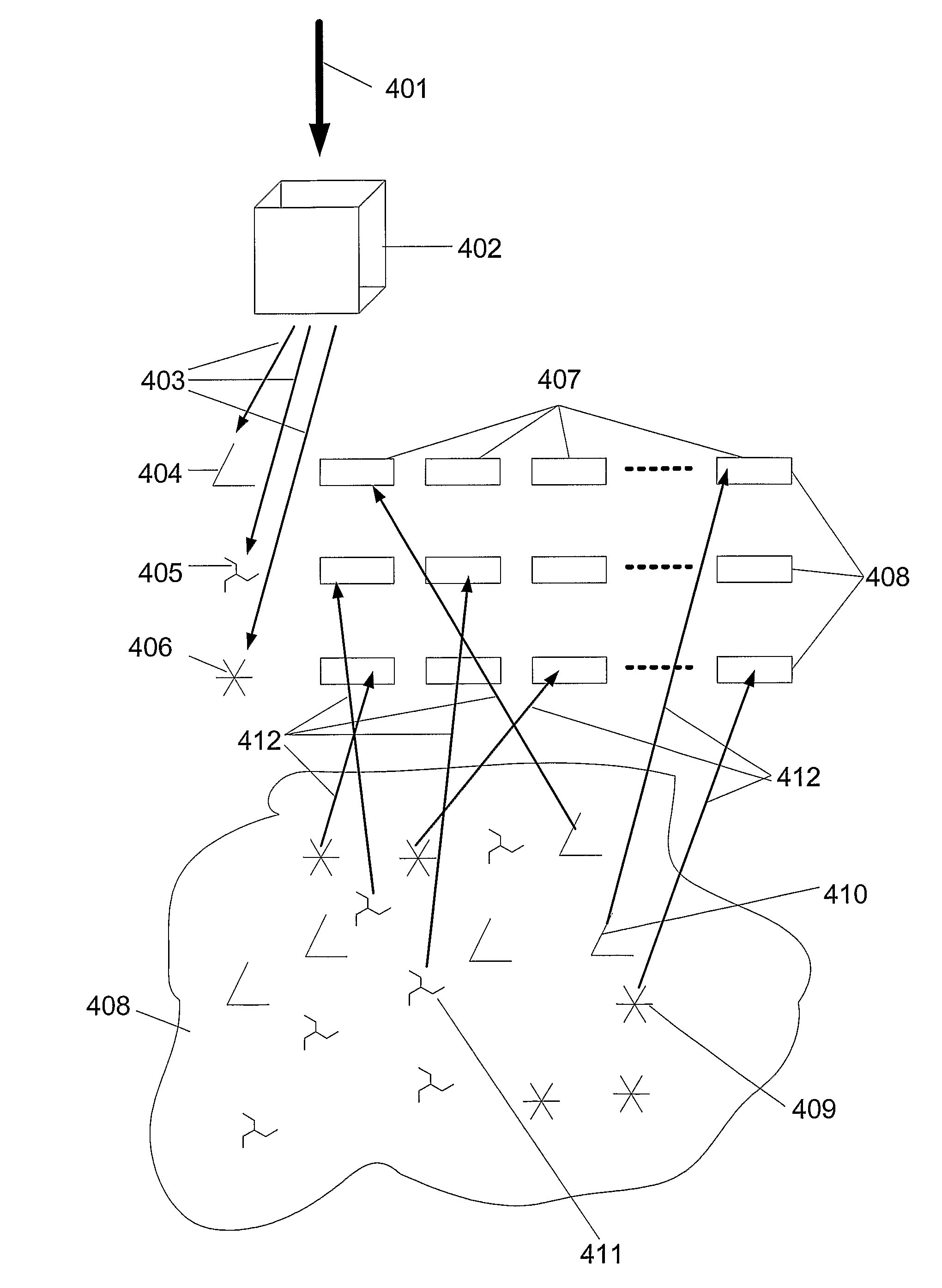
[0047]The present invention combines the 2D (two dimensional) image information of individual molecules of interest taken with low dose electron microscopy from several directions, e.g. by tilting the sample and acquiring images in a tilt series, used for determining the three dimensional structure, with image information in 2D taken at higher dose electron microscopy. The two image information sets may be correlated to each other and used in improving the 3D image information from the 2D set. The 3D “experimentally determined” structures of the molecule using low dose electron microscopy are used to provide simulated views of different conformations of the molecular structures. The simulation is provided with different angles of view of the molecule, different electron beam dosage, different molecules of interest, and so on. These simulated conformations are used in the determination of conformations present in the 2D image material.

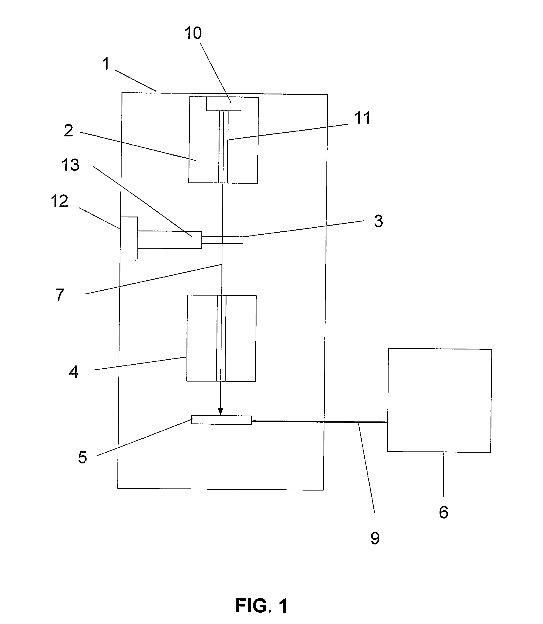
[0048]In FIG. 1 reference numeral 1 generally den...


PUM


Abstract

A method for improving image resolution of a three dimensional structure of at least one molecule conformation includes: determining a three dimensional structure of at least one conformation of a molecule in a sample from a first data set obtained from a series of 2D measurements of different geometrical projections of the molecule at a low electron beam dose in an electron microscope; producing a second data set including calculated two dimensional projections of the determined three dimensional structure of the at least one conformation of the same molecule; correlating data from a third data set obtained from at least one measurement of the same molecule using a higher electron beam dose with the second data set; and using the correlated data to improve the resolution of the three dimensional structure of the at least one conformation of the molecule by increasing the first data set with the correlated data and re-determining a three dimensional structure.

Description

TECHNICAL FIELD[0001]The present invention relates to electron tomography in an electron microscope and in particular to a method, computer program, and electron microscope using at least one experimentally determined three dimensional conformation as a tool for selecting and orienting particles from 2D EM images for improving the 3D conformation image resolution.BACKGROUND OF THE INVENTION[0002]Today cryo electron microscopy can be used to determine the 3 dimensional structures of macromolecules at near atomic resolution. EM can be used to get diffraction from 2D crystals or images in 2D of macromolecules that can be reconstructed into 3D by using 2D images from different angles. The first method is usually referred to as EM crystallography and the latter can be divided into electron tomography and single-particle electron microscopy. The electron tomography approach requires many pictures of the same particle tilted into different orientations. The single particle technique, colle...

Claims


Application Information

Patent Timeline
IPC IPC(8): G01N23/04G06K9/00
CPCG01N23/046G01N2223/419G06K2209/403G06K9/00134G06V20/693G06V2201/122
Inventor SKOGLUND, ULF
Owner OKINAWA INST OF SCI & TECH PROMOTION CORP
Why Eureka
Try Eureka