Hyoid suspension for obstructive sleep apnea

a technology of obstructive sleep apnea and hyoid suspension, which is applied in the field of treating sleep disorders, can solve the problems of excess and/or collapse of soft tissue, obstruction of the upper airway,

Inactive Publication Date: 2009-12-24
BIOMERIX CORP
View PDF3 Cites 34 Cited by
  • Summary
  • Abstract
  • Description
  • Claims
  • Application Information

AI Technical Summary

Benefits of technology

[0026]In another aspect of the method of the invention, the implant is attached to one or more hyoid bone segments with one or more staples, another mechanical means, or adhesive.

Problems solved by technology

Upper airway obstruction results from excess and / or collapse of soft tissue in the soft palate, tonsillar pillars, tongue, tongue base, and hypopharyngeal walls.

Method used


Image

  • Hyoid suspension for obstructive sleep apnea
  • Hyoid suspension for obstructive sleep apnea
  • Hyoid suspension for obstructive sleep apnea

Examples


example 1

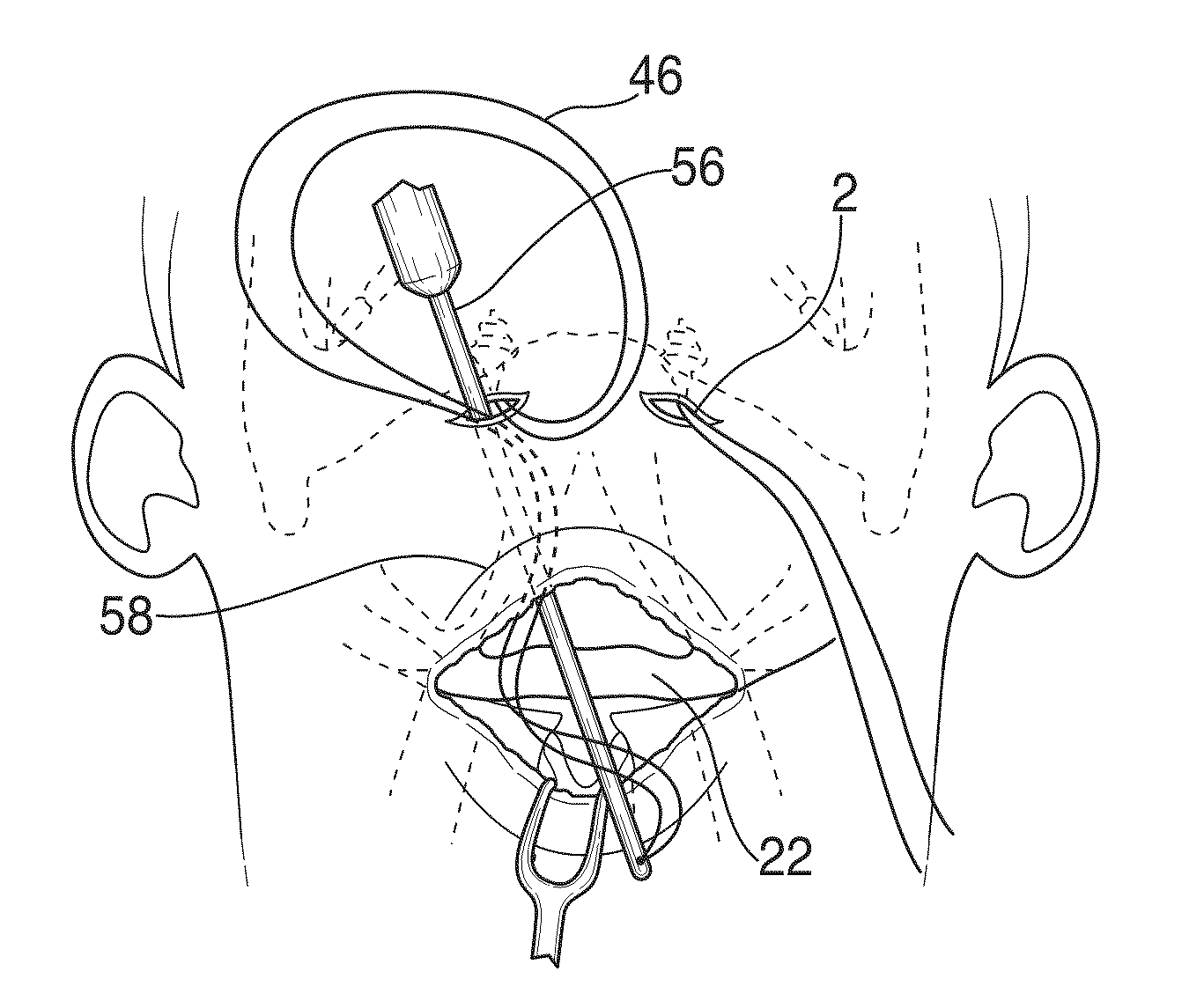
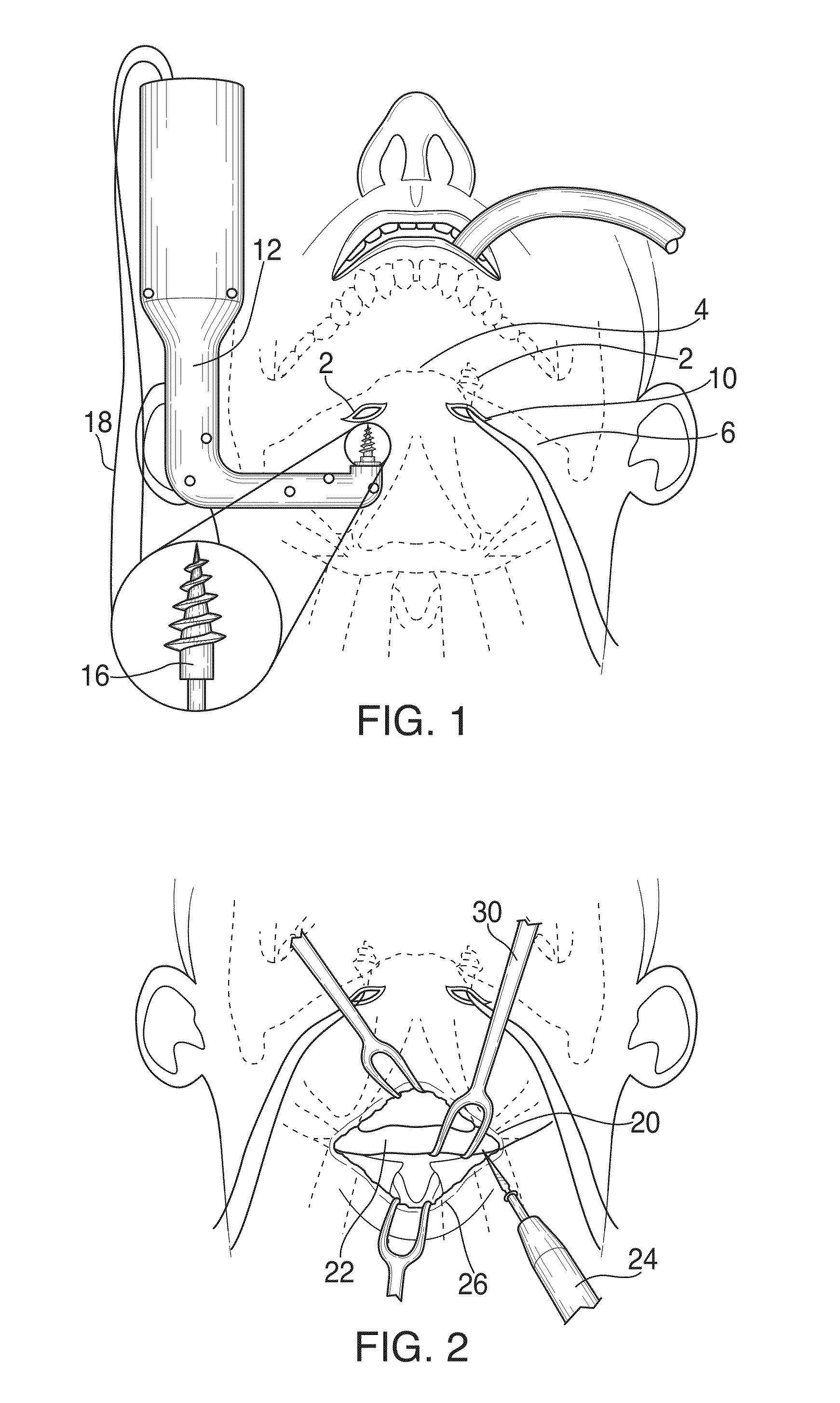
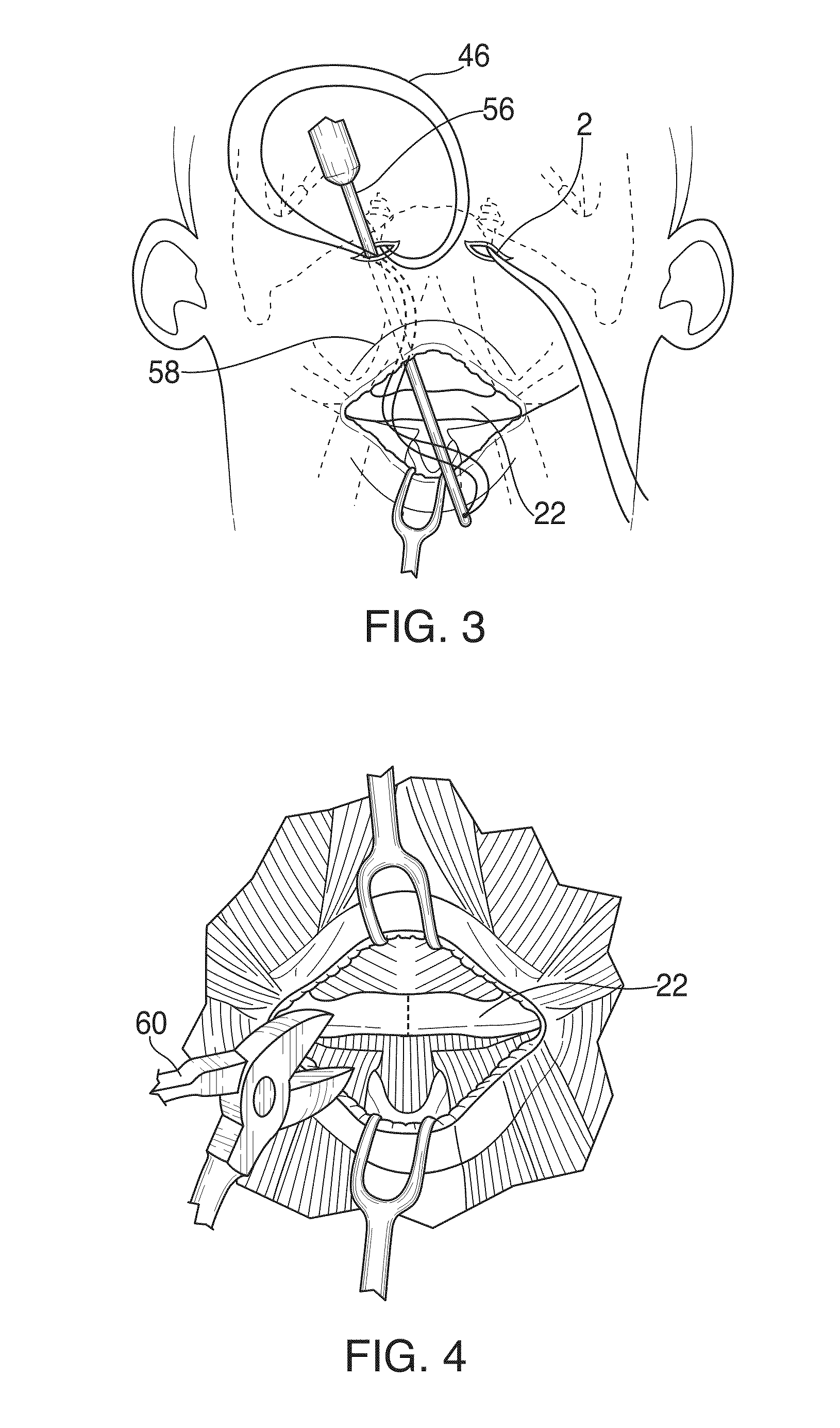
[0079]Patients were treated as described above with regard to FIGS. 1 to 6. The most common postoperative patient complaints included dysphagia, odynophagia and surgical wound pain. All patients were able to tolerate liquids and a soft diet at the time of discharge. No patients requested or required removal of the suspension sutures.

[0080]After 3 to 12 months of follow-up postoperatively, over 90% of the patients were subjectively improved by patient and / or spouse history. Symptomatic improvement included decrease in snoring, improved quality of sleep, and decreased daytime somnolence. Follow-up polysomnographic studies were performed 3 months to 1 year postoperatively in 52 patients. The patients that underwent hyo-mandibular suspension with or without hyoid expansion showed significant improvement in the mean RDI (Respiratory Disturbance Index) from 71.2 (±18.0) preoperatively to 28.4 (±16.8) postoperatively.

example 2

[0081]An incision was made in the anterior portion of the neck of a dog, and an implant comprised of reticulated elastomeric polycarbonate polyurethane-urea matrix was inserted into musculature at the base of the dog's tongue. A cavity was created, and the implant was secured with sutures in that position and left in place for three months. Fibrovascular tissue cells grew into the elastomeric matrix of the implant, as is shown in the micrograph slides shown in FIGS. 11 and 12. The implant comprises a netlike structure of unstained, short, narrow, birefringent synthetic fibers, and these foreign fibers are surrounded by individual histiocytes and (mmGCs) of foreign body type. These cells are between the device material and many small (˜0.2 mm to ˜0.4 mm) irregularly round masses of fibroblasts and collagen. Within these fibrous foci are arterioles, veins and capillaries of varied size and number. The fibrocytic reaction at the perimeter of the device extends into the interstitial tis...


PUM


Abstract

In an improved method for surgically treating a sleep-related breathing disorder, comprising hyoid distruction/expansion and hyo-mandibular suspension, an implant comprises a polypropylene and/or reticulated elastomeric biostable polyurethane matrix. The matrix is positioned between two or more segments of the distructed hyoid bone. A single or multiple midline or paramedian incisions are made under a patient's chin; soft tissues overlying the mandible are cleaned; a first screw is inserted into the edge of the mandible; a second screw is inserted into the inferior edge of the mandible; a second horizontal incision is made over the body of the hyoid; the infrahyoid muscles are separated from the body of the hyoid bone; the hyoid is refracted and stabilized; the sternohyoid and thyrohyoid muscles are detached from the body of the hyoid between the lesser cornuae; the hyoid bone is divided midline or paramedian into two or more segments; and an absorbable or nonabsorbable, rigid or semirigid, implant is positioned between segments of the hyoid bone. The hyoid bone is stabilized by the implant; and sutures or portions of polypropylene and/or polyurethane matrix are passed around the hyoid bone segments and implant to connect to muscle still attached to the upper portion of the hyoid bone, wherein the hypopharyngeal airway is expanded.

Description

FIELD OF THE INVENTION[0001]This invention is directed to a method of treating sleep disorders. More particularly, this invention is directed to a method of treating sleep apena comprising hyoid expansion and hyo-mandibular suspension.BACKGROUND OF THE INVENTION[0002]Upper airway obstruction results from excess and / or collapse of soft tissue in the soft palate, tonsillar pillars, tongue, tongue base, and hypopharyngeal walls. Specific anatomic risk factors predisposing to obstructive sleep apnea / hypopnea syndrome (OSAHS) include long soft palate, shallow palatal arches, large tongue base, narrow mandibular arches, and mandibular hypoplasia. [Rojewski et al., 1984] Other general anatomic factors often associated with OSAHS include obesity, large body mass index (BMI), shortened thick neck, anterior larynx location, enlarged tonsils and / or adenoids, lingual tonsils, thickened pharyngeal walls, elongated uvula, redundant soft palate, large tongue volume, deviated nasal septum, turbinat...

Claims


Application Information

Patent Timeline
Patent Type & Authority Applications(United States)
IPC IPC(8): A61F2/28A61B17/56
CPCA61F5/56
Inventor KRESPI, YOSEF P.FRIEDMAN, CRAIG D.DATTA, ARINDAMLAVELLE, LAWRENCE P.
Owner BIOMERIX CORP
Why Eureka
Try Eureka