Automatic interpretation of 3-d medicine images of the brain and methods for producing intermediate results

a technology of brain and image, applied in the field of processing and interpreting medical images, can solve the problems of difficult and time-consuming, difficult and time-consuming, and long time-consuming to achieve the effect of a large amount of time and effor

Active Publication Date: 2010-03-18
EXINI DIAGNOSTICS
View PDF6 Cites 37 Cited by
  • Summary
  • Abstract
  • Description
  • Claims
  • Application Information

AI Technical Summary

Benefits of technology

[0005]It is also an object of the present invention to provide a method for creating such a normal image, and it is a furth...

Problems solved by technology

Diagnosing based on 3-D nuclear images of the brain, such as SPECT images of cerebral blood flow (CBF) and DaTSCAN images, is difficult and time dema...

Method used


Image

  • Automatic interpretation of 3-d medicine images of the brain and methods for producing intermediate results
  • Automatic interpretation of 3-d medicine images of the brain and methods for producing intermediate results
  • Automatic interpretation of 3-d medicine images of the brain and methods for producing intermediate results

Examples


examples

Overall Method

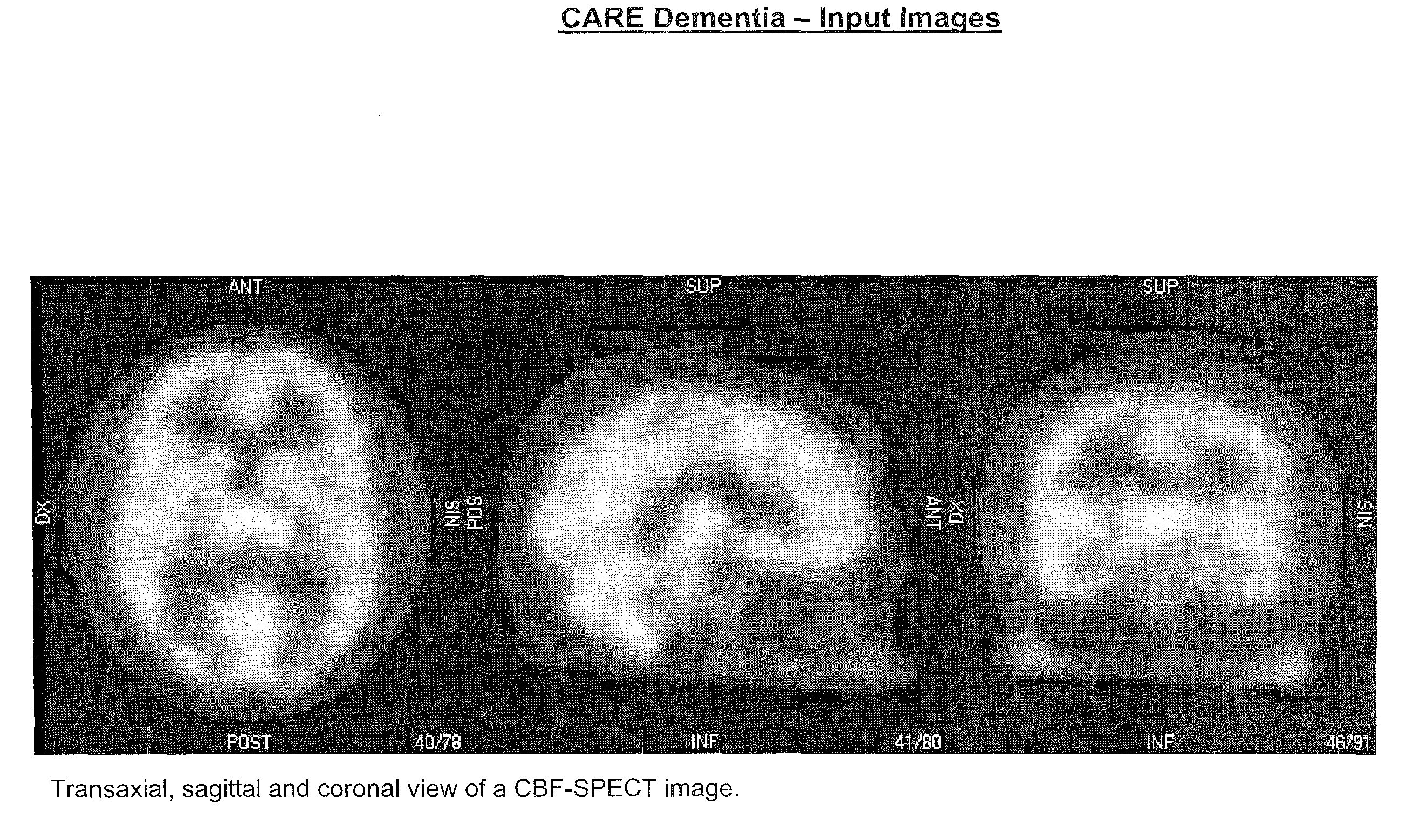
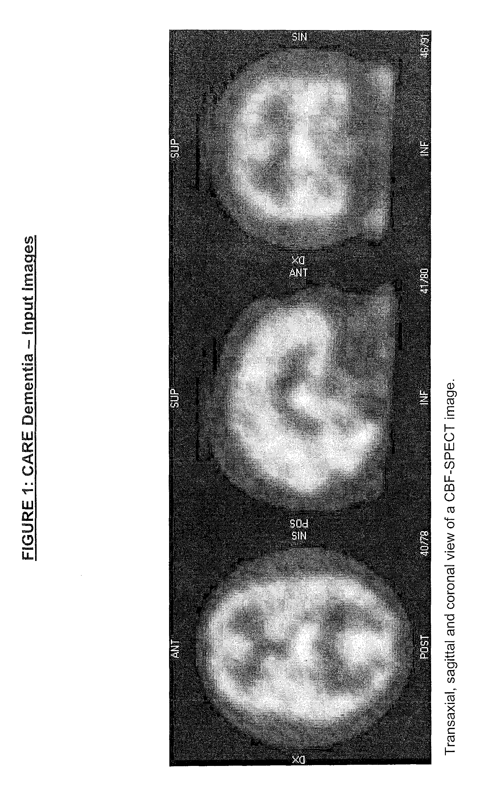
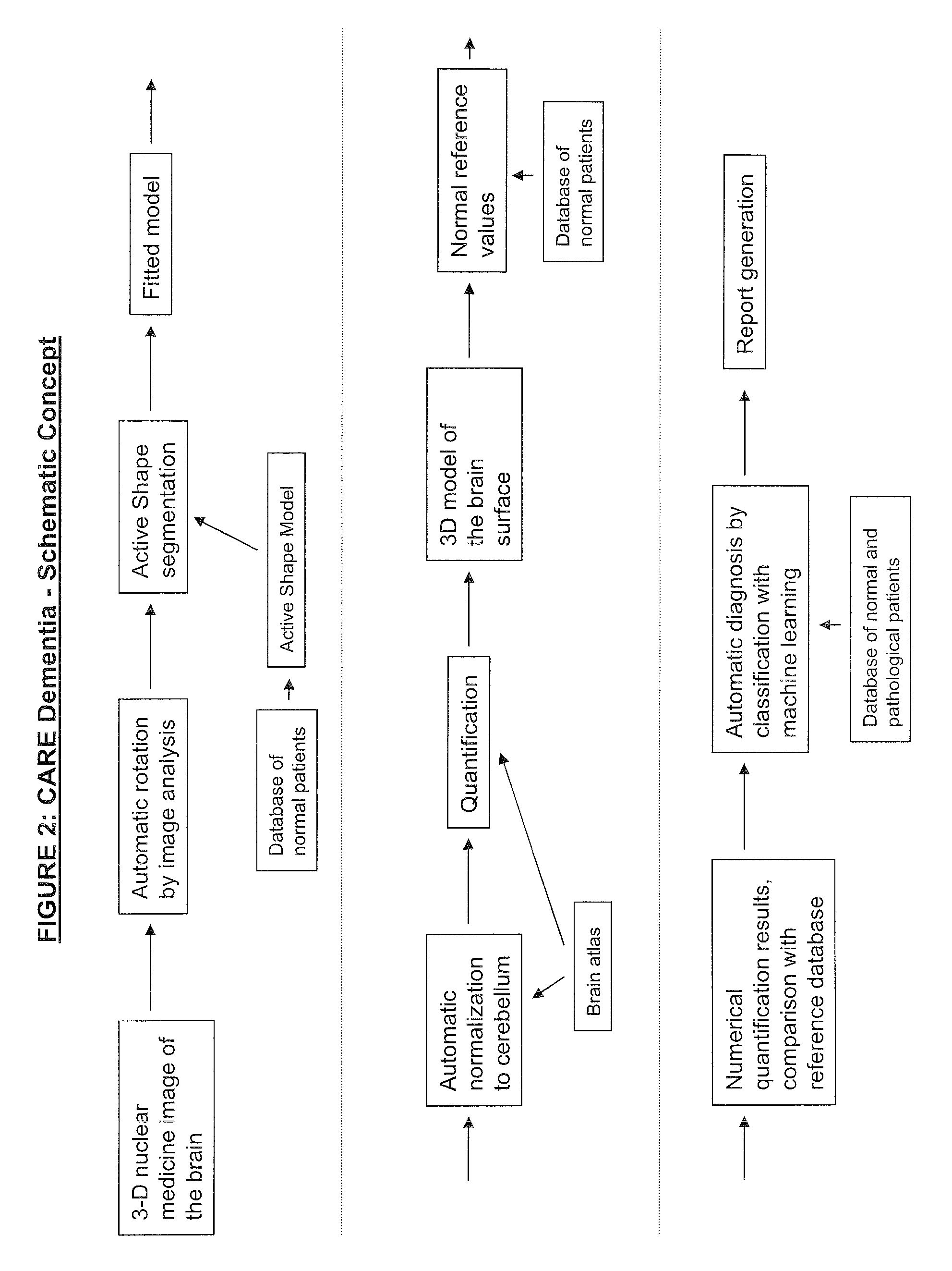
[0095]FIG. 7 shows an overview flowchart of a method for computer aided diagnosis of 3D images of the brain. The method comprising the following steps:

[0096]Automatically rotating 710 three-dimensional image to align to conventional views;

[0097]Outlining 715;

[0098]Normalisation 720;

[0099]Quantification 725, point-by-point, and region-by-region;

[0100]Comparison 730 with reference database;

[0101]Displaying 735 of results;

[0102]Extracting 740 of features for input to artificial neural network (ANN);

[0103]Automatic interpretation 745;

[0104]Automatic generation 750 of report.

[0105]Also provided is a method for the automatic rotation, mentioned above, of a numerical representation of a three dimensional object. The representation comprises a number of 3D pixels, here called voxels, each voxel having a value representing an intensity value corresponding to an amount of some quality of the original object voxel.

[0106]The method comprises the following steps:

[0107]a first step ...


PUM


Abstract

Methods for fully automatic quantification and interpretation of three dimensional images of the brain or other organs. A system for Computer Aided Diagnosis (CAD) of diseases affecting cerebral cortex from SPECT images of the brain, where said images may represent cerebral blood flow (CBF). The methods include image processing, statistical shape models, a virtual brain atlas, reference databases and machine learning.

Description

FIELD OF THE INVENTION[0001]The present invention relates to the field of processing and interpreting medical images.BACKGROUND OF THE INVENTION[0002]Diagnosing based on 3-D nuclear images of the brain, such as SPECT images of cerebral blood flow (CBF) and DaTSCAN images, is difficult and time demanding and relies on visual interpretation and numerical quantification of the images. The existing systems for displaying and quantifying such images bring a lot of manual work for the physician which may have to mark region(s) of interests with geometrical templates. This takes a lot of time and the accuracy of the segmentation is poor.[0003]SE 2020565 discloses a method and a device for determining of a three dimensional contour of an organ in a patients body, starting from an image of said organ. The method comprises a step of adjusting a predefined contour model to the image of said organ.[0004]The object of the present invention is therefore to provide a method for reducing the need f...

Claims


Application Information

Patent Timeline
IPC IPC(8): G06K9/00
CPCG06K9/6209G06K2209/05G06T7/0012G06T7/0083G06T2207/30016G06T2207/10108G06T2207/20124G06T2207/20128G06T7/0089G06T7/12G06T7/149G06V10/7553G06V2201/03
Inventor JAKOBSSON, DAVIDRICHTER, JENSJARUND, ANDREAS
Owner EXINI DIAGNOSTICS
Why Eureka
Try Eureka