Tissue Slicer

a tissue slicer and tissue technology, applied in the field of biological tissue slicers, can solve the problems of affecting pathologists' gross evaluation of surgical tissue specimens, so as to improve the quality of tissue sections, minimize trauma and structural distortion, and optimize processing

Inactive Publication Date: 2010-03-25
UNIV KANSAS MEDICAL CENT
View PDF42 Cites 18 Cited by
  • Summary
  • Abstract
  • Description
  • Claims
  • Application Information

AI Technical Summary

Benefits of technology

[0007]The present invention is directed to a biological tissue slicer device which rapidly produces slices of tissue of identical uniform thickness, while minimizing trauma and structural distortion, and contamination to the tissue, thus standardizing tissue sections for optimal processing, improving quality of tissue sections for optimal microscopic evaluation, and ultimately improving diagnostic accur

Problems solved by technology

Currently, pathologists grossly evaluate surgical tissue specimens and tediously cut the sections with primitive blades, razors, and scalpels slice-by-slice in order to prepare the tissue for analysis and diagnosis.
That process sometimes leads to many unwel

Method used


Image

  • Tissue Slicer
  • Tissue Slicer
  • Tissue Slicer

Examples


first embodiment

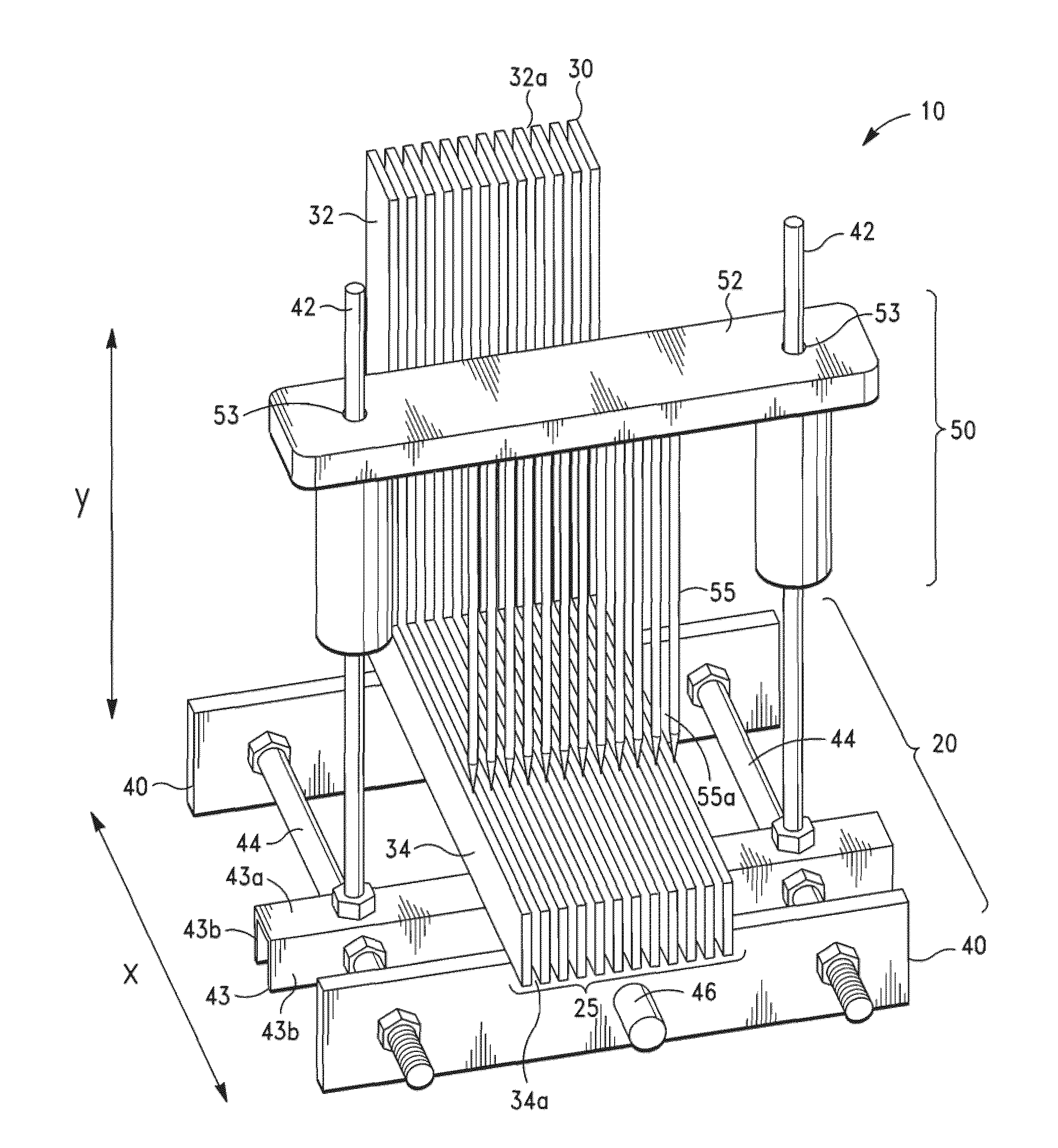
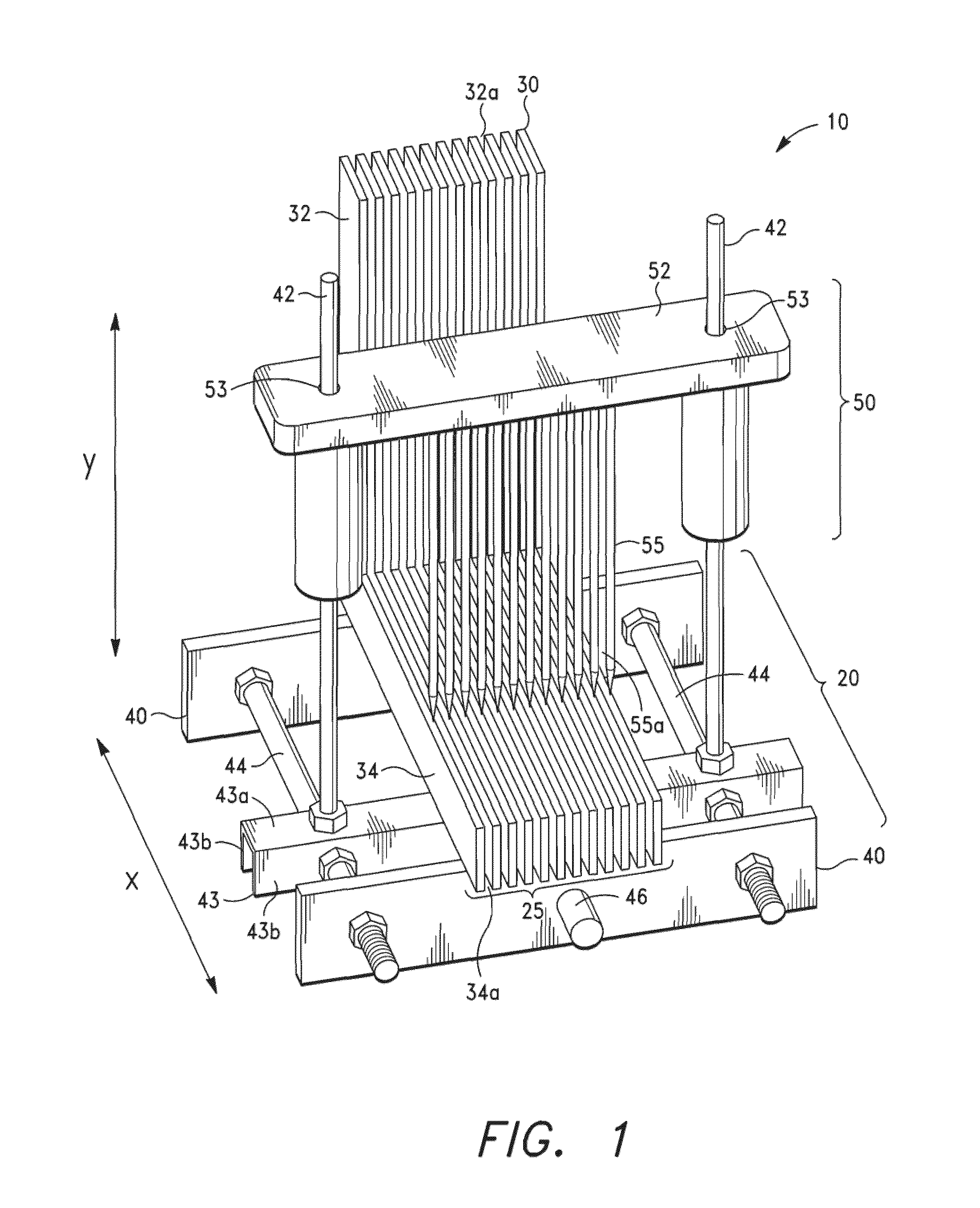
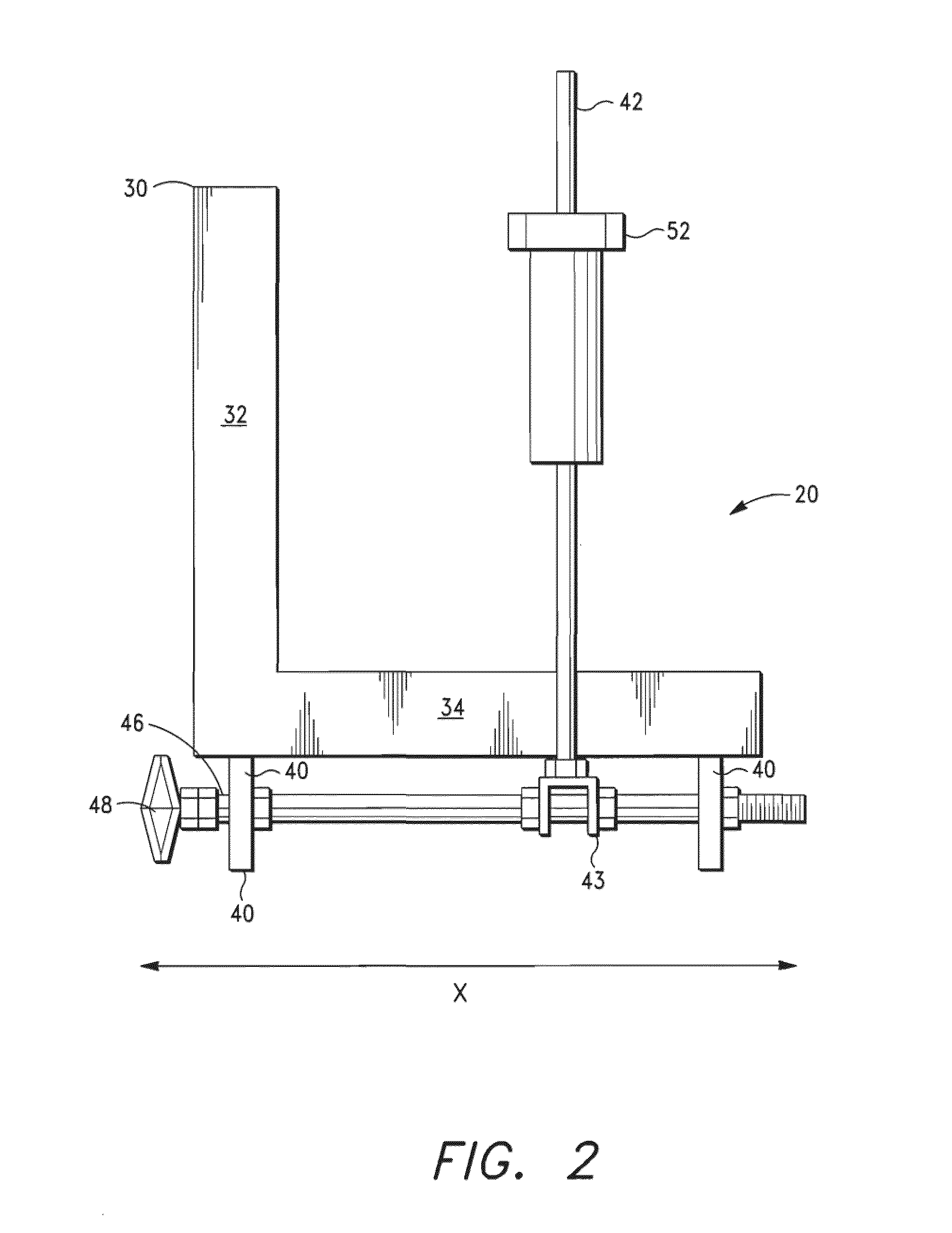
[0040]FIGS. 1 to 7B illustrate a tissue slicer 10 in accordance with the present invention. As shown in FIG. 1, the tissue slicer 10 comprises a base 20 having a partially open horizontal surface 25. The partially open surface 25 has a plurality of openings or slits that permit one or more cutting blade(s) to traverse therethrough. The base 20 is comprised of a plurality of plates 30, preferably angled plates 30, which are aligned to create the openings. The angled plates are aligned in parallel such that there is a gap between the adjacent plates 30. The angled plates 30 preferably comprise metal L-shaped plates, each having a vertical member 32 and a horizontal member 34 spaced about 1 to 10 mm apart (e.g., 1, 2, 3, 4, 5, 6, 7, 8, 9, 10 mm apart, or as needed). The gaps 32a between the vertical members 32 are adapted to receive the blade(s) in the tissue slicer and also function as a guide for precise cutting. The biological tissue specimen may also be positioned against the verti...

second embodiment

[0050]FIGS. 8 to 11 illustrate a biological tissue slicer 210 in accordance with the present invention. The tissue slicer 210 comprises a base plate 220 and at least one vertical member 230 for engaging a dual pivoting member 240 connected to a blade cartridge 260 (as discussed more fully below). The base plate 220 is adapted to receive a removable pin base 252 holding a plurality of pins 255 that forms a partially open horizontal surface 225. The pins are preferably pointed where they engage the tissue specimen, but need not be as shown in the figures. FIG. 8 illustrates the plurality of pins 255 in a linear rectangular configuration; however, the pins may be configured in other geometric and non-geometric configurations. In addition, the pins may be spaced either equidistantly (e.g., about 2 to 6 mm, preferably about 4 mm) or non-equidistantly relative to one another. However, the pins are positioned such that there are gaps 255a that permit the cutting blades 265 in the blade car...

third embodiment

[0055]FIGS. 12 to 16B illustrate a tissue slicer 310 in accordance with the present invention. As shown in FIGS. 12 to 14A, the tissue slicer 310 comprises a base 320 having a partially open horizontal surface 325. The base 320 is comprised of a plurality of horizontal plate members 334 spaced about 1 to 10 mm apart (e.g., 1, 2, 3, 4, 5, 6, 7, 8, 9, 10 mm apart, or as needed). The horizontal plate members 334 function as a partially open horizontal surface 325 where the tissue specimen is positioned. The gaps 334a between the horizontal plate members 34 are adapted to receive the blades(s) in the tissue slicer so that the blade(s) can extend through and cut the entire tissue specimen (and even beyond the thickness of the tissue specimen).

[0056]The base 320 having the horizontal plates 334 is mounted to a frame 340 to maintain the horizontal plate members 334 in an adjustable position. The base 320 includes a means for slidably moving the base 320 relative to the pins 355 along the f...


PUM


Abstract

A tissue slicer having a partially open base for permitting the slicing blades to transverse therethrough, a dual pivoting member for activating the sliding blades in a vertical direction, a blade cartridge with a plurality of blades and a multi-pin specimen holder. The device secures the tissue specimen without distortion in place during the slicing process, protect the user while cutting specimens, standardizes tissue sections for optimal processing, improves the quality of sections for microscopic evaluation, and improves diagnostic accuracy and reliability.

Description

CROSS-REFERENCE TO RELATED APPLICATIONS[0001]This application is based on and claims priority to U.S. Provisional Application Ser. No. 61 / 100,050 filed on Sep. 25, 2008, which is incorporated herein by reference.STATEMENT REGARDING FEDERALLY SPONSORED RESEARCH OR DEVELOPMENT[0002]Not applicable.BACKGROUND OF THE INVENTION[0003]1. Field of the Invention[0004]This invention relates to a biological tissue slicer designed to produce thin, uniform slices of tissue suitable for biochemical, pharmacological, toxicological, pathologic, autopsy, animal tissue research studies, and other applications. The invention produces standardized tissue sections of a defined thickness for optimal tissue processing.[0005]2. Description of Related Art[0006]Specimen sampling is one of the most critical steps in achieving the correct diagnosis. Currently, pathologists grossly evaluate surgical tissue specimens and tediously cut the sections with primitive blades, razors, and scalpels slice-by-slice in orde...

Claims


Application Information

Patent Timeline
IPC IPC(8): A61B17/32
CPCG01N1/06
Inventor TAWFIK, OSSAMAJAIN, SWARAN K.FANOUS, MAGEDMONTGOMERY, BROOKE
Owner UNIV KANSAS MEDICAL CENT
Why Eureka
Try Eureka