Treatment instrument for endoscope

a treatment instrument and endoscope technology, applied in the field of endoscope treatment instruments, can solve the problems of poor visibility of the distal end electrode portion and the axial electrode that are on the extension of the distal end portion of the sheath as well as a treatment site, and it is difficult to insert the distal end electrode portion into the submucosa from the dissected mucosa, so as to achieve safe and easy ablation of mucosa or submu

Active Publication Date: 2010-04-01
JICHI MEDICAL UNIVERSITY +1
View PDF9 Cites 5 Cited by
  • Summary
  • Abstract
  • Description
  • Claims
  • Application Information

AI Technical Summary

Benefits of technology

[0020]According to the treatment instrument for an endoscope of the aspects of the present invention, since a distal end portion of a sheath is formed in a tapered shape, visibility with respect to a distal end electrode portion, an axial electrode, and a treatment site can be improved, and ablation of mucosa or submucosa can be performed safely and with ease.

Problems solved by technology

Thus, there is the drawback that visibility with respect to the distal end electrode portion and the axial electrode that are on an extension of the distal end portion of the sheath as well as a treatment site is poor.
Also, there is the drawback that it is difficult to insert the distal end electrode portion into the submucosa from the dissected mucosa.

Method used


Image

  • Treatment instrument for endoscope
  • Treatment instrument for endoscope
  • Treatment instrument for endoscope

Examples


second embodiment

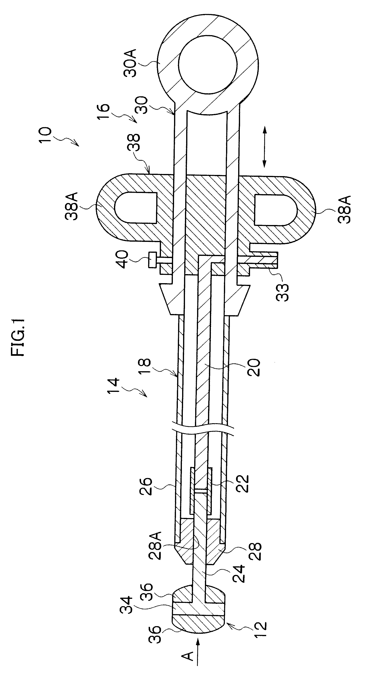
[0076]FIG. 9 is a side view that illustrates a treatment instrument for an endoscope according to the In FIG. 9, members that are the same as, or similar to, members of the treatment instrument 10 for an endoscope shown in FIG. 3 are designated by the same reference numerals, and a description of those members is omitted.

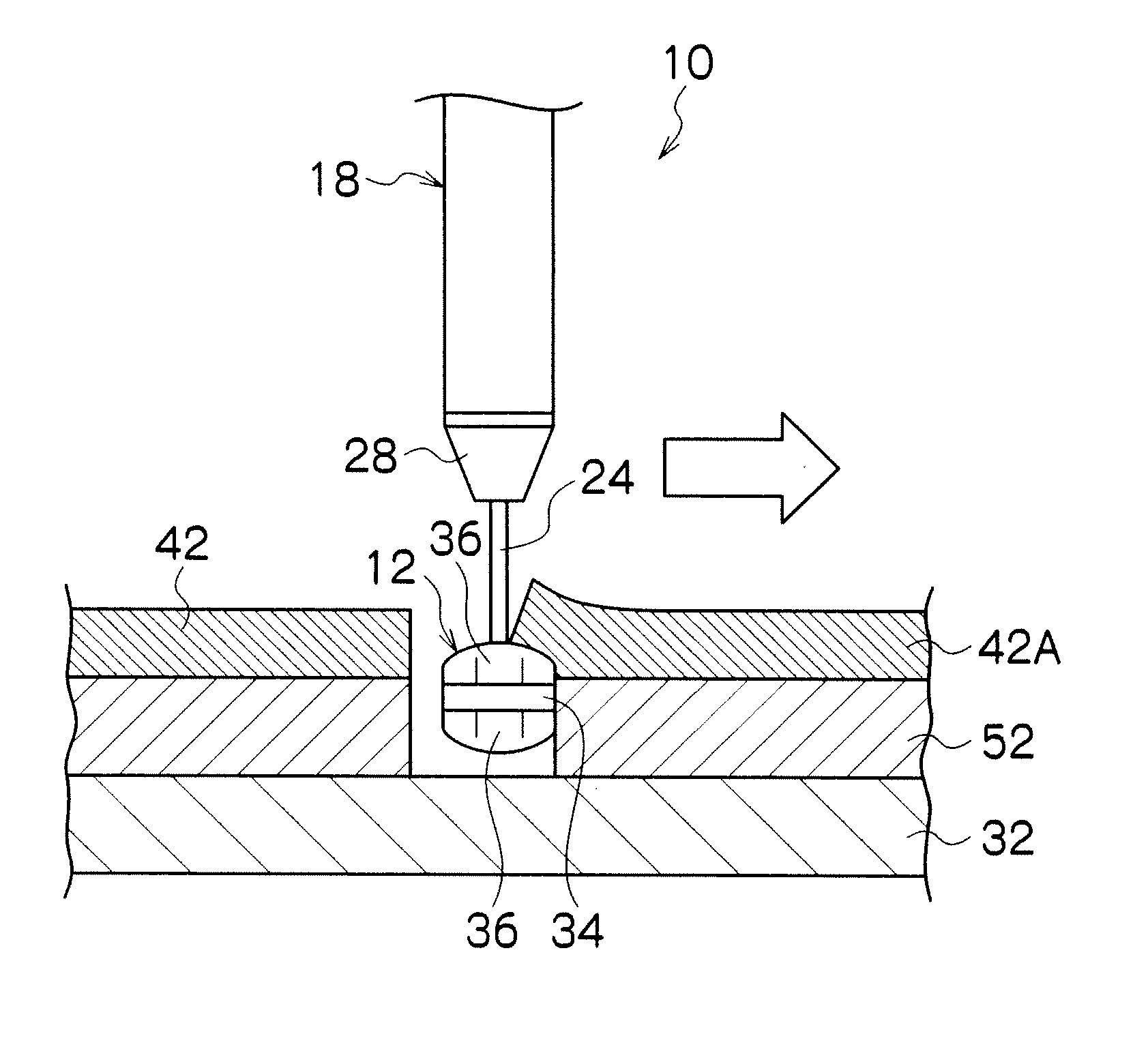
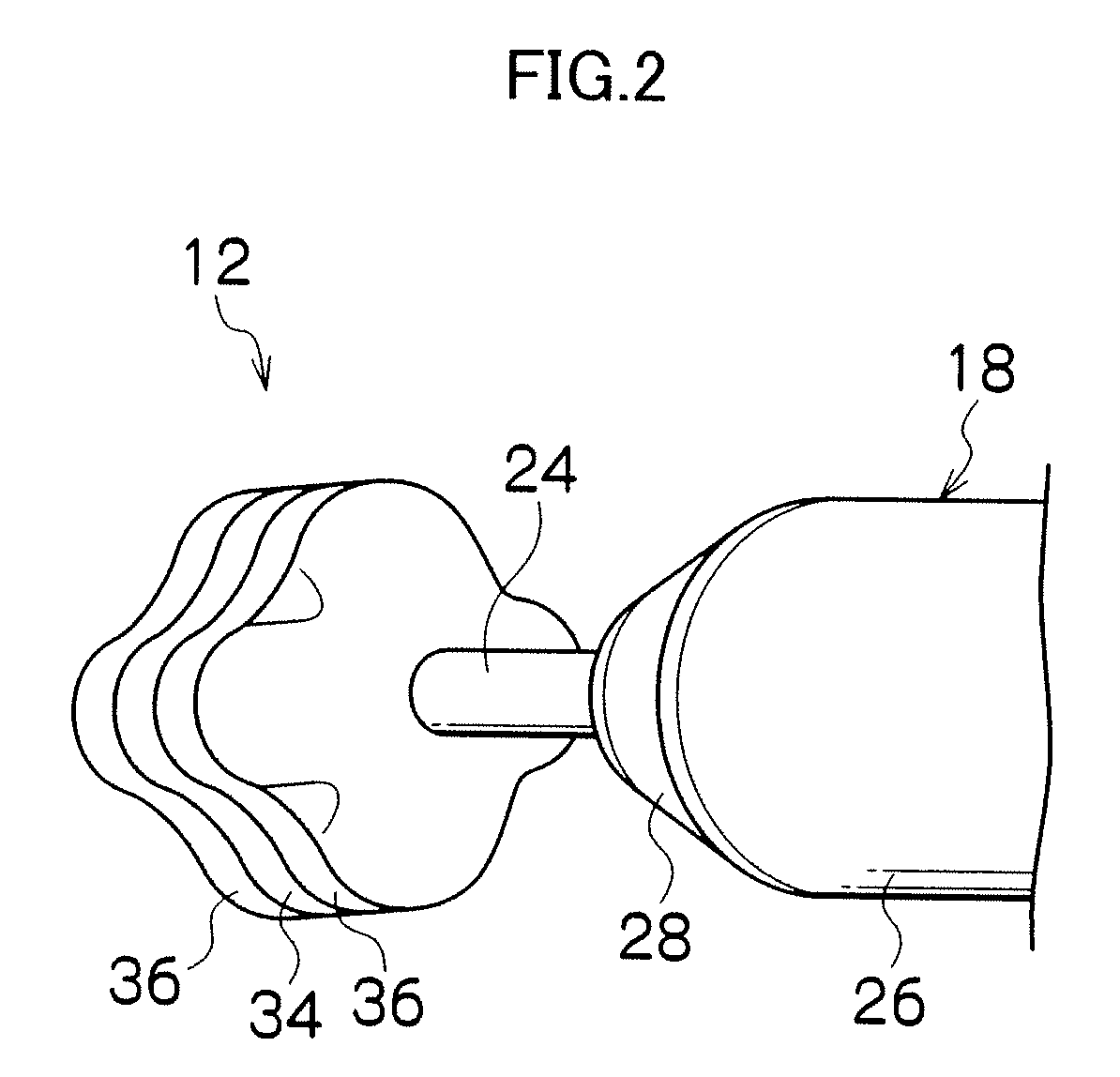
[0077]In the treatment instrument for an endoscope shown in FIG. 9, a flat face 31A that is substantially parallel with the protrusion / retraction direction of the axial electrode 24 is formed at the distal end portion of the sheath 18, that is, at the distal end portion of the collar member 28, and an end face 31B of the flat face 31A is formed in a largish fashion. When the axial electrode 24 is retracted into the sheath 18, a rear end face 36A of the non-conductive portion 36 on the proximal end side of the distal end electrode portion 12 has surface contact with the end face 31B. Therefore, the distal end electrode portion 12 is stably retained by the end face 3...

third embodiment

[0079]For example, a distal end electrode portion 112 shown in FIG. 10 and FIG. 11 which show the third embodiment is formed in a shape in which four plate members are combined in the shape of a cross. More specifically, in a main unit 114 of the distal end electrode portion 112, four non-conductive plate members are disposed at 90° intervals to form a cross shape that connects with a central shaft side of the distal end electrode portion 114.

[0080]On the distal end side of the main unit 114, four protruded portions 114A, 114A, . . . are formed by each plate member protruding to the distal end side, and depressed portions 114B, 114B, . . . are formed at center positions between each of the protruded portions 114A, 114A, . . . . Similarly, on the proximal end side of the main unit 114, protruded portions 114C, 114C, . . . are formed by each plate member protruding to the proximal end side, and depressed portions 114D, 114D, . . . are formed at center positions between each of the pro...


PUM


Abstract

A treatment instrument for an endoscope includes: an operation portion; a cylindrical sheath that is connected to the operation portion and that also comprises an insertion portion; an axial electrode that is protruded from and retracted into a distal end opening portion of the sheath by an operation of the operation portion; and a distal end electrode portion that is provided at a distal end portion of the axial electrode; wherein a tapered portion is formed at a distal end portion of the sheath, the tapered portion being in a tapered shape that a distal end side of the distal end portion is narrower than a proximal end side of the distal end portion.

Description

BACKGROUND OF THE INVENTION[0001]1. Field of the Invention[0002]The present invention relates to a treatment instrument for an endoscope, and more particularly to a treatment instrument for an endoscope that is used in endoscopic submucosal dissection (ESD).[0003]2. Description of the Related Art[0004]Endoscopic mucosal resection is recognized as a minimally invasive and reliable treatment that is useful as a radical operation for neoplastic mucosal lesions such as early esophageal cancer, early gastric cancer and early colorectal cancer. In recent years, a method referred to as endoscopic submucosal dissection (ESD) has been developed and brought into widespread use as a method that applies endoscopic mucosal resection to enable reliable en bloc resection of a lesion that extends over a wider area. In this method, en bloc resection of neoplastic mucosa is performed by cleaving submucosa between the mucosa and muscularis propria, after dissection of peritumoral mucosa.[0005]Internat...

Claims


Application Information

Patent Timeline
Patent Type & Authority Applications(United States)
IPC IPC(8): A61B1/018
CPCA61B1/018A61B18/1492A61B2018/00982A61B2017/00269A61B2018/00482A61B18/1815
Inventor YAMAMOTO, HIRONORIISHIKAWA, RYO
Owner JICHI MEDICAL UNIVERSITY
Why Eureka
Try Eureka