3D medical anatomical image system using 2d images

Active Publication Date: 2010-04-08
SIEMENS HEALTHCARE GMBH
View PDF12 Cites 87 Cited by
  • Summary
  • Abstract
  • Description
  • Claims
  • Application Information

AI Technical Summary

Benefits of technology

[0004]A system reduces X-ray exposure of a patient by eliminating spatially redundant imaging and increasing image quality in 2D image acquisition for 3D image volume reconstruction by eliminating gaps in angular coverage in ECG triggered rotational image acquisition, eliminating redundant acquisition and providing visual feedback of spatial coverage percentage during acquisition. A medical imaging system generates 3D anatomical images from acquired 2D anatomical images. The system includes a synchronization processor for providing a synchronization signal identifying a particular phase of heart operation of a particular patient. An image acquisition device acquires 2D anatomical images

Problems solved by technology

In known imaging systems, during medical imaging examination there are angles at which X-ray exposure is not performed (or not performed using high intensity X-ray beams) and this lack of data may prevent 3D image reconstruction from 2D images or reduce quality of a 3D image reconstruction.
However, duration of sequential heart cycles is not known and v

Method used


Image

  • 3D medical anatomical image system using 2d images
  • 3D medical anatomical image system using 2d images
  • 3D medical anatomical image system using 2d images

Examples


Example

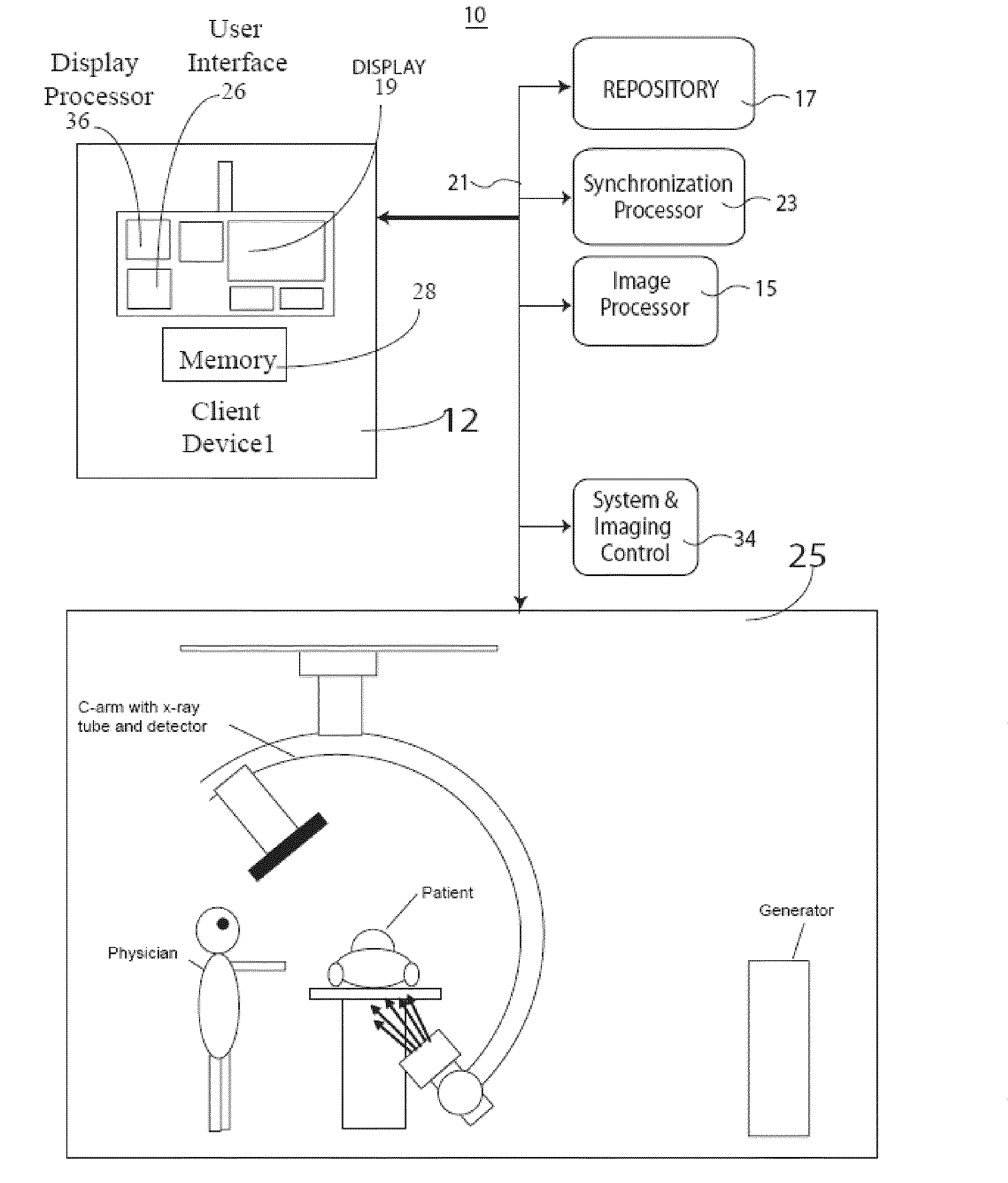
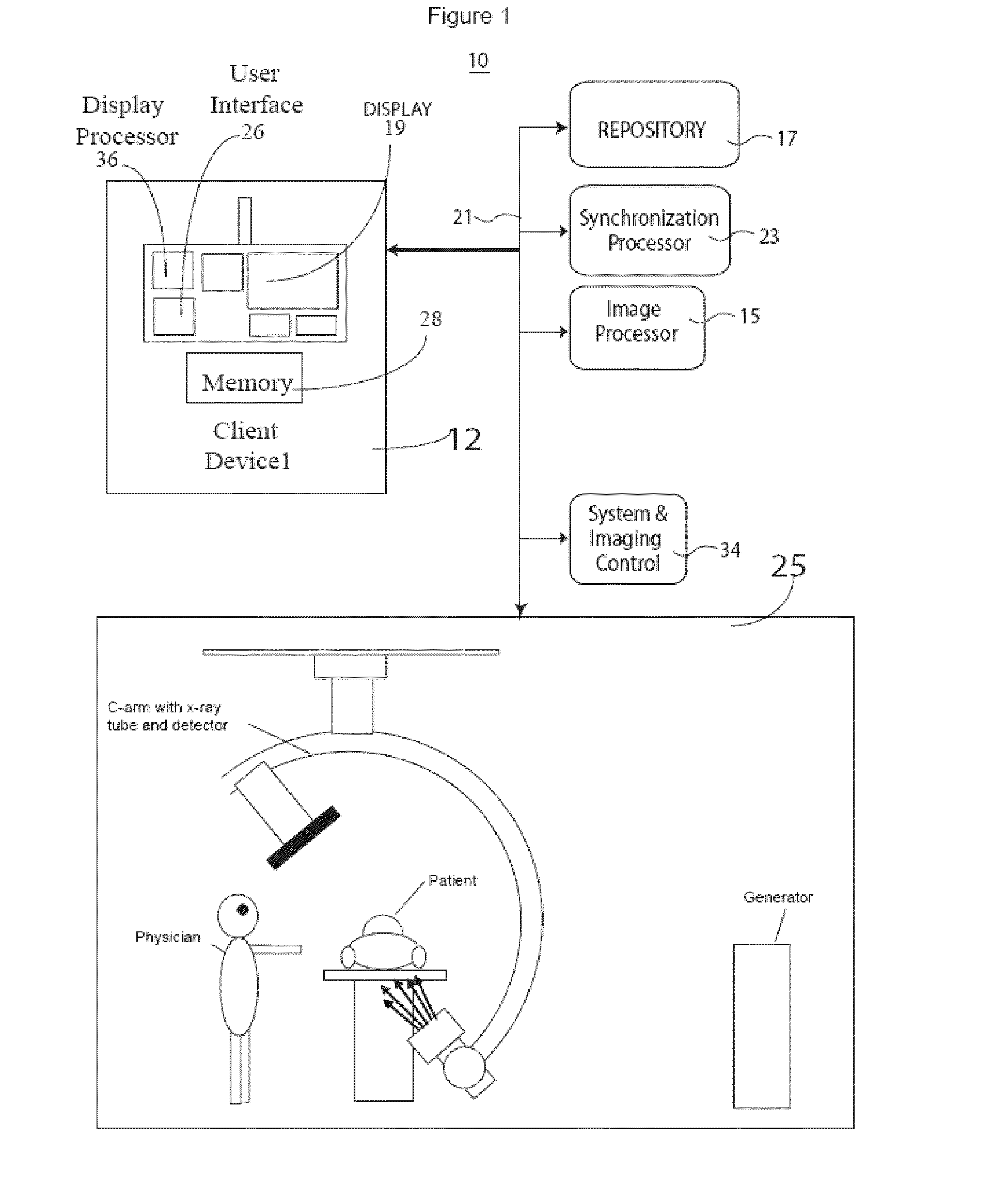
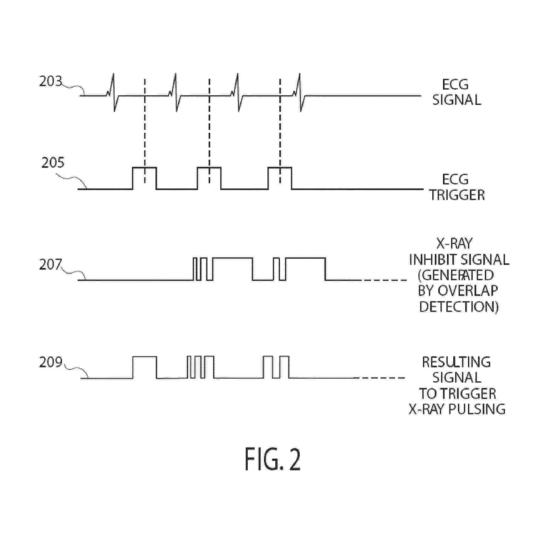
[0017]A system reduces X-ray exposure of a patient by eliminating spatially redundant imaging and increasing image quality in 2D image acquisition for 3D image volume reconstruction by eliminating gaps in angular coverage in ECG triggered rotational image acquisition and eliminating redundant acquisition by monitoring and tracking a set of angles at which exposure has been performed. The system provides visual feedback of spatial coverage percentage during image acquisition. The system eliminates both overlaps and coverage gaps which reduces patient X-ray dosage and improves reconstructed 3D image volume quality. During a portion of an ECG cycle, the heart is relatively still. This fact is used by ECG triggered (or ECG gated) image acquisitions so that high intensity exposures are performed within this period and skipped or performed at lower radiation intensity outside of this period which advantageously reduces motion artifacts in a reconstructed 3D imaging volume dataset. High in...


PUM


Abstract

A medical imaging system generates 3D anatomical images from acquired 2D anatomical images. The system includes a synchronization processor for providing a synchronization signal identifying a particular phase of heart operation of a particular patient. An image acquisition device acquires 2D anatomical images of a patient heart in angularly variable imaging planes over multiple different heart cycles at the particular phase of heart operation in response to the synchronization signal and in response to recorded data indicating imaging previously being performed at particular imaging plane angles. An image processor stores the recorded data to prevent imaging overlap at the particular imaging plane angles and to prevent unnecessary radiation exposure of the patient. The image processor processes 2D images acquired by the image acquisition device of the patient heart in multiple different imaging planes having relative angular separation, to provide a 3D image reconstruction of the patient heart

Description

[0001]This is a non-provisional application of provisional application Ser. No. 61 / 102,387 filed Oct. 3, 2008, by M. Kuzmanovic.FIELD OF THE INVENTION[0002]This invention concerns a medical imaging system for generating 3D anatomical images from acquired 2D anatomical images by adaptively inhibiting imaging radiation emission and image acquisition to prevent imaging overlap at particular imaging plane angles and unnecessary radiation exposure of a patient.BACKGROUND OF THE INVENTION[0003]In known imaging systems, during medical imaging examination there are angles at which X-ray exposure is not performed (or not performed using high intensity X-ray beams) and this lack of data may prevent 3D image reconstruction from 2D images or reduce quality of a 3D image reconstruction. In order to avoid this, multiple imaging passes are performed in order to try to cover spatial and angular gaps and obtain exposure from almost all angles within a desired range. Known systems perform these addit...

Claims


Application Information

Patent Timeline
IPC IPC(8): H05G1/60A61B5/05G06K9/00
CPCA61B6/503A61B6/541G06T2211/436G06T2211/412G06T11/006
Inventor KUZMANOVIC, MILJAN
Owner SIEMENS HEALTHCARE GMBH
Why Eureka
Try Eureka