Method and apparatus of spectral differential phase-contrast cone-beam CT and hybrid cone-beam CT

a phase-contrast cone and phase-contrast technology, applied in tomography, image enhancement, instruments, etc., can solve the problems of relatively low sensitivity of mammography to small breast cancer (under several millimeters), sharp decline in the probability of metastasis, and limited specificity and positive predictive value of mammography. achieve the effect of increasing spatial resolution

Active Publication Date: 2016-06-14
UNIVERSITY OF ROCHESTER
View PDF125 Cites 21 Cited by
  • Summary
  • Abstract
  • Description
  • Claims
  • Application Information

AI Technical Summary

Benefits of technology

[0019]It is therefore another object of the invention to substantially reduce the x-ray radiation dose to a patient without reducing spatial resolution and contrast to noise ratio.
[0020]It is therefore another object of the invention to achieve mechanically rigid and robust implementation for a rotational-gantry system of phase contrast cone beam CT.
[0021]It is therefore another object of the invention to substantially reduce x-ray radiation dose to a patient for grating-based phase contrast cone beam CT imaging.
[0022]To achieve the above and other objects, the present invention is directed to a system and method for breast imaging or other purposes (for example, vascular imaging, pediatric cone beam CT, whole body CT imaging and interventional cone beam CT), using spectral x-ray differential phase-contrast cone beam CT that is able to record and present object information in one or more quasi-monochromatic energy channels. X-ray phase contrast cone beam CT and cone beam CT imaging as an emerging new technology will potentially achieve a spatial resolution level up to 25 lp / mm (20 μm voxel size) while maintaining an x-ray dose similar to that of the current CBBCT and mammography. In addition, since x-ray phase contrast imaging is dependent on the principles of refraction and interference of x-ray waves, more subtle information can be detected by retrieving the phase coefficients than that possible with conventional attenuation-based x-ray imaging techniques retrieving attenuation coefficients.
[0024]In the past decade, various phase-contrast techniques have been developed to manifest the contrast of δ, almost all of which depend on micro-focus x-ray tubes or synchrotron radiation, which are not practical for widespread clinical applications. Recently, a new phase contrast imaging technique called the differential phase-contrast (DPC) technique has been proposed, which is a grating-based interferometry method. A high power hospital-grade x-ray tube with a wide polychromatic spectrum and high output x-ray power can be used to acquire DPC images. To further reduce the x-ray exposure, we propose a spectral DPC-based cone beam CT (DPC-CBCT) approach. One way is to shape the input x-ray as a quasi-monochromatic spectrum and thus greatly improve the dose utilization efficiency, and another way is to use an energy-resolving detector that can differentiate acquired images in several energy channels. However, it has not previously been used in the context of the present invention. This spectral DPC-CBCT approach is novel over the DPC-CBCT method, and it has not previously been used in the context of the present invention.

Problems solved by technology

In addition, as the diameter of the tumor at detection decreases, the probability of metastasis declines sharply.
Although mammography, which on average can detect cancers ˜12 mm in size, is the most effective tool for the early detection of breast cancer currently available, mammography has relatively low sensitivity to small breast cancers (under several millimeters).
Specificity and the positive predictive value of mammography remain limited owing to structure and tissue overlap.
The limited sensitivity and specificity in breast cancer detection of mammography are due to its poor contrast detectability, which is common for all types of projection imaging techniques (projection imaging can only have up to 10% contrast detectability), and mammography initially detects only 65-70% of breast cancers.
The relatively low specificity of mammography leads to biopsy for indeterminate cases, despite the disadvantages of added cost and the stress it imposes on patients.
Nearly 80% of the over one million breast biopsies performed annually in the U.S. to evaluate suspicious mammographic findings are benign, burdening patients with excessive anxiety and the healthcare system with tremendous cost.
US is a low spatial resolution study, has severe limitations in visualizing and characterizing calcifications and is highly dependent on operator skill.
Although CEBMRI has a high sensitivity for invasive cancers, current techniques may be limited in detecting ductal carcinoma in situ (DCIS).
Though a measure of success has been achieved, DBT is fundamentally limited by its constraints in projection geometry; the tomographic slice is not well defined, which can cause a loss of resolution in the axial direction that affects visualization of subtle features, such as amorphous microcalcifications.

Method used


Image

  • Method and apparatus of spectral differential phase-contrast cone-beam CT and hybrid cone-beam CT
  • Method and apparatus of spectral differential phase-contrast cone-beam CT and hybrid cone-beam CT
  • Method and apparatus of spectral differential phase-contrast cone-beam CT and hybrid cone-beam CT

Examples


second embodiment

[0082]It should be noted that the analyzer grating 208 does not have to be an attenuation grating as that for the Instead, it could be a second phase grating that produces significant phase change but negligible amplitude change. A phase-phase grating pair will also produce similar moiré patterns if the detector is placed at an appropriate location, which could be a fractional Talbot distance or an integer Talbot distance.

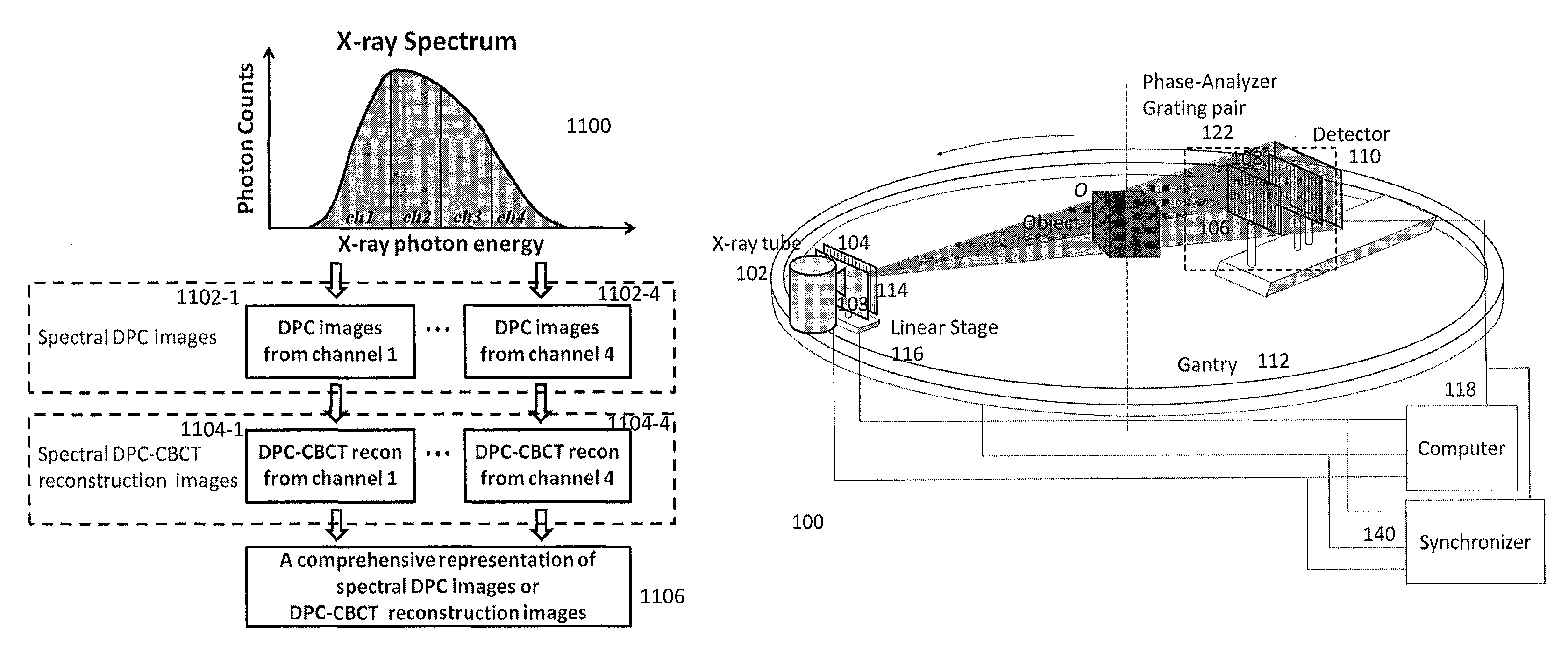
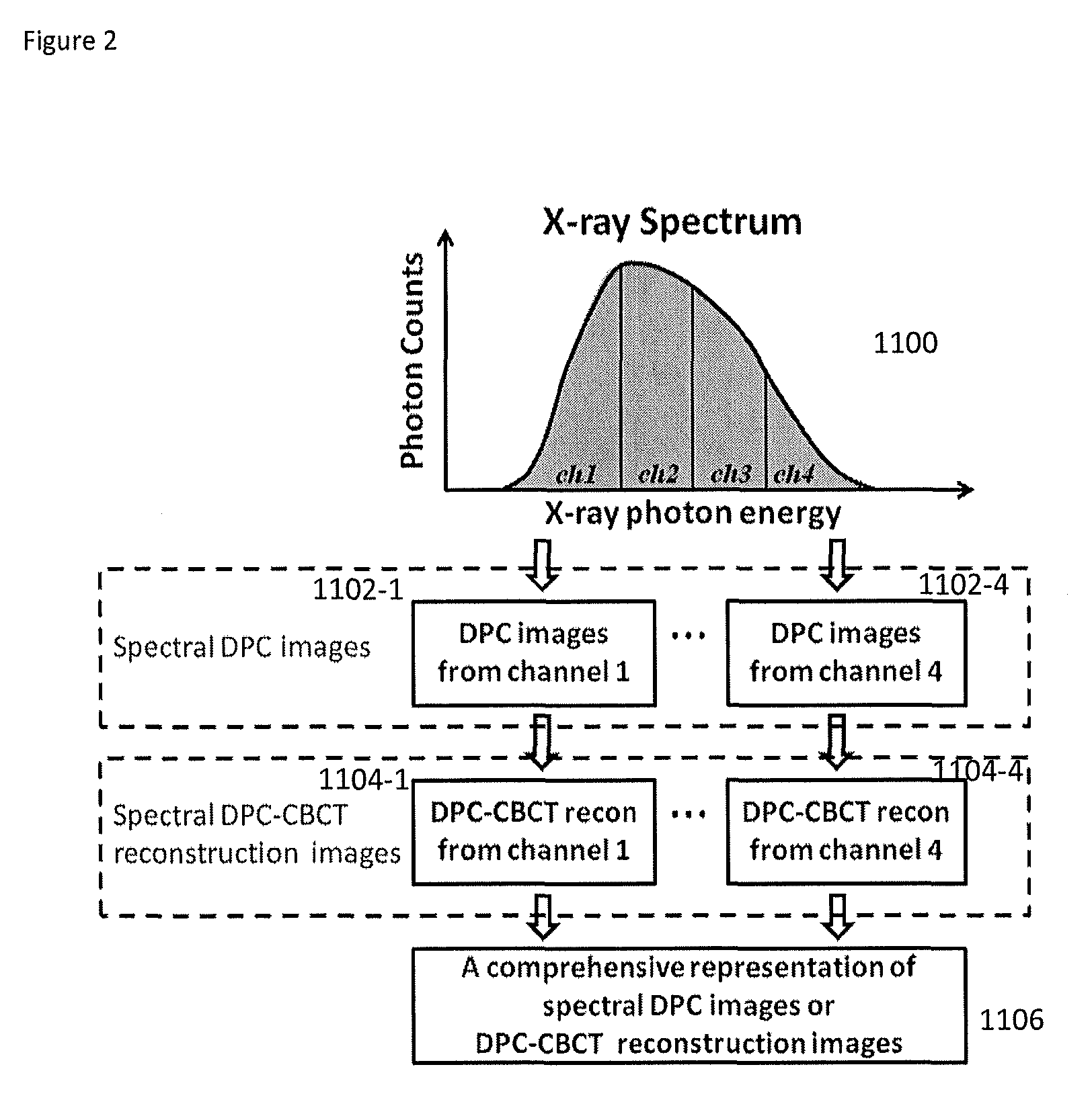
[0083]The third preferred embodiment is a variation of the first preferred embodiment where the beam filter 103 is removed and the detector is an energy-resolving detector 130. Such a system works in the same way as the first preferred embodiment except that by recording images in different energy channels, it can perform spectral DPC and spectral DPC-CBCT imaging as described in the second direction. This embodiment is shown in FIGS. 7A-7D.

[0084]The fourth preferred embodiment is a variation of the second preferred embodiment where the beam filter is removed and ...

embodiment 600

[0097]The sixth preferred embodiment is a variation of the hybrid system as shown in FIGS. 11A and 11B. FIG. 11A shows the cone beam CT breast imaging system as disclosed in U.S. Pat. No. 6,480,565 “Apparatus and method for cone beam volume computed tomography breast imaging” (Ning '565), and the hybrid cone beam CT system is supposed to replace the cone beam CT system beneath the patient table to perform hybrid breast imaging. FIG. 11B is actually a combination of the moiré pattern-based system (second preferred embodiment) and the current CBBCT system. CBBCT system images the breast only using a half-cone geometry in which x-ray radiation radiates the breast only without penetrating chest cavity or other body parts of the patient, resulting in substantially reduced radiation to the patent. It should be noted that as no stepping is required in the system 600, it can perform fast data acquisition, which makes dynamic imaging possible using this system. As illustrated, the embodiment...

embodiment 700

[0098]The seventh preferred embodiment is a variation of the hybrid system as shown in FIGS. 12A-12E. FIG. 12A shows the cone beam CT breast imaging system as disclosed in U.S. Pat. No. 6,480,565 “Apparatus and method for cone beam volume computed tomography breast imaging” (Ning '565), and the hybrid cone beam CT system is supposed to replace the cone beam CT system beneath the patient table to perform hybrid breast imaging. FIGS. 12B, 12C, 12D and 12F are actually combinations of the phase stepping-based spectral DPC-CBCT system using an energy-resolving detector (the third preferred embodiment) and the current CBBCT system. As illustrated, the seventh preferred embodiment 700 includes CBBT X-ray tube 720 and CBBT detector 722, and gantry 724. The CBBCT system images the breast only using a half-cone geometry in which x-ray radiation radiates the breast only without penetrating chest cavity or other body parts of the patient, resulting in substantially reduced radiation to the pat...


PUM

PropertyMeasurementUnit
diameteraaaaaaaaaa
sizeaaaaaaaaaa
sizeaaaaaaaaaa

Abstract

DPC (differential phase contrast) images are acquired for each photon energy channel, which are called spectral DPC images. The final DPC image can be computed by summing up these spectral DPC images or just computed using certain ‘color’ representation algorithms to enhance desired features. In addition, with quasi-monochromatic x-ray source, the required radiation dose is substantially reduced, while the image quality of DPC images remains acceptable.

Description

REFERENCE TO RELATED APPLICATION[0001]The present application claims the benefit of U.S. Provisional Patent Application No. 61 / 763,159, filed Feb. 11, 2013, whose disclosure is hereby incorporated by reference in its entirety into the present disclosure.STATEMENT OF GOVERNMENT INTEREST[0002]This invention was made with government support under Grant No. R01 CA 143050 awarded by National Institutes of Health. The government has certain rights in the invention.FIELD OF THE INVENTION[0003]The present invention is directed to cone-beam computed tomography (CT) imaging and more particularly to phase-contrast cone-beam CT for such uses as breast imaging.DESCRIPTION OF RELATED ART[0004]According to the National Cancer Institute, one out of eight women will be diagnosed with breast cancer in her lifetime. And while a reduction in mortality from breast cancer is evident in published reports, each year 40,000 women will die of the disease.[0005]The optimal breast imaging technique detects tum...

Claims


Application Information

Patent Timeline
Patent Type & Authority Patents(United States)
IPC IPC(8): A61B6/03G01N23/087A61B6/00G06T11/00G01N23/20
CPCA61B6/4241A61B6/482G01N23/20091A61B6/5235G01N23/087A61B6/032A61B6/502A61B6/4035A61B6/4291A61B6/484A61B6/4085G06T11/005G06T2207/10081G06T2207/20212
Inventor NING, RUOLACAI, WEIXING
Owner UNIVERSITY OF ROCHESTER
Why Eureka
Try Eureka