X-ray CT Photographic Apparatus

a technology of x-ray detection and photographic equipment, which is applied in the direction of instruments, diaphragms for radiation diagnostics, applications, etc., can solve the problems of degrading reducing the transfer efficiency of x-ray detection signals, so as to achieve efficient x-ray detection signals.

Active Publication Date: 2013-11-07
MORITA MFG CO LTD
View PDF17 Cites 22 Cited by
  • Summary
  • Abstract
  • Description
  • Claims
  • Application Information

AI Technical Summary

Benefits of technology

[0045]As seen from the above, an object of the present invention is to provide a t...

Problems solved by technology

Therefore, there is a risk of degrading transfer efficiency of the X-ray detection signal.
Accordingly, there is a risk of degrading the transfer efficiency of the X-ray detection signal.
Therefore, in the case tha...

Method used


Image

  • X-ray CT Photographic Apparatus
  • X-ray CT Photographic Apparatus
  • X-ray CT Photographic Apparatus

Examples


Embodiment Construction

[0072]Hereinafter, a preferred embodiment of the present invention will be described with reference to the accompanying drawings. In the accompanying drawings, for the sake convenience, sometimes the size or the number of pieces of each unit is magnified or simplified as needed.

1. First Preferred Embodiment

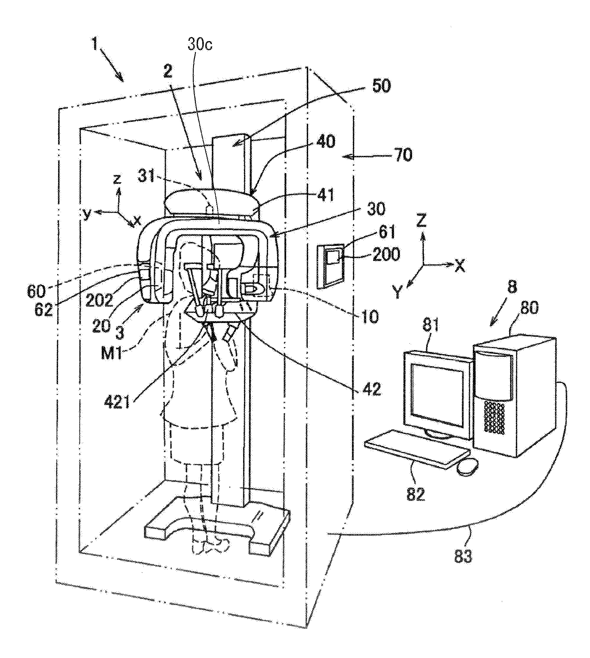
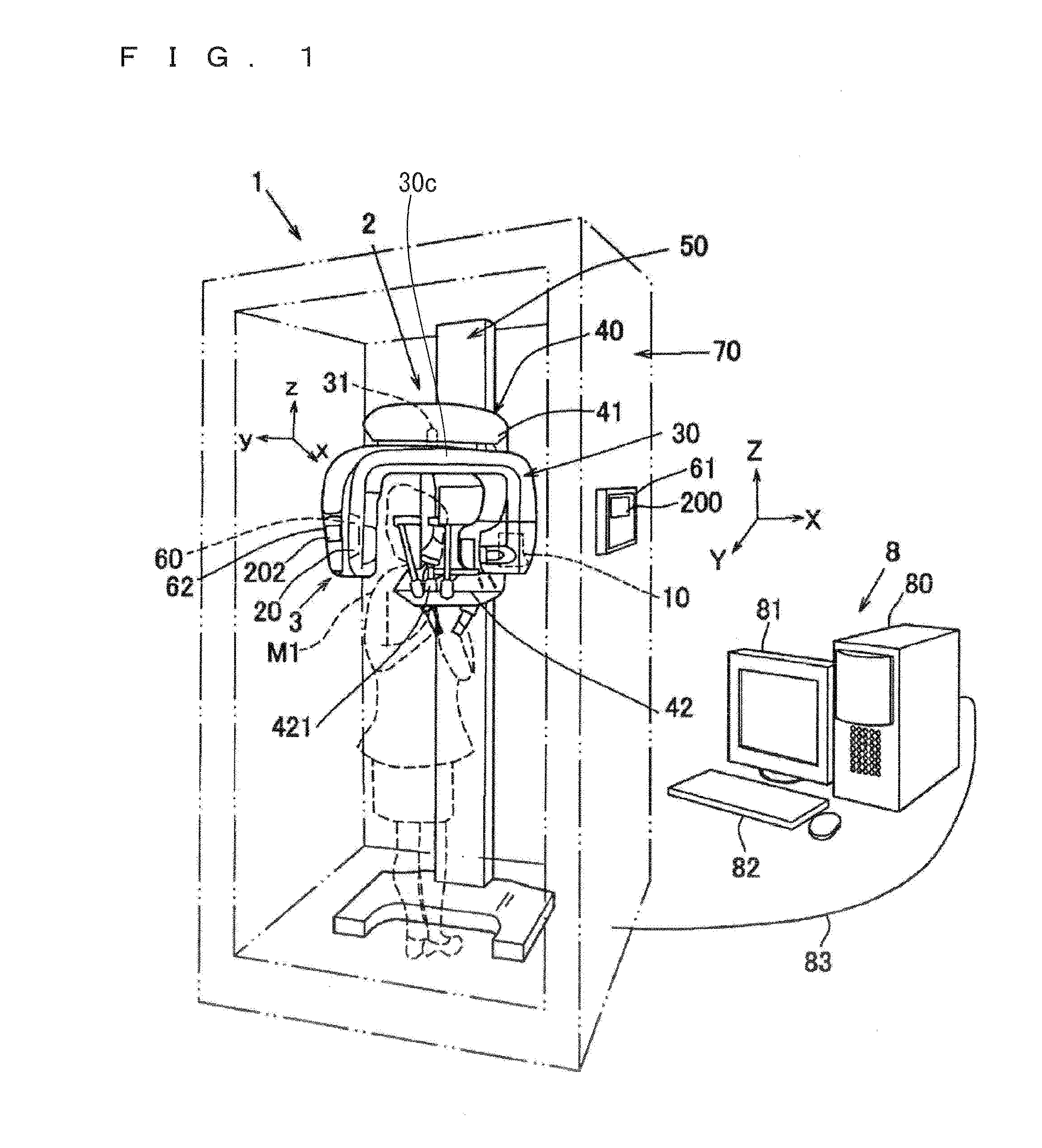
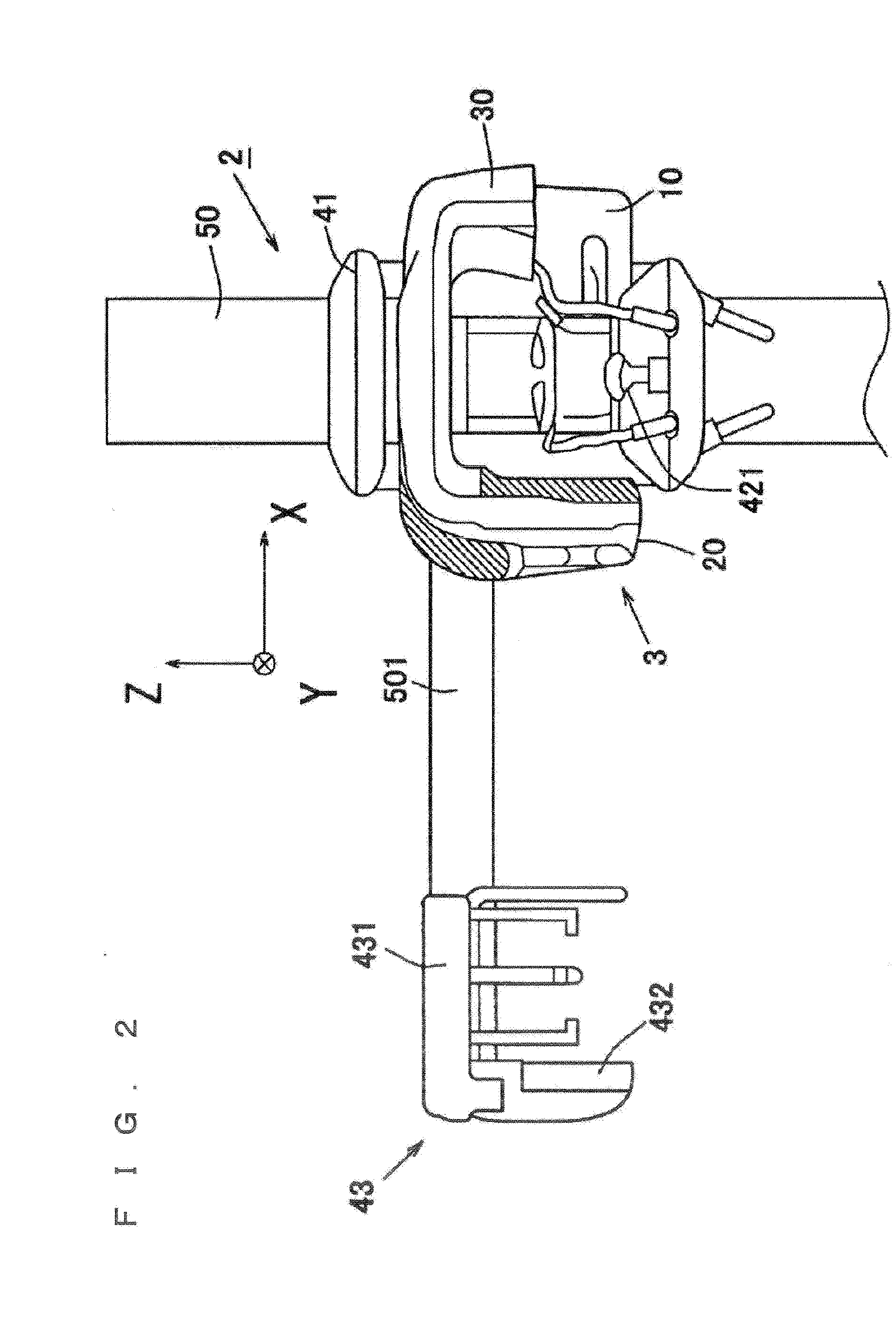
[0073]FIG. 1 is a schematic perspective view of an X-ray CT photographic apparatus 1 according to a first preferred embodiment of the present invention. FIG. 2 is a partial front view of the X-ray CT photographic apparatus 1 on which a cephalostat 43 is mounted. FIG. 3 is a block diagram illustrating a configuration of the X-ray CT photographic apparatus 1.

[0074]FIG. 4 is a schematic perspective view of a beam shaping mechanism 13 (the X-ray regulating unit).

[0075]FIG. 5 (FIGS. 5A and 5B) is a schematic perspective view of an X-ray generation unit 10 that emits an X-ray cone beam BX in which an irradiation range is regulated. Particularly, FIG. 5A is a schematic perspective view o...


PUM


Abstract

An X-ray CT photographic apparatus including: a beam shaping mechanism that regulates an irradiation range of an X-ray generated from an X-ray generator and shapes the X-ray into an X-ray cone beam; and a main body controller that changes a read region, where an X-ray detection signal is read in the X-ray detector, according to the irradiation range of the X-ray cone beam. The main body controller changes the irradiation range of the X-ray cone beam to an x-axis direction during an X-ray CT photography such that only a set CT photographic region is irradiated with the X-ray cone beam according to the set CT photographic region input through a CT photographic region setting unit. The main body controller changes a read region in an X-ray detector with respect to the x-axis direction in a detection surface of the X-ray detector during the X-ray CT photography.

Description

BACKGROUND OF THE INVENTION[0001]1. Field of the Invention[0002]The present invention relates to an apparatus for X-ray CT photography.[0003]2. Description of the Background Art[0004]Conventionally, X-ray CT photography is performed in a medical field and the like. In the X-ray CT photography, a subject is irradiated with an X-ray to collect projection data, and the obtained projection data is reconstructed on a computer to generate a CT (Computed (Computerized) Tomography) image.[0005]In the X-ray CT photography, the subject is disposed between an X-ray generator and an X-ray detector, the X-ray generator and the X-ray detector turn about the subject, and the X-ray generator emits conical X-ray (an X-ray cone beam) to subject. The X-ray detector collects an X-ray detection result (the projection data), and three-dimensional data (volume data) is reconstructed based on the collected X-ray detection result. For example, Japanese Patent Application Laid-Open No. 2010-42121, PCT Applic...

Claims


Application Information

Patent Timeline
IPC IPC(8): A61B6/03A61B6/14
CPCA61B6/035A61B6/14A61B6/032A61B6/06A61B6/405A61B6/4085A61B6/469A61B6/501
Inventor YOSHIKAWA, HIDEKISUZUKI, MASAKAZU
Owner MORITA MFG CO LTD
Why Eureka
Try Eureka