Methods for tissue classification in cervical imagery

a cervical imaging and tissue classification technology, applied in the field of medical imaging, can solve the problems of undesigned reid colposcopic index (rci) and unsupervised approach based on prior medical knowledge, and achieve the effect of low grade dysplasia and low grade dysplasia

Inactive Publication Date: 2010-04-15
CADES SCHUTTE A LIMITED LIABILITY LAW PARTNERSHIP
View PDF26 Cites 31 Cited by
  • Summary
  • Abstract
  • Description
  • Claims
  • Application Information

AI Technical Summary

Benefits of technology

[0029]If the texture region is greater than approximately 1% of the area of the cervix, said opacity parameter is between approximately 4% of the pixel value and approximately 12% of the pixel value, the size of the acetowhite region is greater than 7% of the area of the cervix, and if the size of the acetowhite region near the os is greater than 0% of the area of the cervix, then a final prediction of high grade dysplasia is obtained.
[0030]If the texture region is greater than approximately 1% of the area of the cervix, if said opacity parameter is between approximately 4% of the pixel value and approximately 12% of the pixel value, the size of the acetowhite region is less than 7% of the area of the cervix, and there is no acetowhite region near the os, then a final prediction of low grade dysplasia is obtained.
[0031]If the texture region is less than approximately 1% of the area of the cervix, and if the opacity parameter is less than 5% of the pixel value, the number of coarse punctations is less than 2, and there are no coarse mosaic vessels, then a final prediction of low grade dysplasia is...

Problems solved by technology

Consequently, the Reid Colposcopic Index (RCI) is not designed to discriminate premalignant from malignant cervical neoplasia.
Long linear lesions—those greate...

Method used


Image

  • Methods for tissue classification in cervical imagery
  • Methods for tissue classification in cervical imagery
  • Methods for tissue classification in cervical imagery

Examples


Embodiment Construction

(1) Pre-Method Applications

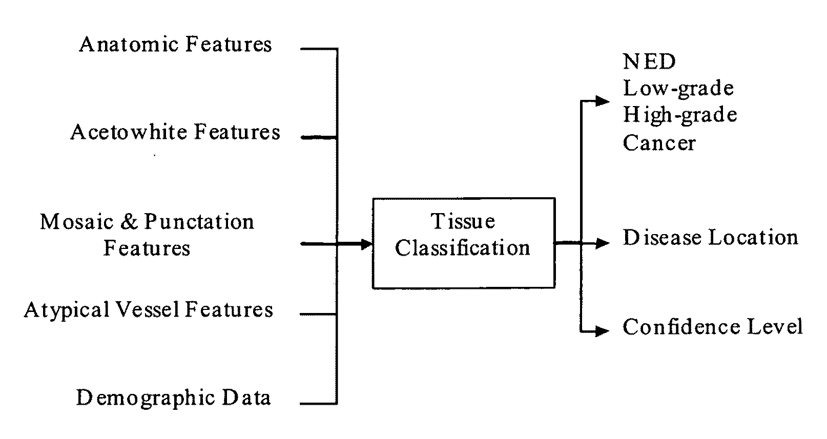
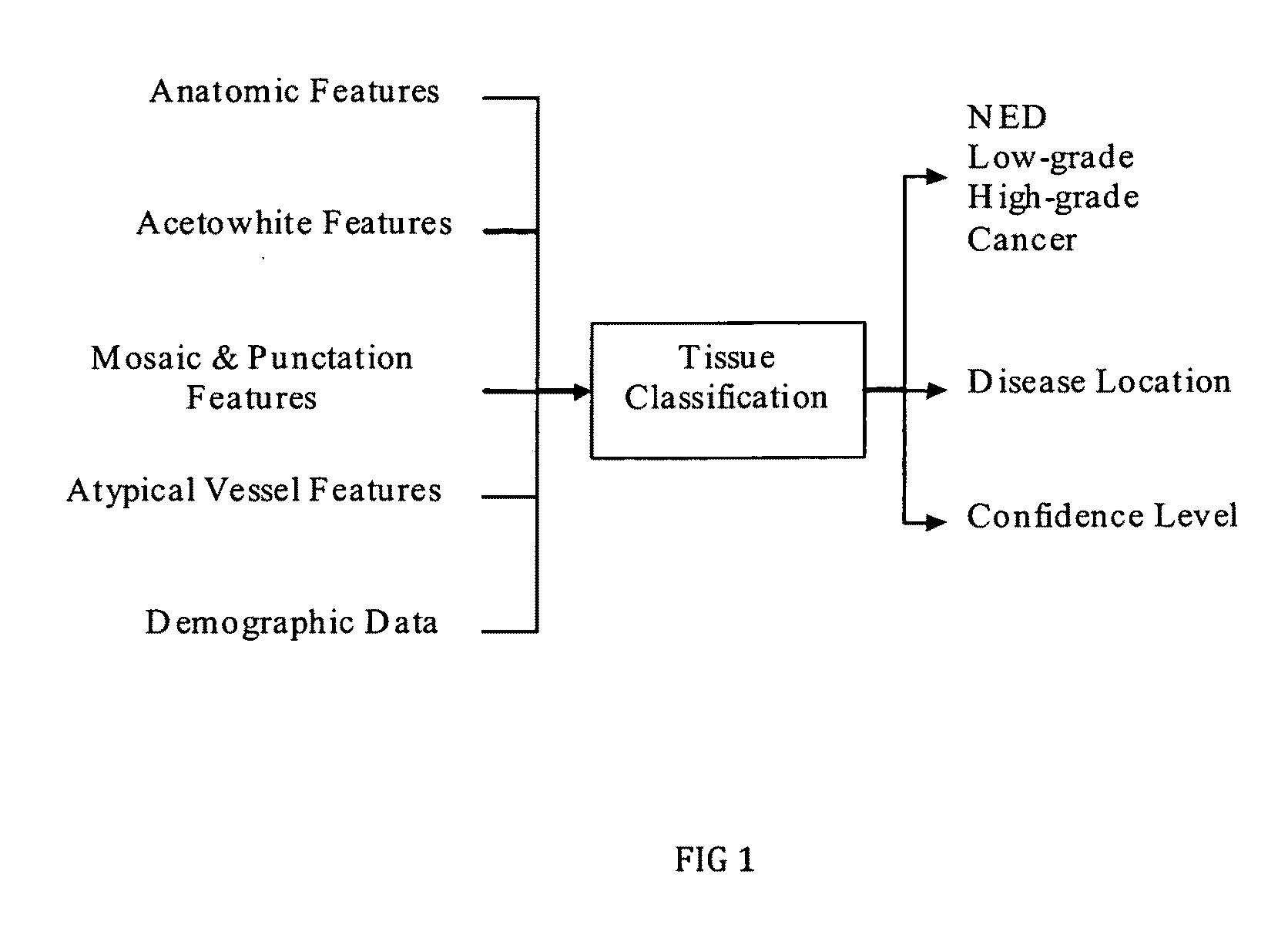
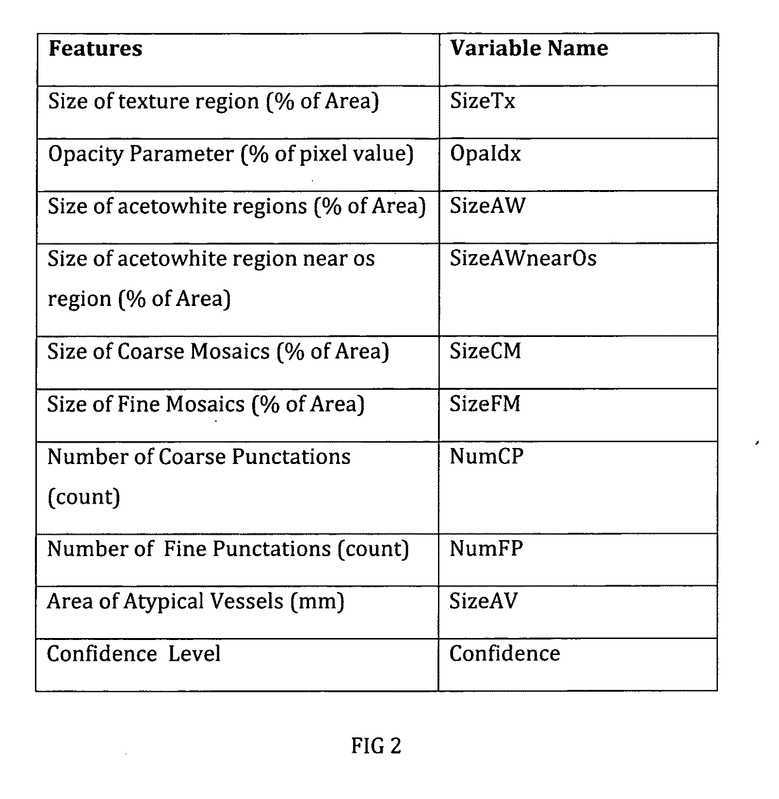
[0048]The presently preferred embodiment of the invention described herein preferably uses the output of several pre-method applications (the output being certain parameters described in FIG. 2) as the input for the tissue classification method. Many of the parameters or variables are described as a percentage of that particular feature's area versus the total area of the cervix in the image. The punctation feature is defined as the number of punctations present in the image. The size of atypical blood vessels is preferably calculated in millimeters (mm).

[0049]The process begins by collecting digital color images (using red, green, and blue channels or RGB) of the cervix—one before and one after the application of acetic acid. Preferably, the image is cross-polarized to suppress specular reflection (glint) and calibrated. Next, several pre-method applications including anatomic feature extraction, acetowhite feature extraction, mosaic and punctation detect...


PUM


Abstract

A rule-based unsupervised process for classifying cervical tissue by serially applying classifiers selected from the group that consists of determining size of texture region, opacity parameter, size of acetowhite regions, number of coarse and fine punctations, size of coarse and fine mosaics, size of atypical blood vessels and demographic data, so that the cervical tissue can be classified into no evidence of disease, low-grade dysplasia, high-grade dysplasia or cancer.

Description

[0001]This application claims the benefit of U.S. Provisional Patent Application No. 61 / 196,159 filed Oct. 10, 2008.TECHNICAL FIELD[0002]The present invention generally relates to medical imaging, and more specifically to the computer aided detection and diagnosis of uterine cervical cancer and pre-cancerous lesions using methods for cervical tissue identification and classification.BACKGROUND ART[0003]This invention is being disclosed in connection with cervical cancer. Uterine cervical cancer is the second most common cancer in women worldwide, with nearly 500,000 new cases and over 270,000 deaths annually (http: / / www-depdb.iarc.fr / globocan2002.htm, incorporated herein by reference) Colposcopy is a diagnostic method used to detect cancer precursors and cancer of the uterine cervix (B. S. Apgar, Brotzman, G. L. and Spitzer, M., Colposcopy: Principles and Practice, W.B. Saunders Company: Philadelphia, 2002, incorporated herein by reference). CAD (“computer aided diagnosis”) for colp...

Claims


Application Information

Patent Timeline
IPC IPC(8): G06K9/00G06V10/764G16Z99/00
CPCA61B5/0075A61B5/0084A61B5/4331A61B5/7264G06F19/3431G06T2207/30101G06T7/0012G06T2207/10024G06T2207/10068G06T2207/20212G06T2207/30096G06K9/6282G16H50/30G16Z99/00G06V10/764G06F18/24323
Inventor LI, WENJING
Owner CADES SCHUTTE A LIMITED LIABILITY LAW PARTNERSHIP
Why Eureka
Try Eureka