Methods of Detecting and Monitoring Cancer Using 3D Analysis of Centromeres

Active Publication Date: 2010-06-10
TELO GENOMICS HLDG CORP
View PDF2 Cites 2 Cited by
  • Summary
  • Abstract
  • Description
  • Claims
  • Application Information

AI Technical Summary

Benefits of technology

[0006]The method disclosed in the present application has advantages over methods in the prior art including methods that assess telomeric and / or chromosome organization in cancer cells. In particular, the methods described herein can detect alterations in centromere organization in cells in earlier stages of the transformation process, such as precancerous cells.

Problems solved by technology

However, centromeric changes during cellular immortalization and transformation have not been previously examined.

Method used


Image

  • Methods of Detecting and Monitoring Cancer Using 3D Analysis of Centromeres
  • Methods of Detecting and Monitoring Cancer Using 3D Analysis of Centromeres
  • Methods of Detecting and Monitoring Cancer Using 3D Analysis of Centromeres

Examples


example 1

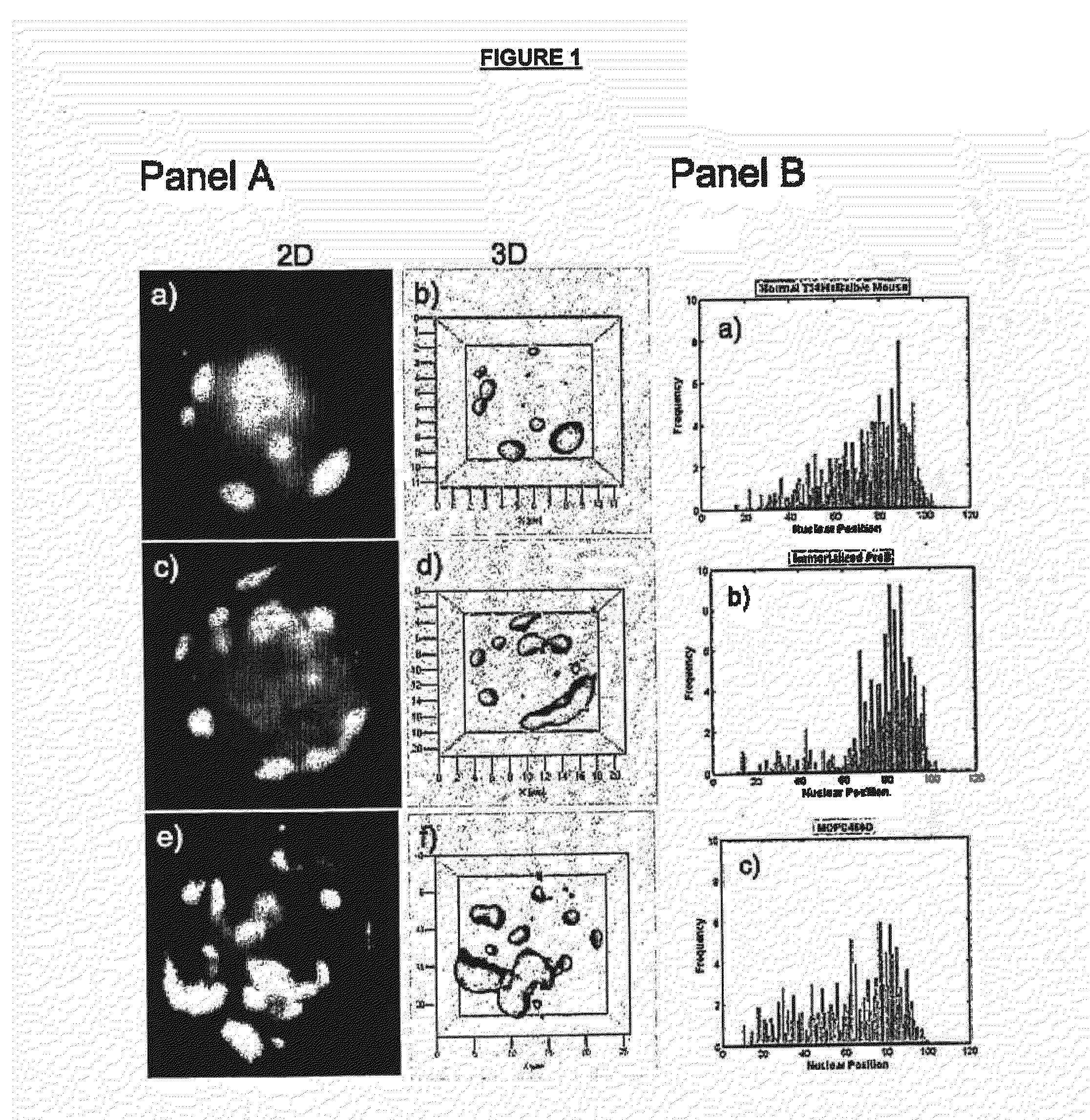
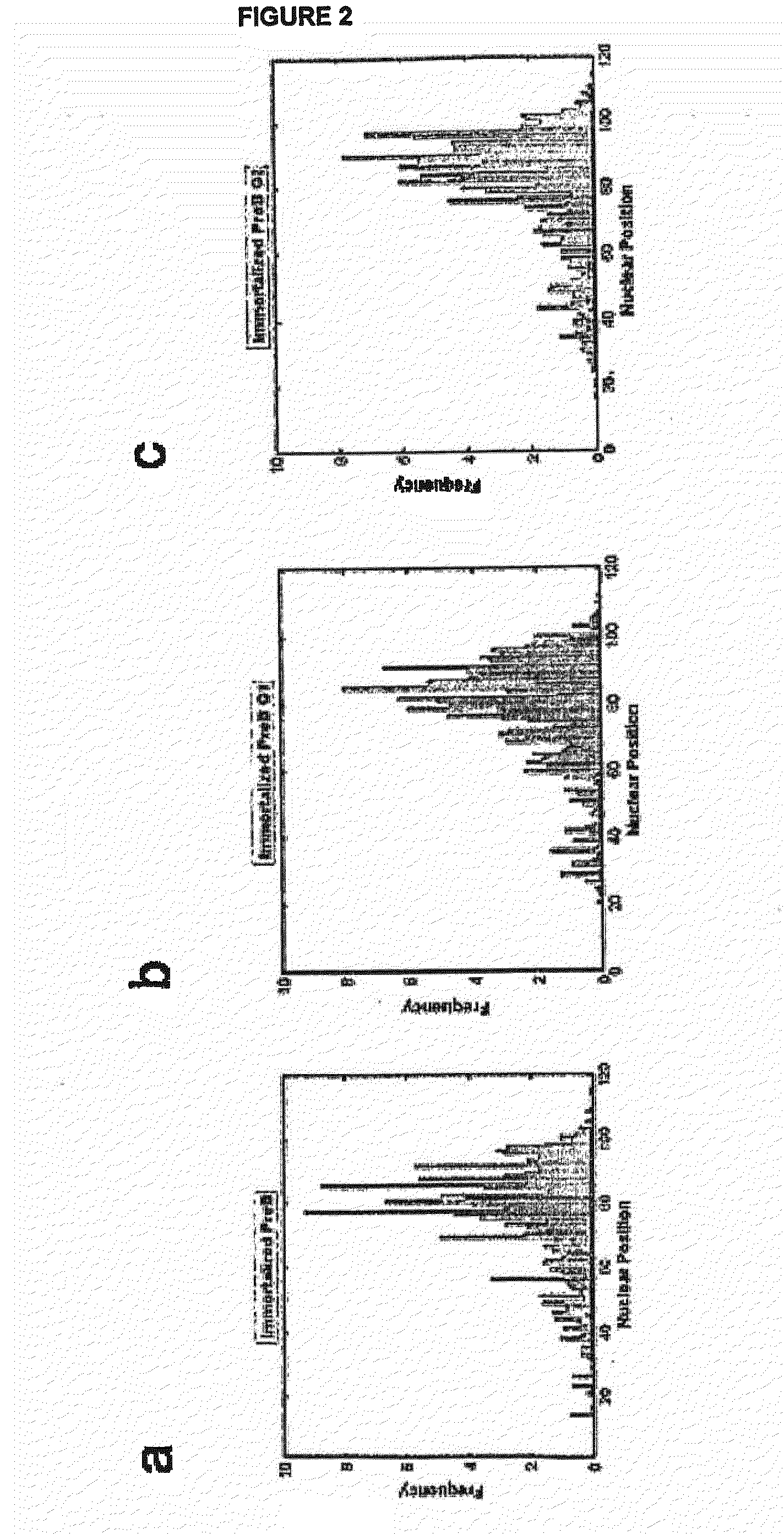
Centromere Positions in Nuclei of Normal, Immortalized, and Malignant B Cells

[0059]The three-dimensional (3D) positions of centromeres have been studied in several cell systems. However, data on centromere positions during cellular transformation remain elusive. This study has focused on B lineage cells and investigated the centromere positions in primary, immortalized and tumor cells.

[0060]Using CentroView™, a program the inventors developed to measure nuclear centromere positions, the positions of centromeres in primary, immortalized and malignant mouse B cells were determined. The results show that centromeres exhibit altered nuclear positions in immortalized and malignant B cells. These changes are independent of previously described cell cycle-dependent centromere dynamics.

[0061]In summary, the inventors have shown that the 3D positions of centromeres are altered during cellular transformation. These nuclear changes reflect structural remodeling of mammalian nuclei during oncog...

example 2

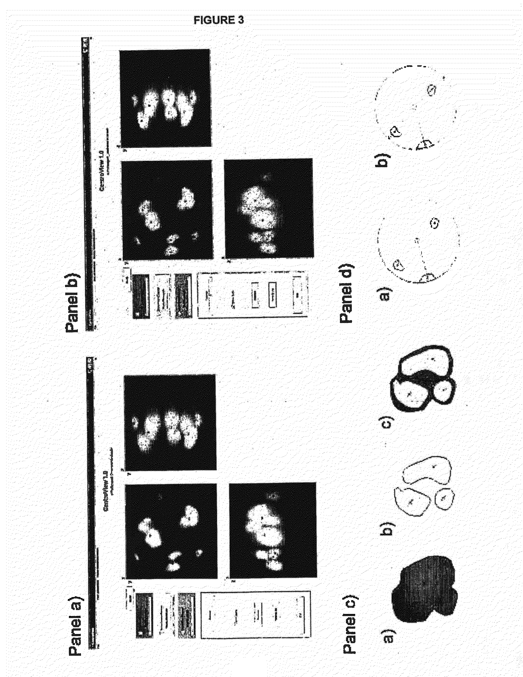
Mouse Robertsonian Chromosome Formation Following c-Myc Deregulation

[0084]Robertsonian (Rb) chromosomes occur in human and murine cancers. Mechanisms involved in their generation remain elusive. The inventors here report on a novel mechanism of c-Myc oncogene-mediated Rb chromosome formation. The results show that Rb chromosomes are generated during nuclear remodeling of centromere positions in mouse interphase nuclei in a c-Myc oncogene- and myc boxII-dependent manner via telomere fusions at centromeric termini of acrocentric chromosomes.

Materials and Methods

Abbreviations

[0085]Q-FISH: quantitative fluorescent in situ hybridization; SKY: spectral karyotyping; Rb chromosomes: Robertsonian chromosome; 3D: three-dimensional; 2D: two-dimensional; BBF: breakage-bridge-fusion; TCT: telomere-centromere-telomere; CTC: centromere-telomere-centromere.

Cells

[0086]All cells used are listed in Table 1. Primary splenic lymphocytes and primary plasmacytoma (PCT1G1) were directly isolated without an...


PUM

PropertyMeasurementUnit
Distanceaaaaaaaaaa

Abstract

The present application relates to a method of detecting and monitoring cancer or precancer in a cell using three-dimensional analysis to assess centromere organization. In addition, the application relates to a method and system for characterizing the 3D organization of centromeres.

Description

FIELD OF THE INVENTION[0001]The present application relates to methods of detecting and monitoring cancer in a cell, in particular the use of three-dimensional (3D) analysis to characterize the organization of centromeres in a cell.BACKGROUND OF THE INVENTION[0002]The three-dimensional (3D) organization of the nucleus has been shown to be important in genomic stability [Zink D, Fischer A H, Nickerson J A: Nuclear structure in cancer cells. (2004)Nat Rev Cancer, 4:677-687], and any change(s) in this organization potentially cause genomic instability [Mai S, Garini Y: Oncogenic remodeling of the three-dimensional organization of the interphase nucleus: c-Myc induces telomeric aggregates whose formation precedes chromosomal rearrangements. (2005) Cell Cycle, 4:1327-1331. Epub Oct. 5, 2005; Mai S and Garini Y: The significance of telomeric aggregates in the interphase nuclei of tumor cells. (2006) J Cell Biochem, 97:904-915. Review].[0003]At present, there is inexact knowledge of about ...

Claims


Application Information

Patent Timeline
IPC IPC(8): C12Q1/68
CPCC12Q1/6886G01N15/1475G01N33/5035G06F19/345G01N33/574G01N33/57426G06K9/0014G01N33/5091G16H50/20G06V20/695G01N15/1433
Inventor MAI, SABINEGARINI, YUVALSARKAR, RAHULVERMOLEN, BARTHOLOMEUS J.
Owner TELO GENOMICS HLDG CORP
Why Eureka
Try Eureka