

Method and apparatus for the non-invasive imaging of anatomic tissue structures
a tissue structure and non-invasive technology, applied in the direction of image enhancement, instruments, measurements using nmr, etc., can solve the problems of difficult to determine the location of the lesion, recovery period, nerve damage, etc., and achieve the effect of significant clinical diagnostic valu
- Summary
- Abstract
- Description
- Claims
- Application Information
AI Technical Summary
Benefits of technology
Problems solved by technology
Method used
Image

Examples
Embodiment Construction
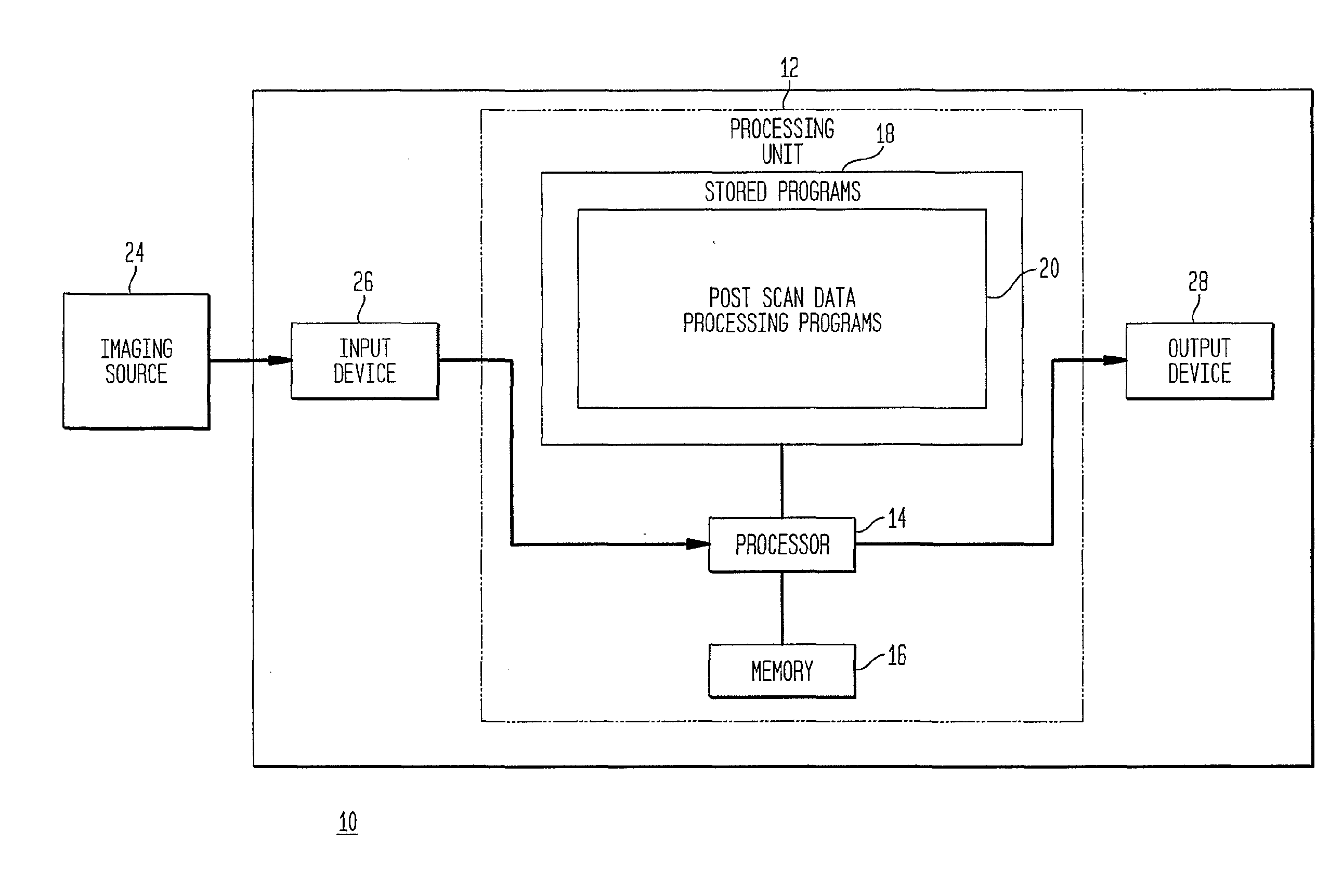
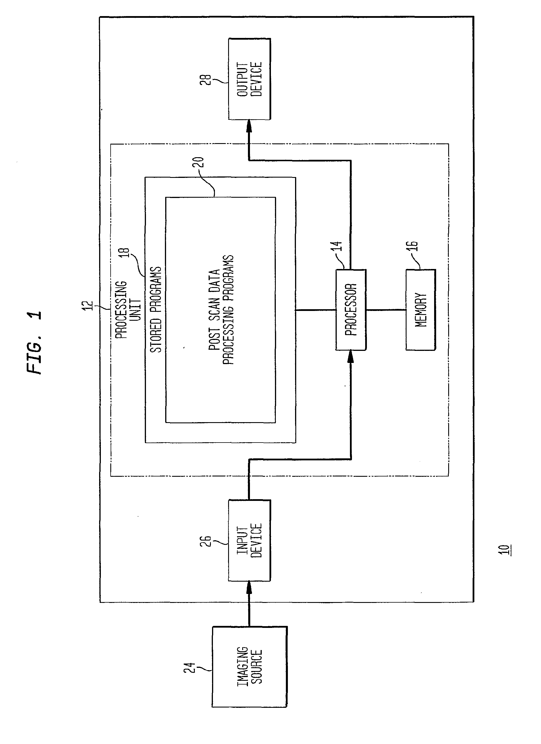
[0080]Referring to the drawings, in which like reference numerals identify similar or identical elements, as shown in FIG. 1, the present disclosure describes a method and apparatus implementing an imaging device 10, which includes a processing unit 12 having processor 14, memory 16 and stored programs 18, including post-scan data processing programs 20. The processing device also includes an input device 26 and an output device 28.
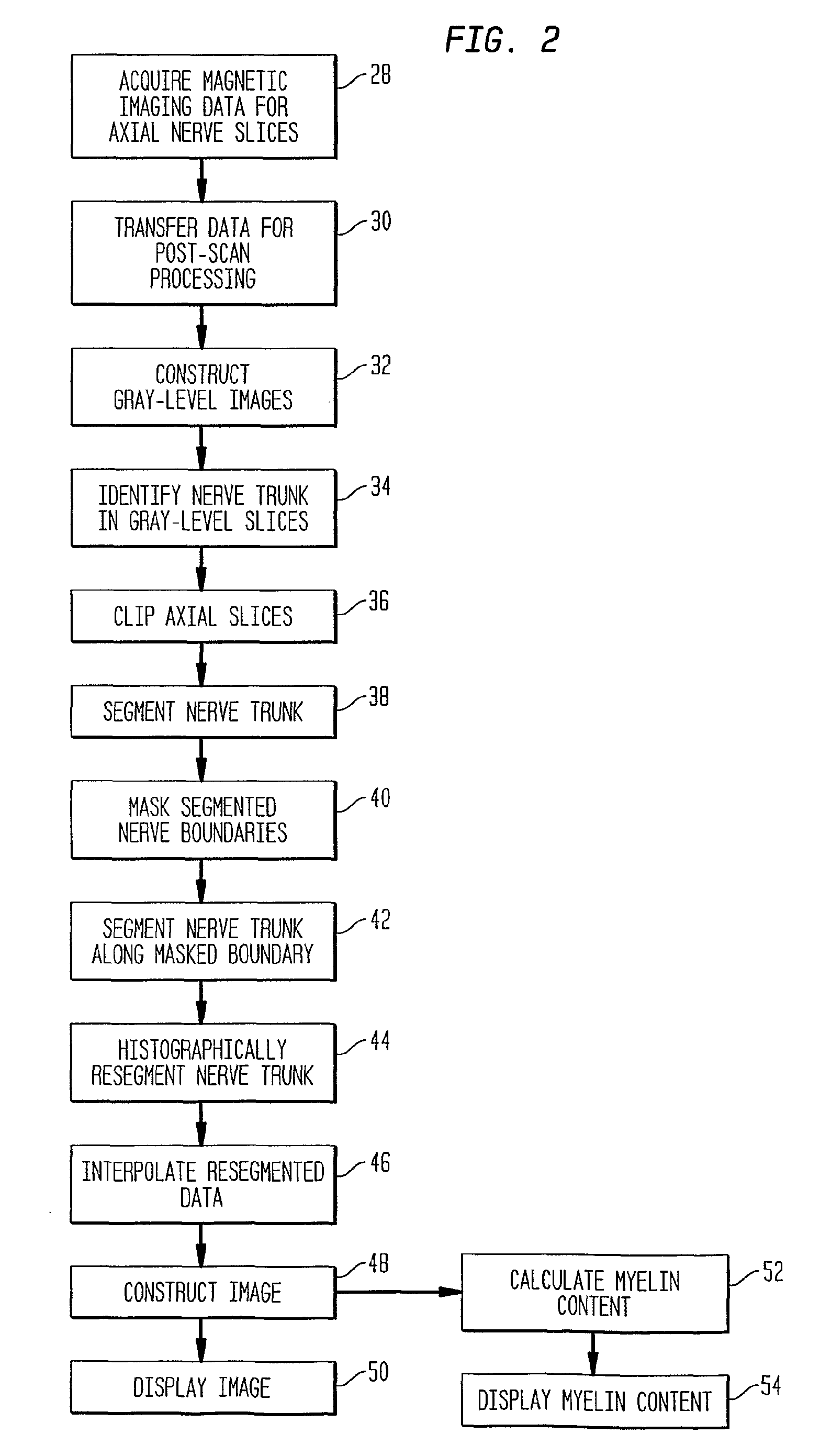
[0081]The present invention is based on the discovery that by first segmenting imaging data to isolate anatomic tissue structures from surrounding tissues before histographically resegmenting the imaging data, it is possible to construct images of the tissue microstructures having exceptional clarity.
[0082]The new methods and apparatus disclosed herein are particularly adapted for use in the diagnosis of neuropathy, especially the neuropathy resulting from compression and the associated demyelination, such as occurs with CTS.
[0083]In an exemplary embodime...
PUM
 Abstract
Description
Claims
Application Information