
Integrated Breast X-Ray and Molecular Imaging System

a breast x-ray and molecular imaging technology, applied in the field of breast imaging, can solve the problems of increasing the difficulty of distinguishing cancerous lesions from breast tissue, affecting the diagnosis of patients, and affecting so as to facilitate the registration of images, increase and improve the diagnosis speed and accuracy

Active Publication Date: 2010-10-14
HOLOGIC INC
View PDF6 Cites 54 Cited by
  • Summary
  • Abstract
  • Description
  • Claims
  • Application Information

AI Technical Summary

Benefits of technology

The present invention is a breast imaging device that combines tomosynthesis and molecular imaging capabilities. This integration allows the radiologist to choose the most appropriate imaging method based on the patient's needs and obtain additional information as needed. The device enables the viewing of tomosynthesis and molecular images together on a single display, which facilitates registration of the images, comparison of anatomical and physiological information, and increases the speed and accuracy of diagnosis, reducing unnecessary biopsies and patient anxiety.

Problems solved by technology

The technical problem addressed in this patent text is the need for a breast cancer imaging technique that has both sensitivity and specificity, meaning it can detect cancerous lesions accurately while also being able to distinguish them from other structures in the breast. The current standard for breast cancer screening, mammography, suffers from sensitivity issues due to the two-dimensional representation of a three-dimensional structure and the difficulty of differentiating cancerous lesions from breast tissue. While breast tomosynthesis systems offer a solution to some of these issues, they still struggle with specificity issues associated with dense breasts and are expensive. Molecular Resonance Imaging (MRI) is a sensitive modality but has poor specificity and is costly. Molecular Breast Imaging (MBI) has advanced in recent years but is still in development and has lower sensitivity than MRI.

Method used


Image

  • Integrated Breast X-Ray and Molecular Imaging System
  • Integrated Breast X-Ray and Molecular Imaging System
  • Integrated Breast X-Ray and Molecular Imaging System

Examples


Embodiment Construction

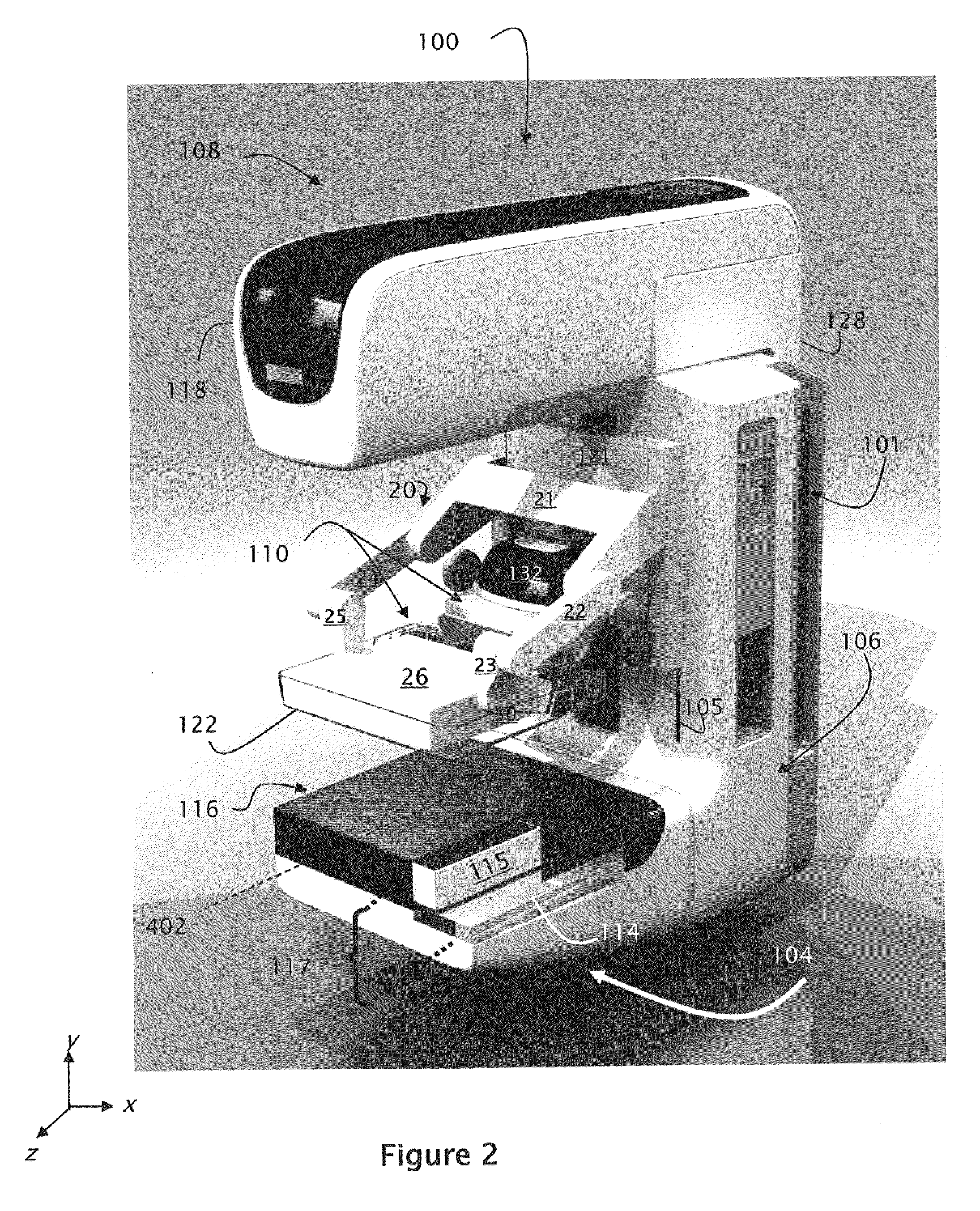
[0030]FIG. 2 illustrates an exemplary embodiment of an integrated Tomosynthesis / Molecular Breast Imaging (T / MBI) device 100 of the present invention. The T / MBI device 100 integrates x-ray components with molecular imaging components to provide a breast imaging system having increased sensitivity and specificity.

[0031]The T / MBI device 100 of FIG. 2 is shown to include a generally C shaped gantry comprised of an x-ray tube assembly 108, a gantry base 106 and a receptor housing 104, each of which is described in detail below. The C-shaped gantry is slideably mounted on a stand 140 (FIG. 12) via tracks 101 for movement along a Y axis to selectively position the gantry for breast imaging.

[0032]X-Ray Tube Assembly 108

[0033]The x-ray tube assembly 108 includes an x-ray tube head 118 and a x-ray support arm 128. The x-ray support arm is pivotably mounted on the gantry base 106 to enable movement of the x-ray tube head 118 about a horizontal axis 402 for tomosynthesis imaging. For example, d...


PUM


Abstract

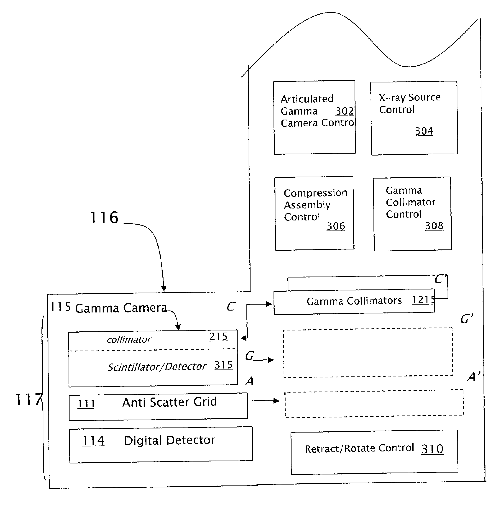
An integrated tomosynthesis/molecular breast imaging device having improved sensitivity includes tomosynthesis imaging components and molecular breast imaging components. The imaging components may be used individually or in combination to provide a system with improved sensitivity and specificity. Molecular imaging components may be smoothly advanced or withdrawn depending upon the desired imaging mode. The system supports both PET and SPECT imaging and enables SPECT collimation to be modified in accordance with image capture requirements.

Description


Claims


Application Information

Patent Timeline
Owner HOLOGIC INC
Learn More