
Medical device for modification of left atrial appendage and related systems and methods

a technology of atrial appendage and medical device, which is applied in the field of modification of atrial appendages, can solve the problems of ischemic damage of other organs of the body, blood pooling and thrombosis inside of the appendages, and affecting the function of the atrial appendage,

Active Publication Date: 2011-01-27
COHEREX MEDICAL
View PDF100 Cites 102 Cited by
  • Summary
  • Abstract
  • Description
  • Claims
  • Application Information

AI Technical Summary

Benefits of technology

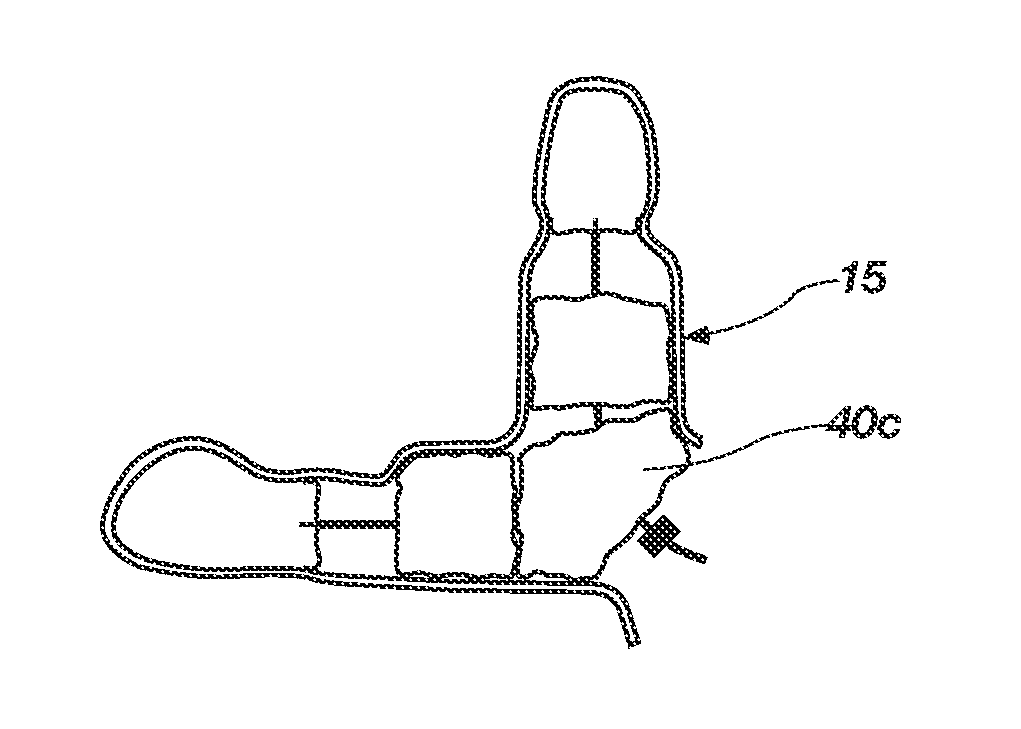
The present invention provides medical devices, systems, and methods for modifying an atrial appendage. The invention includes a catheter, tether, and anchoring member for positioning a tissue growth member within the left atrial appendage. The tissue growth member may be a foam material or multiple members that are placed in the left atrial appendage and maintained there with a locking element. The method may also include deploying the anchoring member in a constrained configuration and then expanding it to anchor it in the left atrial appendage. The invention can be used to occlude the left atrial appendage to prevent blood clots from forming there.

Problems solved by technology

However, in cases of atrial fibrillation, when the atria go into arrhythmia, blood may pool and thrombose inside of the appendages.
Among other things, this can pose a stroke risk when it occurs in the left appendage since the thrombus may be pumped out of the heart and into the cranial circulation.
Such can also lead to ischemic damage of other organs of the body.
However, due to the wide variability of the size of the ostium and the volume of an atrial appendage, implant devices that are currently used typically include structure that cannot meet such variability, resulting in inadequate devices for many left atrial appendages.
Further, such implant devices are substantially limited by the orientation by which they can successfully be deployed.
Thus, successful placement and deployment of such devices becomes limited.

Method used


Image

  • Medical device for modification of left atrial appendage and related systems and methods
  • Medical device for modification of left atrial appendage and related systems and methods
  • Medical device for modification of left atrial appendage and related systems and methods

Examples


Embodiment Construction

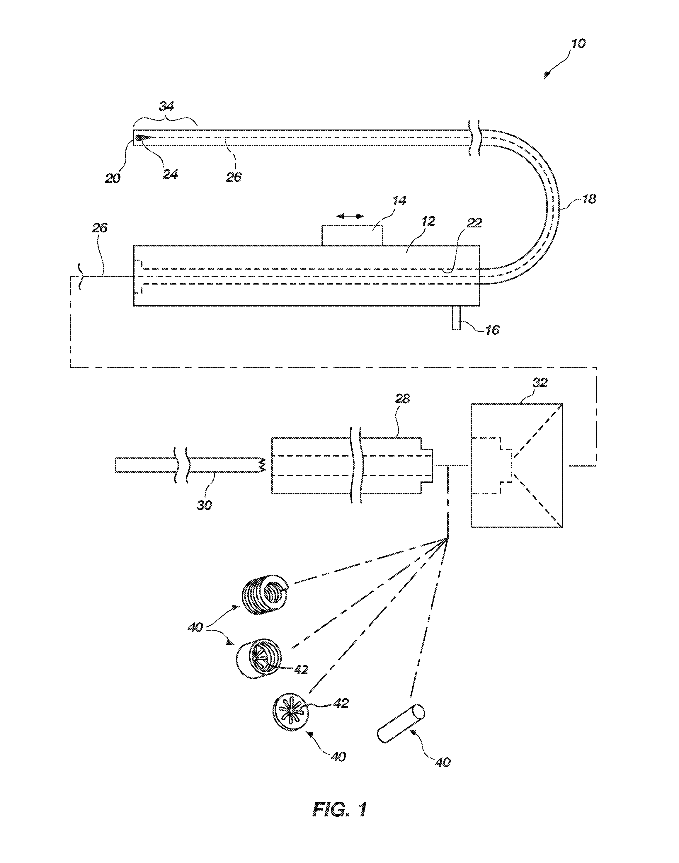
Referring to FIG. 1, a medical device system 10 is shown that may be used to occlude or modify an opening or cavity such as, for example, a left atrial appendage (LAA). In one embodiment, the medical device system 10 may include a handle 12 with an actuator 14 and fluid port 16. The fluid port 16 may be used to flush out the catheter when in use as will be appreciated by those of ordinary skill in the art. In addition, the system 10 includes a catheter 18 with a catheter lumen 20 extending longitudinally therethrough and attached to a distal end of the handle 12. The catheter lumen 20 may coincide and communicate with a handle lumen 22 as well as communicate with the fluid port 16. The actuator 14 may be configured to actuate or move the catheter 18 proximally and distally, relative to an associated tether 26, to deploy and capture, respectively, an anchoring member 24 disposed at a distal end of the tether 26. The tether 26 may be configured to extend through and be positioned with...


PUM

PropertyMeasurementUnit
sizesaaaaaaaaaa
shapesaaaaaaaaaa
imagingaaaaaaaaaa

Abstract

A medical device, system and method for modifying a left atrial appendage (“LAA”). The medical device system includes a tether anchored within the LAA and one or more tissue growth members or occluders configured to be slid over the tether and lodged within the LAA. With this arrangement, a physician may close-off the LAA with a selective number of tissue growth members to meet the varying LAA sizes as determined from imaging information while conducting the procedure.

Description

TECHNICAL FIELDThe present invention relates generally to the modification of an atrial appendage and, more specifically, to devices, systems and methods for occluding or otherwise structurally altering such appendages.BACKGROUNDThe atrial appendage is a feature of all human hearts. The upper chambers of the heart, the atria, have this appendage attached to each of them. The physiologic function of such appendages is not completely understood, but they do act as a filling reservoir during the normal pumping of the heart. The appendages typically protrude from the atria and cover an external portion of the atria. Atrial appendages differ substantially from one to another in size, shape and specific location with respect to the atria. For example, one atrial appendage may be configured as a tapered protrusion while another atrial appendage may be configured as axe-entrant, sock-like hole. The inner surface of an appendage is conventionally trabeculated with cords of muscular cardiac t...

Claims


Application Information

Patent Timeline
Patent Type & Authority Applications(United States)
IPC IPC(8): A61B17/03
CPCA61B17/0057Y10T29/49826A61B17/0487A61B17/12022A61B17/12122A61B17/12159A61B17/12172A61B17/122A61B2017/00243A61B2017/00575A61B2017/00579A61B2017/00588A61B2017/00592A61B2017/00597A61B2017/00601A61B2017/0061A61B2017/00632A61B2017/00867A61B2017/0409A61B2017/0412A61B2017/0414A61B2017/0437A61B2017/0446A61B2017/0448A61B2017/0459A61B2017/0464A61B17/12131A61B17/00234A61B17/1215A61B2017/00526A61B2017/00884A61B2017/1205A61B17/0401A61B17/06166A61B17/12031A61B17/1204
Inventor MILES, SCOTT D.WHISENANT, BRIAN K.EDMISTON, DARYL R.DAVIS, CLARK C.
Owner COHEREX MEDICAL
Learn More