
Medical Device and Delivery System for Modification of Left Atrial Appendage and Methods Thereof

Active Publication Date: 2012-12-13
COHEREX MEDICAL
View PDF10 Cites 93 Cited by
  • Summary
  • Abstract
  • Description
  • Claims
  • Application Information

AI Technical Summary

Benefits of technology

[0010]In another embodiment, the actuation assembly includes a mode switch movable by the actuation member. The mode switch is moveable between a mode switch first position and a mode switch second position such that the mode switch first position facilitates actuation of the anchor operator by the actuation member and the mode switch second position facilitates actuation of the float operator by the actuation member. Further, in one embodiment, the mode switch first position locks-out actuation of the float operator. In another embodiment, the mode switch second position locks-out actuation of the anchor operator.
[0024]In another embodiment, the advancing step includes exposing a distal end of the occluder portion from a distal tip of the sheath to expose a portion of the occluder portion to provide a cushion tip without substantially exposing any metallic portions of a frame of the medical device. Further, the advancing step includes atraumatically positioning the distal tip of the sheath in the left atrial appendage with the cushion tip of the occluder portion exposed at the distal tip of the sheath.

Problems solved by technology

However, in cases of atrial fibrillation, when the atria go into arrhythmia, blood may pool and thrombose inside of the appendages.
Among other things, this can pose a stroke risk when it occurs in the left appendage since the thrombus may be pumped out of the heart and into the cranial circulation once normal sinus rhythm is restored following arrhythmia events.
However, due to the wide variability of the ostium size and volume of the left atrial appendage, current implantable devices conventionally include a structure that cannot meet such variability, resulting in inadequate devices for many left atrial appendage anatomies.
Further, such implantable devices are substantially limited by the orientation by which they can successfully be deployed.

Method used


Image

  • Medical Device and Delivery System for Modification of Left Atrial Appendage and Methods Thereof
  • Medical Device and Delivery System for Modification of Left Atrial Appendage and Methods Thereof
  • Medical Device and Delivery System for Modification of Left Atrial Appendage and Methods Thereof

Examples


Embodiment Construction

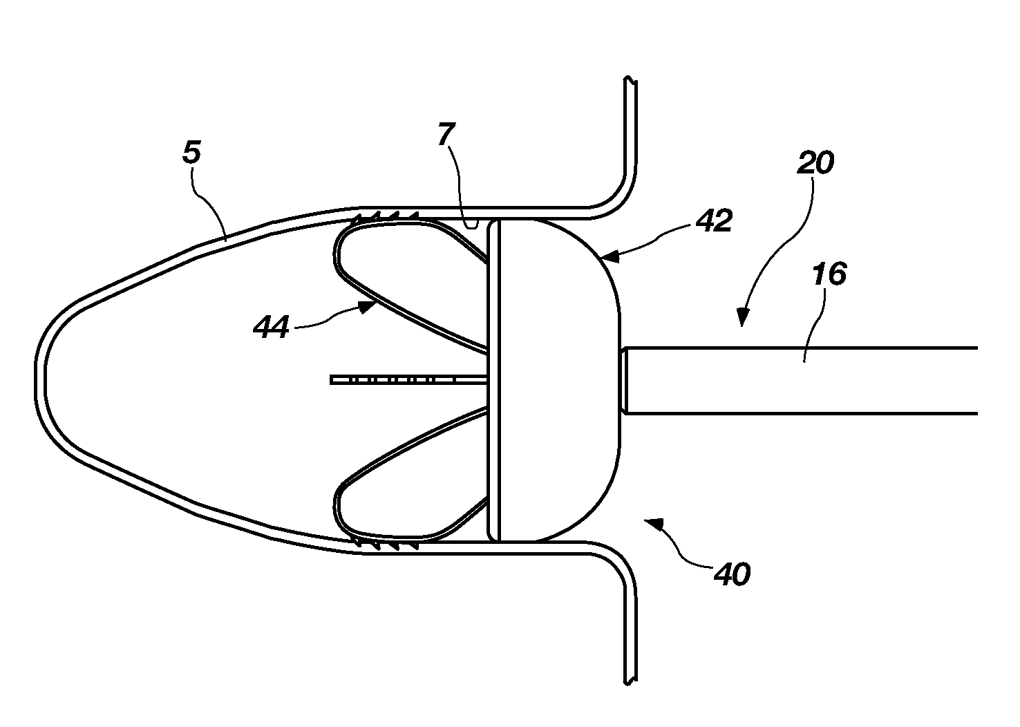
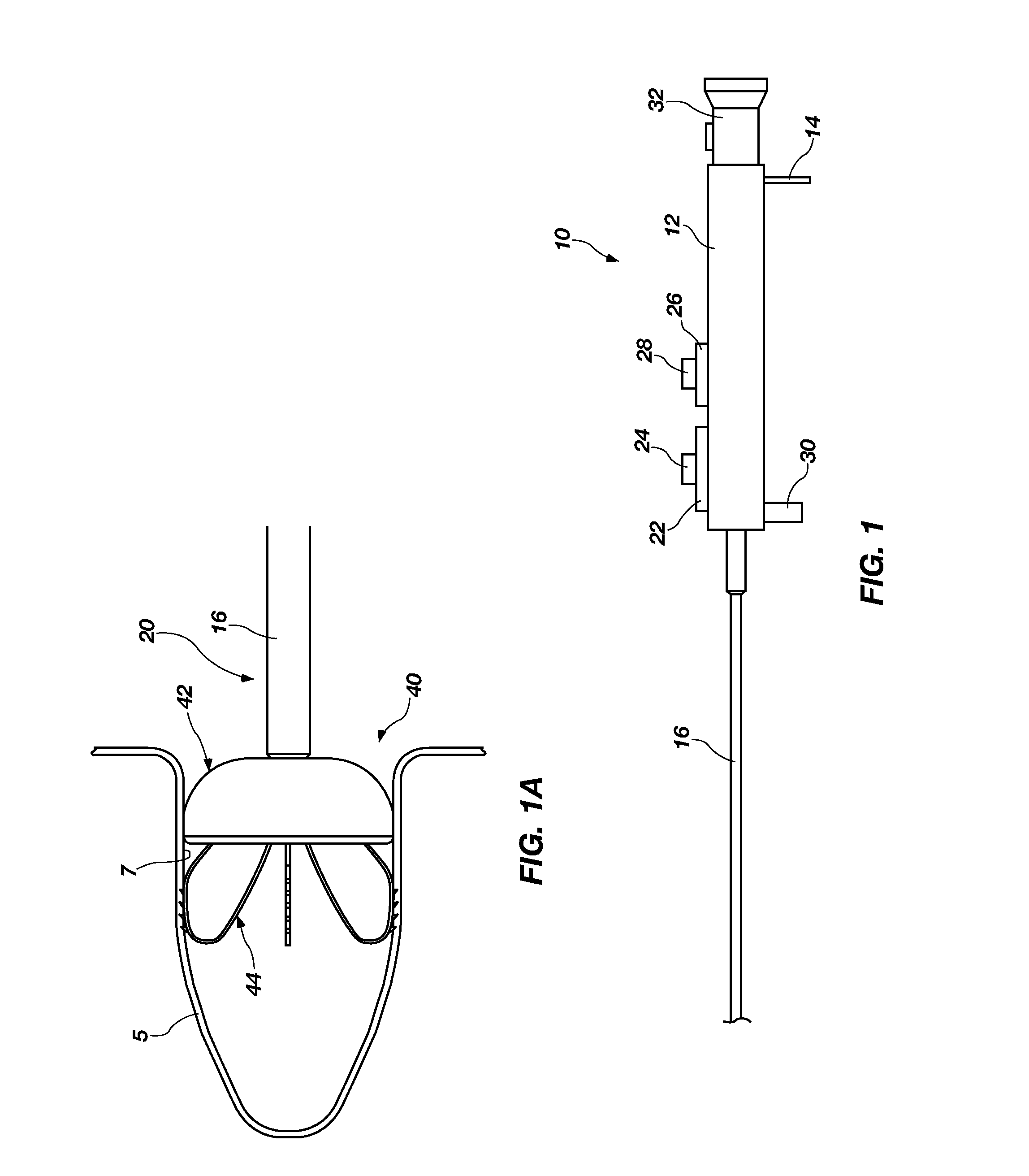
[0133]Referring to FIGS. 1 and 1A, a medical device system 10 is disclosed that may be used to occlude or modify an opening or cavity 5 such as, for example, a left atrial appendage (LAA). In one embodiment, the medical device system 10 may include a handle 12 with one or more actuators and a fluid port 14. In addition, the system 10 may include a catheter 16 with a catheter lumen extending longitudinally therethrough and attached to a distal end of the handle 12. Such a catheter lumen may coincide and communicate with a handle lumen as well as communicate with the fluid port 14.

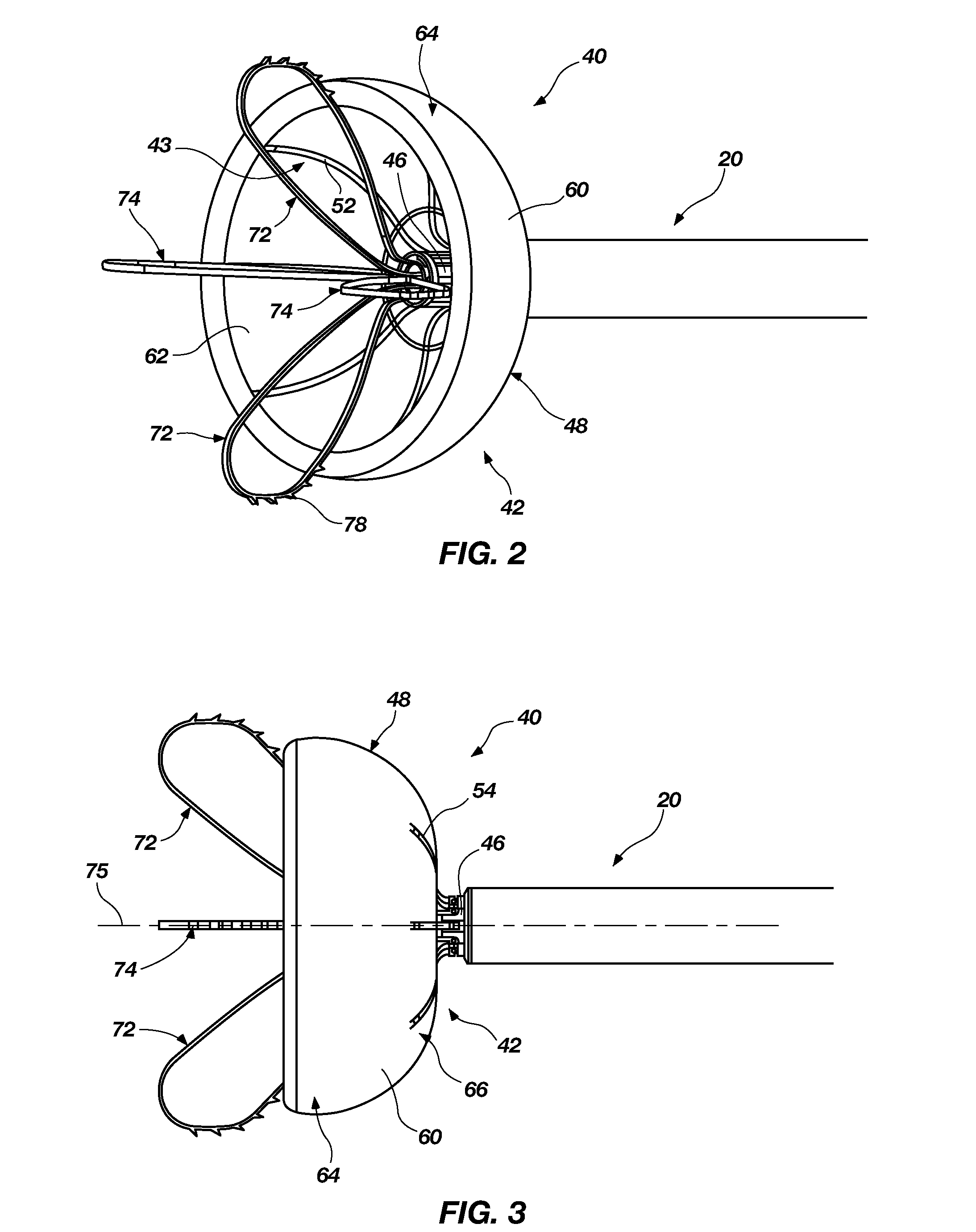
[0134]The actuators associated with the handle may be configured to actuate or move a medical device 40 disposed within a distal portion 20 of the catheter 16 to deploy the medical device 40 from or within the distal portion 20 of the catheter 16, to capture (or recapture) the medical device 40 within the distal portion 20 of the catheter, or to do both. Such a medical device 40 can be interconnected to the ...


PUM


Abstract

Devices, methods and systems are provided for occluding an opening within the tissue of a body, such as a left atrial appendage. In one embodiment, a delivery system for use in occluding an opening in the tissue of a body includes an actuation assembly operatively coupled to a medical device including an anchor portion and an occluder portion. The actuation assembly is configured to move the anchor portion between a deployed state and retracted state while the occluder portion maintains a deployed state. With this arrangement, the deployed occluder portion can be visualized via imaging at a preferred position prior to deploying the anchor portion of the medical device.

Description

CROSS-REFERENCE TO RELATED APPLICATIONS[0001]This application claims benefit to U.S. Provisional Patent Application No. 61 / 477,075, filed on Apr. 19, 2011. This application also claims benefit to, and is a continuation-in-part of, U.S. patent application Ser. No. 12 / 818,059, filed on Jun. 17, 2010, which in turn claims benefit to the following U.S. Provisional patent applications: U.S. Provisional Application No. 61 / 345,514, filed on May 17, 2010; U.S. Provisional Application No. 61 / 325,230, filed on Apr. 16, 2010; U.S. Provisional Application No. 61 / 320,635, filed on Apr. 2, 2010; U.S. Provisional Application No. 61 / 294,058, filed on Jan. 11, 2010; and U.S. Provisional Application No. 61 / 218,018, filed on Jun. 17, 2009. The disclosures of each application listed above are incorporated by reference herein in their entireties.TECHNICAL FIELD[0002]The present invention relates generally to the occlusion or modification of tissue openings or appendages and, more specifically, to device...

Claims


Application Information

Patent Timeline
IPC IPC(8): A61B17/10
CPCA61B17/0057A61B17/12022A61B17/12122A61B17/12172A61B17/12177A61B2017/12054A61B2017/00327A61B2017/0053A61B2017/00575A61B2017/0069A61B2017/00309A61B17/00234A61B2017/00243
Inventor MILES, SCOTT D.DAVIS, CLARK C.EDMISTON, DARYL R.LINDER, RICHARD J.
Owner COHEREX MEDICAL
Learn More