
System and method for determining airway diameter using endoscope

a technology of endoscopy and airway diameter, which is applied in the field of minimally invasive medical procedures, can solve problems such as complicated determination

Active Publication Date: 2011-03-17
BRONCUS MEDICAL
View PDF7 Cites 12 Cited by
  • Summary
  • Abstract
  • Description
  • Claims
  • Application Information

AI Technical Summary

Benefits of technology

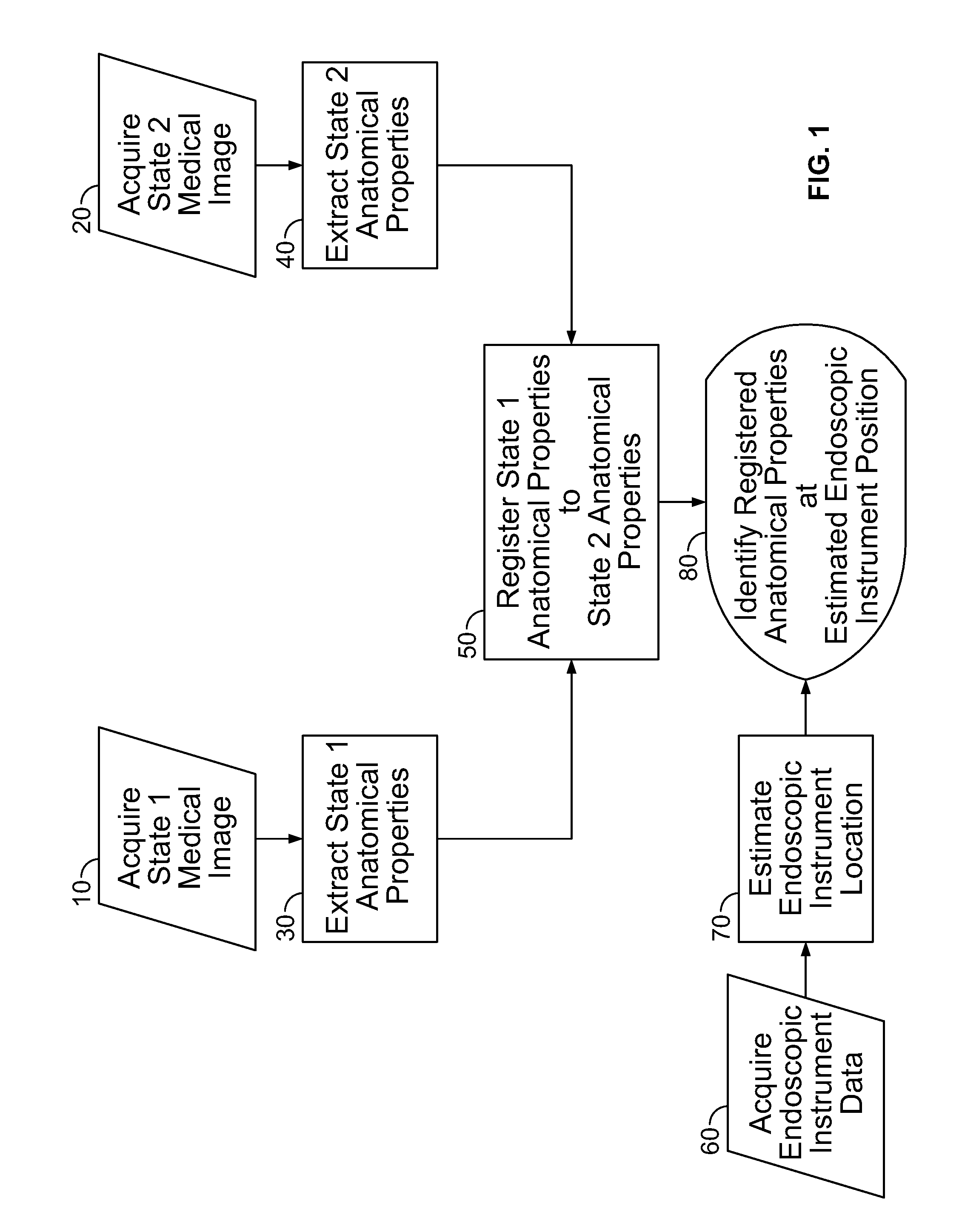
[0009]The invention may utilize a real endoscope, with the property identification being performed during surgery in real time. The properties may also be derived from segmented 3D model data arising from CT scans of the body lumen. The step of estimating the location of the endoscope may be carried out using an image to image based registration approach. This estimating step may be performed prior to, or subsequent to, the registering step.

Problems solved by technology

The determination, however, is complicated by a number of factors including the tidal motion of the lungs, i.e., inhalation and expiration of the lungs modifies the dimension of the lumens.

Method used


Image

  • System and method for determining airway diameter using endoscope
  • System and method for determining airway diameter using endoscope
  • System and method for determining airway diameter using endoscope

Examples


Embodiment Construction

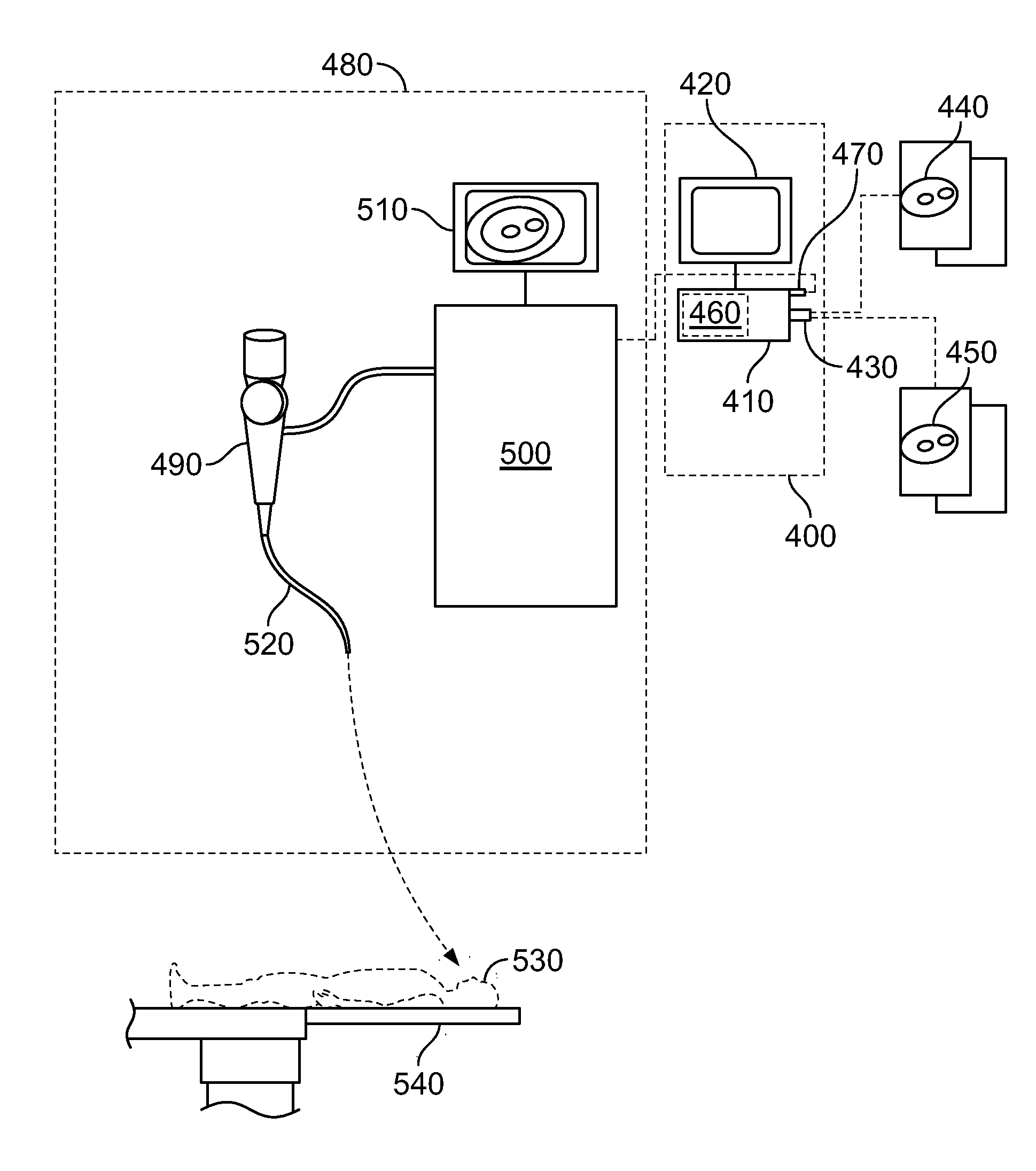
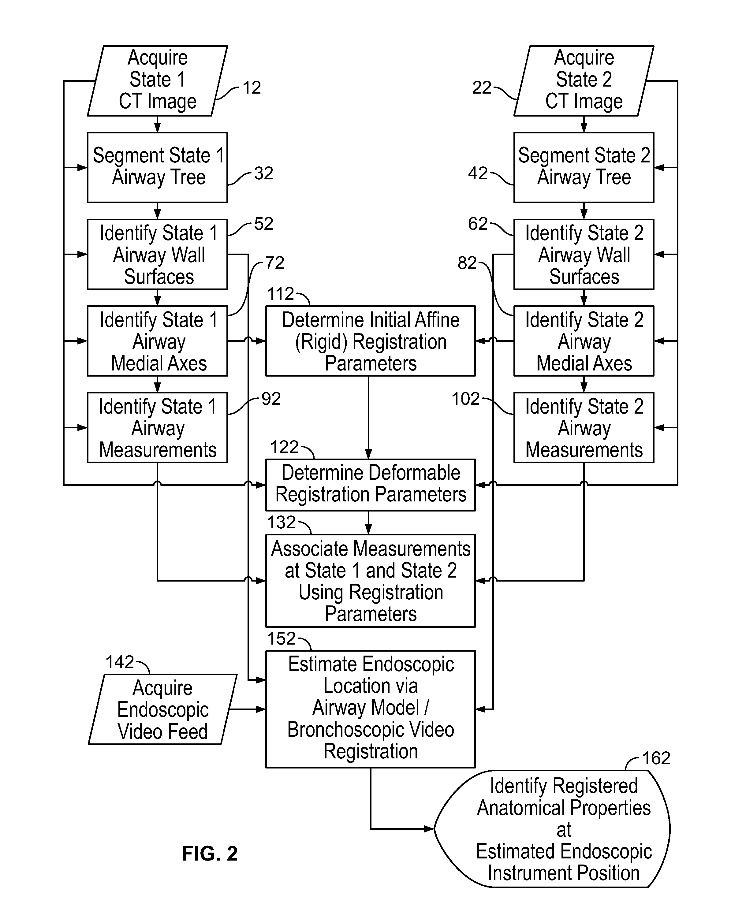
[0016]A minimally invasive method and system for determining various properties of a body lumen, e.g., an airway, is described herein. FIG. 1 shows an embodiment of the present invention including a sequence of image processing and registration steps to identify one or more anatomical properties of the body lumen at a specific location during an endoscopic procedure. In particular, and as explained in more detail below, one embodiment of the invention includes: (1) acquiring medical image data of a lumen while in at least two different states; (2) segmenting the lumen from the image data; (3) determining anatomical properties of the lumen in each state; (3) registering the anatomical properties corresponding to the lumen in the first state to the anatomical properties corresponding to the lumen in the second state; (4) estimating a location of the endoscope; and (5) identifying the properties of the lumen in the first state and the second state corresponding to the location of the e...


PUM


Abstract

A method and system for use with an endoscopic instrument determines anatomical properties of body lumen at various states. Lumen properties such as lumen diameter are identified in two or more states corresponding to, for example, an inflated or deflated state. The lumen states are registered with one another and the anatomical properties are identified in real time at the location of an endoscope or endoscopic instrument used with the endoscope. In one embodiment a diametrical range of an airway is identified in real time at the location of a bronchoscope.

Description

CROSS REFERENCE TO RELATED APPLICATIONS[0001]This application claims priority to provisional application No. 61 / 243,310 filed Sep. 17, 2009 and entitled “System and Method For Determining Airway Diameter Using Endoscope”.BACKGROUND OF THE INVENTION[0002]Endoscopy is a minimally invasive medical procedure that allows a physician to interrogate the interior of the body through an endoscope, which provides a light source to illuminate the anatomy and a method for viewing the anatomy. Typically, the endoscope includes a set of fiber optic bundles connected to a viewing lens or a camera that provides video output. Examples of endoscopes include colonoscopes for examination and therapeutic use in the colon and bronchoscopes for the trachea and branching airways in the lungs. Such devices allow physicians to reach deep into the body through natural orifices, minimizing the trauma that would be required if more invasive procedures were performed.[0003]Endoscopic procedures are often perform...

Claims


Application Information

Patent Timeline
IPC IPC(8): A61B1/00
CPCA61B1/2676A61B5/1076A61B6/12A61B5/418A61B6/03A61B5/415
Inventor WIBOWO, HENKYGIBBS, JASON DAVID
Owner BRONCUS MEDICAL
Learn More