Implantable Devices and Related Methods for Heart Failure Monitoring

a heart failure and implantable device technology, applied in the field of implantable devices for heart failure monitoring, can solve the problems of increased blood pressure in the lungs, direct medical costs, and buildup of fluid in the vascular system,

Active Publication Date: 2019-01-31
FOUNDRY INNOVATION & RES 1 LTD
View PDF15 Cites 14 Cited by
  • Summary
  • Abstract
  • Description
  • Claims
  • Application Information

AI Technical Summary

Benefits of technology

[0028]Another method disclosed herein is a method of monitoring a cardiac condition of a patient, comprising implanting an intravascular device in a vena cava with an anchor element at an anchoring location, the intravascular device further having a pair of markers or sensors, the pair of markers or sensors being positioned in engagement with opposing walls of the vena cava at a monitoring location; receiving a signal from the pair of markers or sensors; and determining a diameter of the vena cava based on the signal; wherein the implanting comprises positioning the anchor element at the anchoring location sufficiently distant from the monitoring location such that diameter determinations based on the signal are not substantially affected by distortion of the vena cava wall by the anchor element at the anchor location.
[0029]In another disclosed embodiment, a monitoring system comprises an intravascular device configured for implantation in a vessel in a body cavity and having first and second markers biased or secured against opposing interior walls of the vessel; at least one anchor element configured to secure and position the intravascular device on the body cavity wall; an anchor isolation structure disposed between the at least one anchor element and the first and second markers, the anchor isolation structure having a length specifically configured to position the first and second markers sufficiently distant from the monitoring location such that diameter calculations derived from the markers are not substantially affected by distortion of the vena cava wall by the anchor element at; and an external device adapted to receive a first signal from the intravascular device and to calculate a distance between the first and second markers based on the signal.

Problems solved by technology

Twenty billion of this cost is direct medical costs.
ADHF is very often a result of some combination of a downturn in the heart's performance and excessive intake of fluids and / or salt.
This leads to a buildup of fluid in the vascular system.
Increased blood volume in the left atrium at higher pressure means higher blood pressure in the lungs, which eventually leads to fluid filling the lungs and an inability to breathe.
Managing these patients to prevent the need for re-hospitalization is extremely challenging.
Congestive heart failure is so-named because additional blood volume backing up into the lungs causes fluid to seep out of the pulmonary circulation into the airway passages of the lungs, causing congestion of the lungs.
This happens because the left ventricle is not able to pump all of the volume of blood returning to the heart from the lungs.
The pulmonary artery hypertension can worsen pre-existing right ventricular dysfunction and exacerbate tricuspid valve regurgitation, leading to systemic venous congestion.
During inspiration, intrathoracic pressure decreases, thereby increasing venous return and causing collapse of the IVC.
While vessel dimensions may be measurable using external ultrasound, magnetic resonance imaging, computerized axial tomography, or other technologies, these imaging procedures must be administered in a hospital or other specialized facility, do not permit continuous monitoring, and do not allow for monitoring of the patient at their home or other remote location.
As a result, the condition of a heart failure patient can worsen into a critical state before care providers become aware of it, dramatically increasing the mortality risk and cost of treatment for the patient.
This is not something that heart failure patients or their caregivers could typically be trained to do predictably and accurately with existing equipment.
Moreover, these systems typically include large, complex, and expensive pieces of equipment which are not suitable for use outside of a specialized medical facility.
However, in spite of the widespread use of IVC filters over many years, heretofore it has not been suggested that an implant in the IVC could be utilized for purposes of monitoring fluid volume or IVC dimensions.
Moreover, even if a suggestion were made to equip an IVC filter with a sensor for monitoring vascular dimensions, such filters would be unsuited to the purpose.
The anchoring structures used to secure IVC filters constrain the vessel from natural size and shape changes in response to changes in fluid volume and would thus limit the usefulness or accuracy of such a device.

Method used


Image

  • Implantable Devices and Related Methods for Heart Failure Monitoring
  • Implantable Devices and Related Methods for Heart Failure Monitoring
  • Implantable Devices and Related Methods for Heart Failure Monitoring

Examples


Embodiment Construction

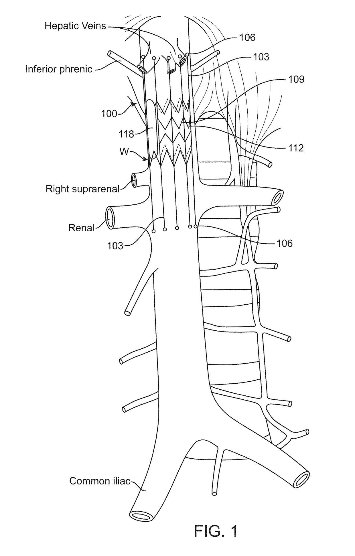
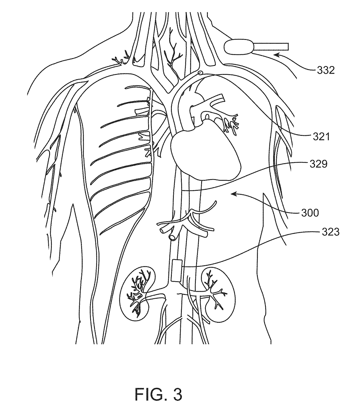
[0083]Various embodiments disclosed herein are intended to monitor for and detect variations in volume and / or pressure of the inferior vena cava (IVC) as an early warning signal of the acute severity of heart failure. Implantable IVC monitors, markers and related systems, devices and methods as described herein may enable the patient and physician to take proactive steps in time to prevent acute decompensation requiring hospitalization. Such devices and methods also may be helpful in managing hemodialysis patients, in whom volume management is a chronic challenge. The present disclosure thus describes methods and devices for measuring IVC volume and / or pressure more or less continuously, depending on clinical need, using various forms of implantable devices.

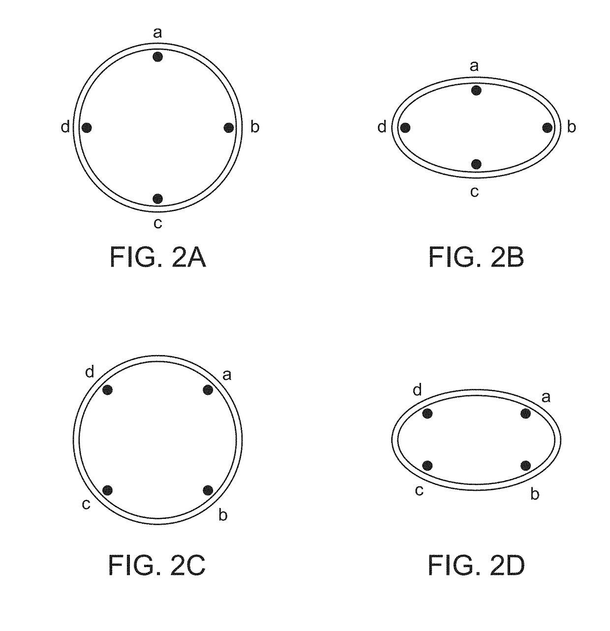
[0084]In order to measure changes in IVC dimension or volume accurately, the devices of the invention must be configured to be secured at the desired location in or on the vessel without affecting the natural dilation and constri...


PUM


Abstract

Implantable devices for continuously monitoring vascular lumen dimensions, in particular in the inferior vena cava (IVC) for determining heart failure status of a patient. Related therapy systems as well as monitoring and therapy methods are also disclosed. Devices include active or passive marker elements placed in contact with, adhered to or injected into the vessel wall to generate or reflect signals from which lumen diameter may be determined. Disclosed devices may be fully implantable and self-contained including capabilities for wirelessly communication monitored parameters.

Description

RELATED APPLICATION DATA[0001]This application is a continuation of U.S. Nonprovisional patent application Ser. No. 15 / 549,042, filed Aug. 4, 2017, which application is a U.S. national phase of International Patent Application No. PCT / US16 / 17902, filed on Feb. 12, 2016, and titled “Implantable Devices and Related Methods For Heart Failure Monitoring.” International Patent Application No. PCT / US16 / 17902 claims the benefit of priority of U.S. Provisional Patent Application No. 62 / 172,516, filed Jun. 8, 2015, and titled “Methods and Apparatus for Monitoring Patient Physiological Status Based On Inferior Vena Cava Volume,” claims the benefit of priority of U.S. Provisional Patent Application Ser. No. 62 / 157,331, filed May 5, 2015, and titled “Heart Failure Monitoring System and Method,” and also claims the benefit of priority of U.S. Provisional Patent Application Ser. No. 62 / 115,435, filed Feb. 12, 2015, and titled “Implantable Device and Related Methods for Heart Failure Monitoring.” ...

Claims


Application Information

Patent Timeline
Patent Type & Authority Applications(United States)
IPC IPC(8): A61B8/08A61B90/00A61B8/00A61B5/00
CPCA61B90/39A61B8/565A61B5/6882A61B2090/3987A61B8/0891A61F2/01A61B5/1076A61B5/4839A61B2090/3929A61B5/4836A61B2090/3991A61B8/12A61F2/848A61F2/82A61M25/04G06T7/11G06T7/337G06T2207/10072G06T2207/10136G06T2207/30101A61B8/4227
Inventor GIFFORD, III, HANSON S.DEEM, MARK E.SUTTON, DOUGLAS S.RAJASEKHAR, VIJAYKUMAR
Owner FOUNDRY INNOVATION & RES 1 LTD
Why Eureka
Try Eureka