
Endoscope system

a technology of endoscope and endoscope, which is applied in the field of endoscope system, can solve the problems of difficulty in introducing a distal end near the target region, and achieve the effect of improving the accuracy of image enhancemen

Active Publication Date: 2015-10-01
OLYMPUS CORP
View PDF2 Cites 15 Cited by
  • Summary
  • Abstract
  • Description
  • Claims
  • Application Information

AI Technical Summary

Benefits of technology

The present invention is an endoscope system that can create a virtual endoscopic image based on three-dimensional image information recorded by a medical diagnosis apparatus. It can also detect changes in the position of the endoscope and record it, along with the virtual endoscopic image. This system can improve the accuracy and efficiency of medical diagnostic procedures involving the use of an endoscope.

Problems solved by technology

On the other hand, when the endoscope is inserted into a lumen organ complexly branching like a bronchus in the body cavity to examine (a diseased tissue of) a target region of the periphery side of the lumen organ or perform a biopsy or treatment by a treatment instrument, it is sometimes difficult to introduce an insertion section distal end near the target region using only an endoscopic image obtained when the endoscope is inserted.

Method used


Image

  • Endoscope system
  • Endoscope system
  • Endoscope system

Examples


first embodiment

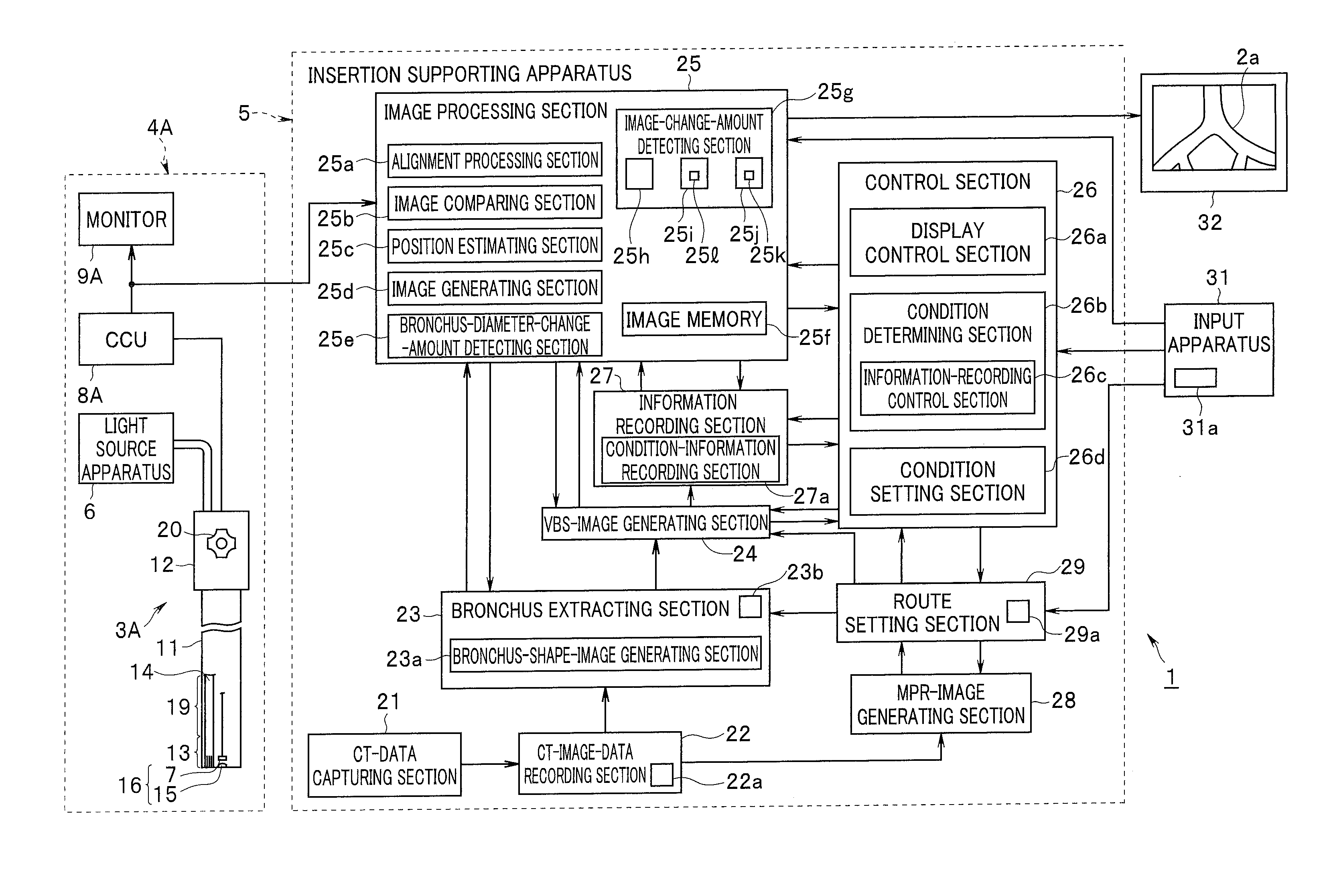
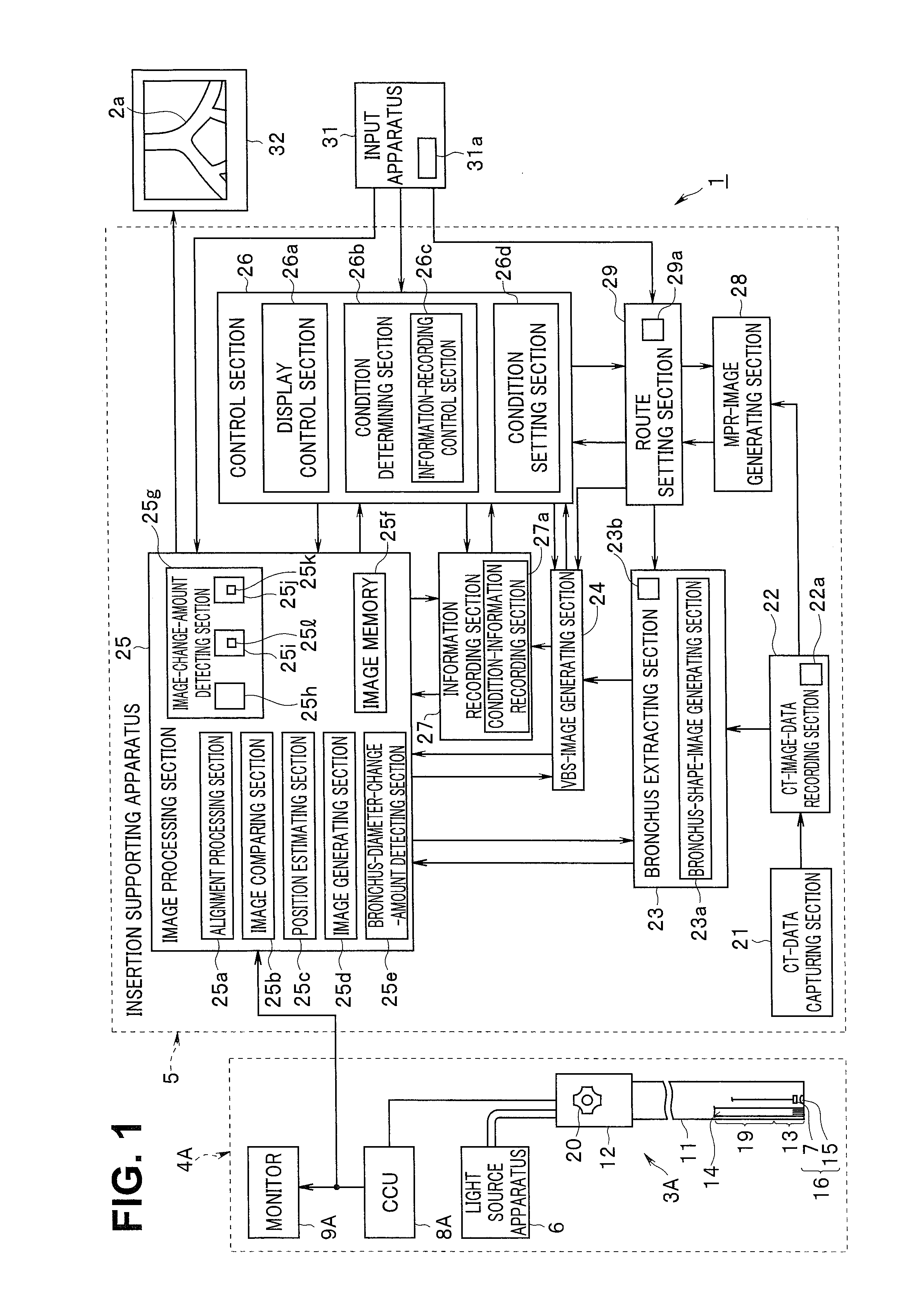
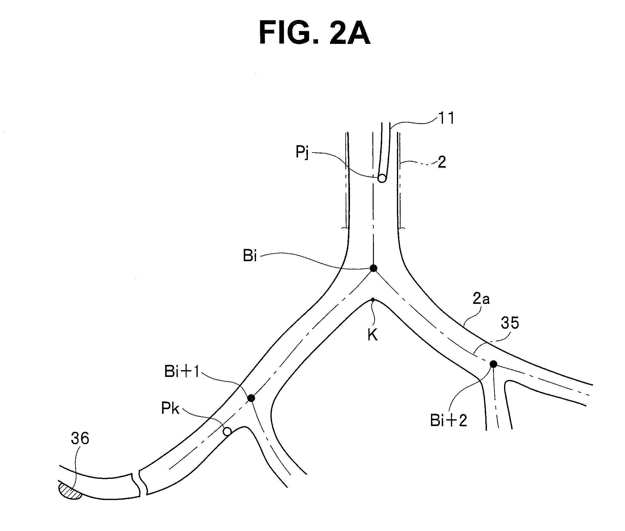
[0039]As shown in FIG. 1, an endoscope system 1 in a first embodiment of the present invention is mainly configured from an endoscope apparatus 4A including an endoscope 3A inserted into a bronchus 2 (FIG. 2A) set as a predetermined lumen organ in a patient treated as a subject, which is a test target, and an insertion supporting apparatus 5 used together with the endoscope apparatus 4A to perform insertion support of the endoscope 3A.

[0040]The endoscope apparatus 4A includes the endoscope 3A, a light source apparatus 6 that supplies illumination light to the endoscope 3A, a camera control unit (abbreviated as CCU) 8A functioning as a signal processing apparatus that performs signal processing for an image pickup device 7, which configures image pickup means mounted on the endoscope 3A, and a monitor 9A that displays an endoscopic image generated by the CCU 8A.

[0041]The endoscope 3A includes an elongated insertion section (or an endoscope insertion section) 11 having flexibility and...


PUM


Abstract

An endoscope system includes an endoscope including an image pickup section that picks up an image of a subject lumen organ, a recording section that records three-dimensional image information of a lumen organ acquired from a medical diagnosis apparatus, a position estimating section that estimates a distal end position of the endoscope, a lumen-diameter acquiring section that acquires a lumen diameter of the organ in the estimated position of the distal end, a condition determining section that determines whether the acquired lumen diameter is smaller than the reference lumen diameter, an image-change-amount detecting section that detects a change amount of a predetermined parameter in an endoscopic image picked up by the image pickup section, a virtual-endoscopic-image generating section that generates a virtual endoscopic image from any visual point position, and an information recording section that records a position of a distal end of an insertion section and the virtual endoscopic image.

Description

CROSS REFERENCE TO RELATED APPLICATION[0001]This application is a continuation application of PCT / JP2014 / 053876 filed on Feb. 19, 2014 and claims benefit of Japanese Application No. 2013-067408 filed in Japan on Mar. 27, 2013, the entire contents of which are incorporated herein by this reference.BACKGROUND OF THE INVENTION[0002]1. Field of the Invention[0003]The present invention relates to an endoscope system that picks up an image of an inside of a subject with image pickup means.[0004]2. Description of the Related Art[0005]In recent years, an endoscope including an insertion section insertable into a body cavity and the like has been widely used in a medical field and the like.[0006]On the other hand, when the endoscope is inserted into a lumen organ complexly branching like a bronchus in the body cavity to examine (a diseased tissue of) a target region of the periphery side of the lumen organ or perform a biopsy or treatment by a treatment instrument, it is sometimes difficult ...

Claims


Application Information

Patent Timeline
IPC IPC(8): A61B1/00A61B1/267A61B1/04A61B5/00A61B5/107
CPCA61B1/00009A61B5/0084A61B5/1076A61B5/1079A61B1/04A61B5/7282A61B1/00055A61B5/7246A61B1/0005A61B1/2676A61B1/0002A61B1/00A61B1/00193A61B6/12A61B5/065A61B6/5247A61B5/066A61B2034/2065A61B6/032A61B6/5223A61B5/7207A61B1/000094A61B1/000095A61B1/00194G06T19/003G06T2207/10068G06T2210/41
Inventor ITO, MITSUHIROAKIMOTO, SYUNYAONISHI, JUNICHI
Owner OLYMPUS CORP
Learn More