Ultrasonic imaging apparatus

a technology of ultrasonic imaging and ultrasonic radiation, applied in ultrasonic/sonic/infrasonic diagnostics, instruments, applications, etc., can solve the problems of limited viewing field and region capable of being imaged

Active Publication Date: 2011-03-31
FUJIFILM HEALTHCARE CORP
View PDF5 Cites 13 Cited by
  • Summary
  • Abstract
  • Description
  • Claims
  • Application Information

AI Technical Summary

Benefits of technology

[0019]According to the present invention, a difference in hemodynamics can be visualized, and a vessel in a contrast-enhancement phase necessary to diagnosis can be specifically displayed.
[0020]Anothe

Problems solved by technology

Contrast enhancement based on ultrasonic waves has such features that a contrast medium itself lacks both toxicity and critical invasiveness such as exposure, and that per

Method used


Image

  • Ultrasonic imaging apparatus
  • Ultrasonic imaging apparatus
  • Ultrasonic imaging apparatus

Examples


embodiment 1

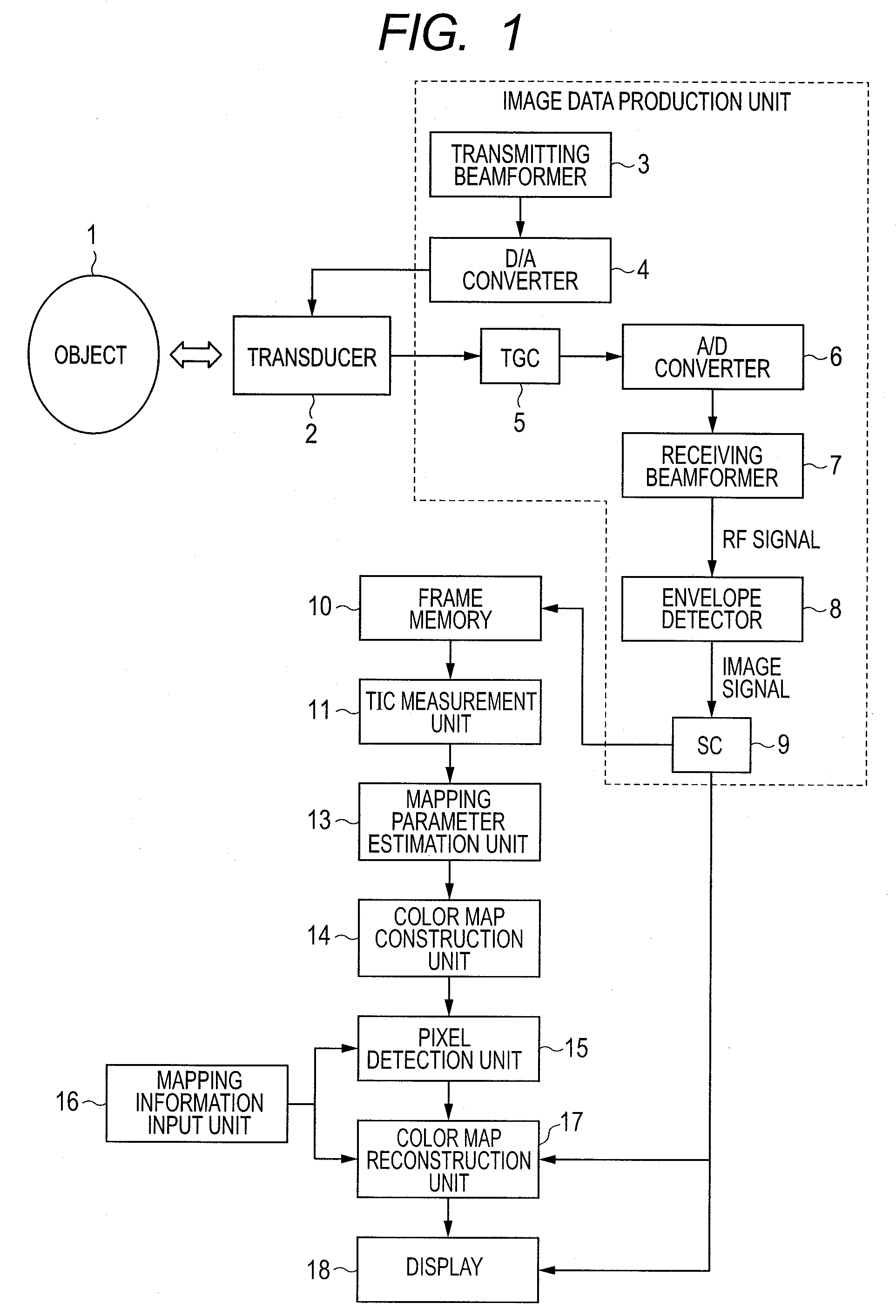
[0065]FIG. 1 is a block diagram showing a constitution of an ultrasonic imaging apparatus in accordance with an embodiment 1 of the present invention.

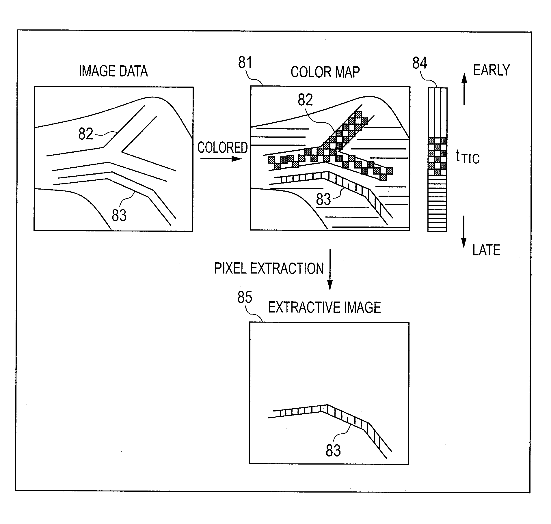
[0066]The ultrasonic imaging apparatus includes: a transducer 2 that transmits or receives ultrasonic waves to or from an object 1; a transmitting beam former 3 and a receiving beam former 7 that give a predetermined time delay, during which a predetermined transmitting / receiving beam is formed, to piezoelectric elements constituting the transducer 2; an analog-to-digital (A / D) converter 6 that analog-to-digital converts a transmitting / receiving signal, and a digital-to-analog (D / A) converter 4; a TGC 5 that compensates an amplitude fading occurring in the process of propagation through the inside of a living body; an envelope detector 8 that detects a received radiofrequency signal and converts it into an image signal; a scan converter 9 that constructs a two-dimensional image from the image signal; a frame memory 10 that preserves th...


PUM


Abstract

Provided is an ultrasonic imaging apparatus including: a time-gain controller (TGC) that compensates an amplitude fading occurring in the process of propagation inside a living body; a scan converter (SC) that constructs image data; a TIC measurement unit that measures a TIC of each pixel; an evaluation index input unit that inputs an index for evaluating hemodynamics on the basis of a TIC; a mapping parameter estimation unit that estimates a mapping parameter comparable to an evaluation index; a TIC image construction unit that constructs a two-dimensional image on the basis of the mapping parameter; and a pixel detection unit that extracts a region corresponding to a color map from a TIC image, and utilizing a TIC measured with each pixel so as to measure a difference in hemodynamics.

Description

INCORPORATION BY REFERENCE[0001]The present application claims the priority of Japanese Patent Application No. 2008-057167 filed on Mar. 7, 2008, the contents of which are incorporated herein by reference.TECHNICAL FIELD[0002]The present invention relates to a technology for transmitting or receiving ultrasonic waves to or from the inside of a living body, and visualizing the inside thereof, in particular, to an ultrasonic imaging apparatus that performs imaging by utilizing a brightness change in an in vivo tissue due to a contrast medium.BACKGROUND ART[0003]An ultrasonic imaging apparatus that transmits pulsating ultrasonic waves to the inside of a living body and visualizing internal information on the living body using a receiving signal has the features of a compact apparatus and real-time image display, and is one of imaging apparatuses used versatilely in the field of medicine along with X ray and MRI.[0004]Vascular contrast enhancement is generally known as a technique of ob...

Claims


Application Information

Patent Timeline
IPC IPC(8): A61B8/00
CPCA61B8/06G01S7/52074G01S7/52071G01S7/52033
Inventor YOSHIKAWA, HIDEKIAZUMA, TAKASHI
Owner FUJIFILM HEALTHCARE CORP
Why Eureka
Try Eureka