System and method for cardiac segmentation in mr-cine data using inverse consistent non-rigid registration

Inactive Publication Date: 2011-04-07
SIEMENS HEALTHCARE GMBH
View PDF2 Cites 33 Cited by
  • Summary
  • Abstract
  • Description
  • Claims
  • Application Information

AI Technical Summary

Benefits of technology

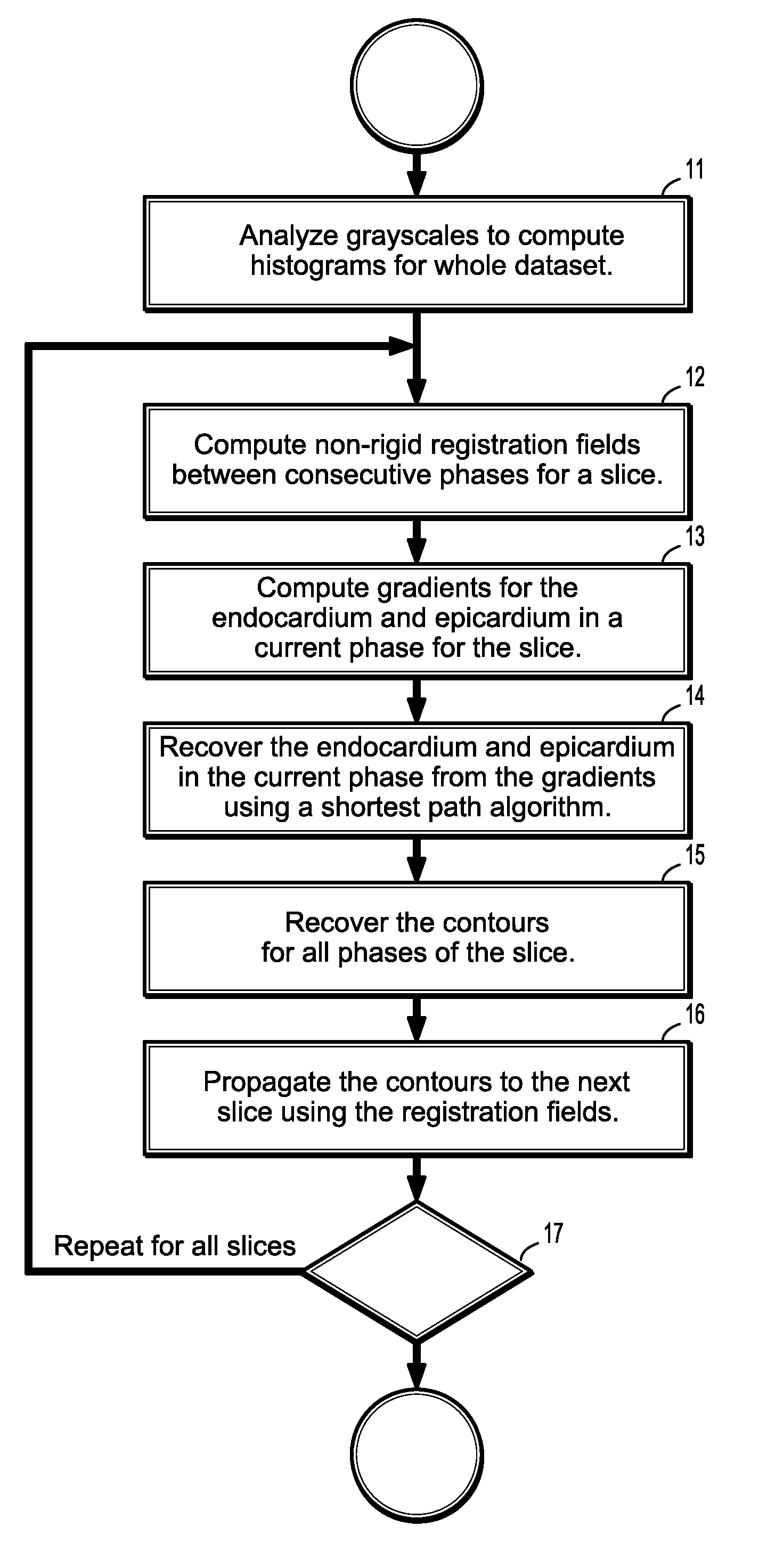
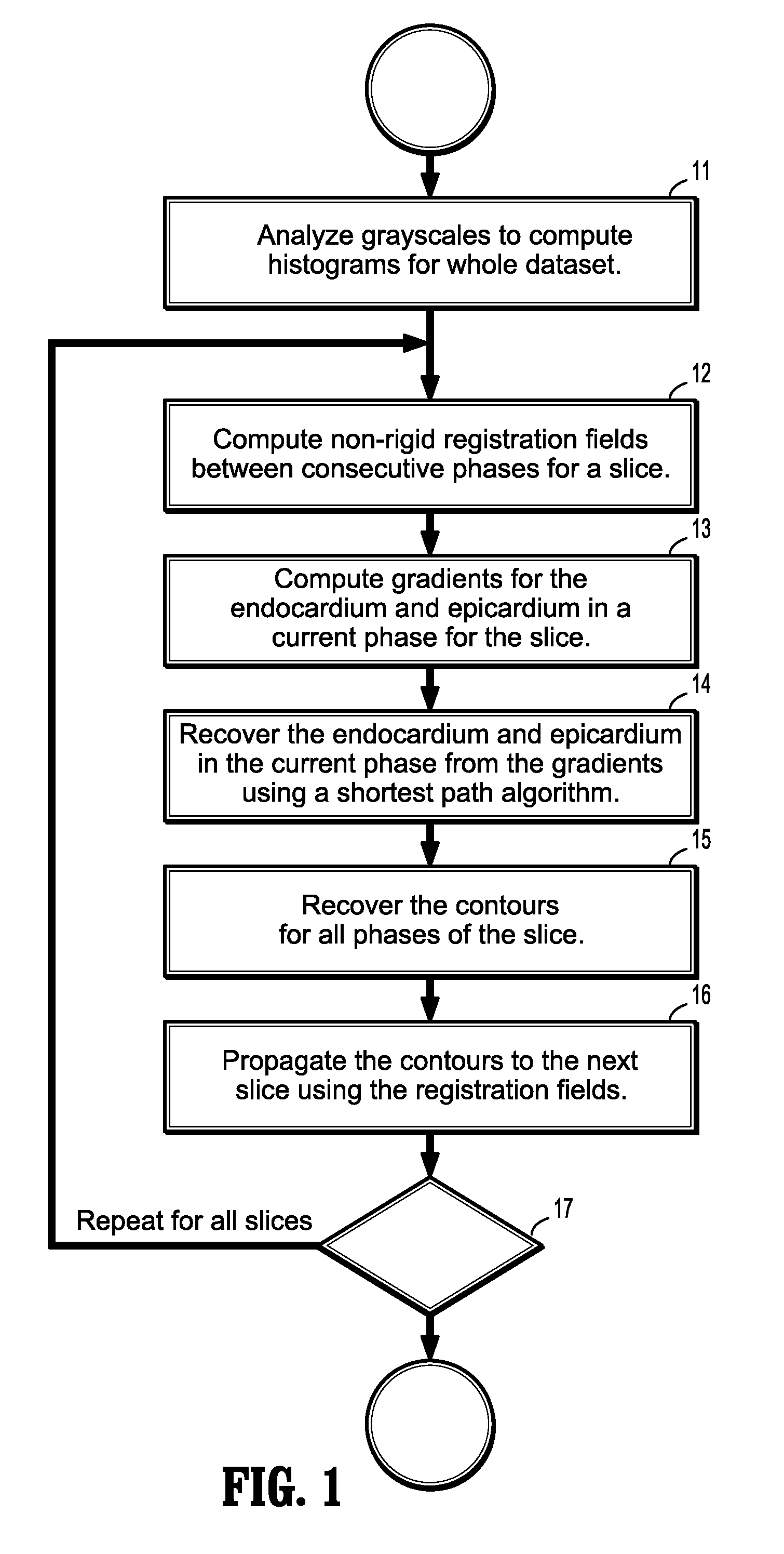
[0007]Exemplary embodiments of the invention as described herein generally include methods and systems for a registration-based segmentation to fully automatically segment the left ventricle in cardiac cine magnetic resonance studies. A method according to an embodiment of the invention segments one slice at a time using non-rigid registration, taking advantage of the strong temporal correlation between phases, and propagate the segmentation between slices. A method according to an embodiment of the invention uses an inverse consistent non-rigid registration algorithm to recover one set of forward and backward deformation fields that allows accessing the deformation from any frame to any other frame in the cardiac sequence. Cardiac phases are segmente

Problems solved by technology

This can be done because the non-rigid registration is inverse consistent, as otherwise, one would

Method used


Image

  • System and method for cardiac segmentation in mr-cine data using inverse consistent non-rigid registration
  • System and method for cardiac segmentation in mr-cine data using inverse consistent non-rigid registration
  • System and method for cardiac segmentation in mr-cine data using inverse consistent non-rigid registration

Examples


Embodiment Construction

[0031]Exemplary embodiments of the invention as described herein generally include systems and methods for segmenting the myocardium in the left ventricle of the heart from cardiac cine magnetic resonance (MR) images. Accordingly, while the invention is susceptible to various modifications and alternative forms, specific embodiments thereof are shown by way of example in the drawings and will herein be described in detail. It should be understood, however, that there is no intent to limit the invention to the particular forms disclosed, but on the contrary, the invention is to cover all modifications, equivalents, and alternatives falling within the spirit and scope of the invention.

[0032]As used herein, the term “image” refers to multi-dimensional data composed of discrete image elements (e.g., pixels for 2-D images and voxels for 3-D images). The image may be, for example, a medical image of a subject collected by computer tomography, magnetic resonance imaging, ultrasound, or any...


PUM


Abstract

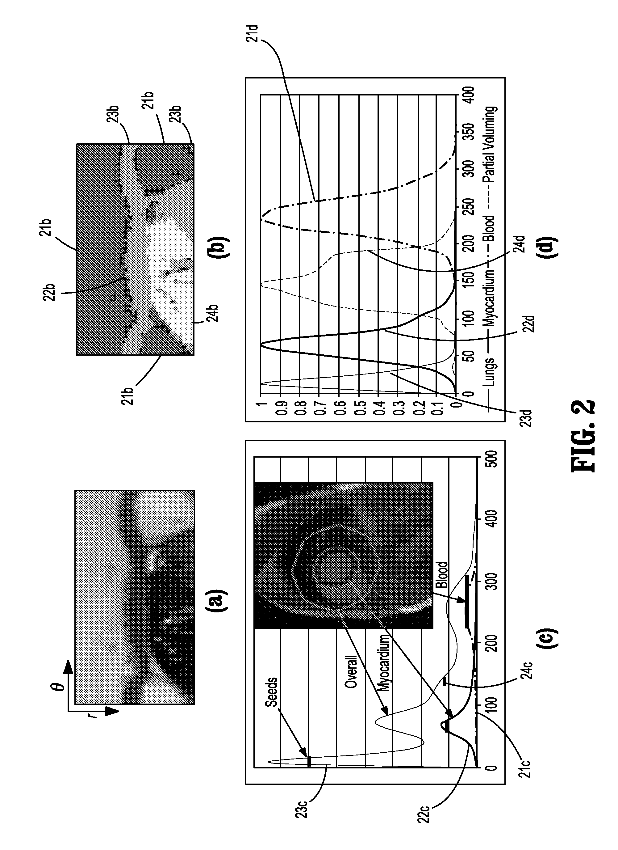
A method for cardiac segmentation in magnetic resonance (MR) cine data, includes providing a time series of 3D cardiac MR images acquired at a plurality of phases over at least one cardiac cycle, in which each 3D image includes a plurality of 2D slices, and a heart and blood pool has been detected in each image. Gray scales of each image are analyzed to compute histograms of the blood pool and myocardium. Non-rigid registration deformation fields are calculated to register a selected image slice with corresponding slices in each phase. Endocardium and epicardium gradients are calculated for one phase of the selected image slice. Contours for the endocardium and epicardium are computed from the gradients in the one phase, and the endocardium and epicardium contours are recovered in all phases of the selected image slice. The recovered endocardium and epicardium contours segment the heart in the selected image slice.

Description

CROSS REFERENCE TO RELATED UNITED STATES APPLICATIONS[0001]This application claims priority from “Cardiac Segmentation in MR Cine Data Using Inverse Consistent Deformable Registration”, U.S. Provisional Application No. 61 / 249,273 of Jolly, et al., filed Oct. 7, 2009, the contents of which are herein incorporated by reference in their entirety.TECHNICAL FIELD[0002]This disclosure is directed to the segmentation of the myocardium (endocardium and epicardium) in the left ventricle of the heart from cardiac cine magnetic resonance (MR) images.DISCUSSION OF THE RELATED ART[0003]Cardiovascular disease is an important health concern in the western world. Physicians use non invasive technologies such as magnetic resonance imaging (MRI) to observe the left ventricle (LV), which pumps oxygenated blood out to the rest of the body. They are interested in measuring quantities such as the ejection fraction, myocardial mass, blood volume over time, and myocardial thickening that can all be estimat...

Claims


Application Information

Patent Timeline
IPC IPC(8): G06K9/00
CPCG06K2209/051G06T7/0083G06T2207/30048G06T2207/10076G06T7/0097G06T7/12G06T7/174G06V2201/031
Inventor JOLLY, MARIE-PIERREGUETTER, CHRISTOPHGUEHRING, JENS
Owner SIEMENS HEALTHCARE GMBH
Why Eureka
Try Eureka