System and method for assessing the formation of a lesion in tissue

a tissue and lesion technology, applied in the field of tissue lesion formation system and method, can solve the problems of varying ailments and even death, difficult to evaluate, assess and/or determine the depth of a lesion in the tissue, and difficult to determine whether the tissue has been sufficiently or acceptably ablated, or at least whether the lesion has reached the desired depth

Active Publication Date: 2011-05-19
ST JUDE MEDICAL ATRIAL FIBRILLATION DIV
View PDF98 Cites 114 Cited by
  • Summary
  • Abstract
  • Description
  • Claims
  • Application Information

AI Technical Summary

Benefits of technology

[0009]The present invention is directed to a method and system for assessing the formation of a lesion in a tissue as a result of an ablation procedure being performed thereon. The system according to the present teachings includes an electronic control unit. The electronic control unit is configured to acquire a magnitude for at least one component of a complex impedance between an electrode and the tissue. The electronic control unit is further configured to acquire a magnitude for the power applied to the tissue during the formation of the lesion therein. The electronic control unit is still further configured to calculate a value responsive to the magnitudes of the at least one complex impedance component and the power. The value is indicative of one of a predicted depth of the lesion formed in the tissue, a likelihood the lesion has reached a predetermined depth, and a predicted temperature of the tissue.

Problems solved by technology

Atrial arrhythmias can create a variety of dangerous conditions including irregular heart rates, loss of synchronous atrioventricular contractions and stasis of blood flow which can lead to a variety of ailments and even death.
One challenge with ablation procedures is in the assessment of the lesion formation as a result of the application of ablative energy to the tissue.
For example, it is difficult to evaluate, assess, and / or determine the depth of a lesion in the tissue.
As such, it is difficult to determine whether the tissue has been sufficiently or acceptably ablated, or at least whether a lesion has reached a desired depth.
However, conventional techniques have proved to be less than optimal as none of the conventional techniques provide an accurate means by which the depth of a lesion, for example, can be predicted with any real certainty.

Method used


Image

  • System and method for assessing the formation of a lesion in tissue
  • System and method for assessing the formation of a lesion in tissue
  • System and method for assessing the formation of a lesion in tissue

Examples


Embodiment Construction

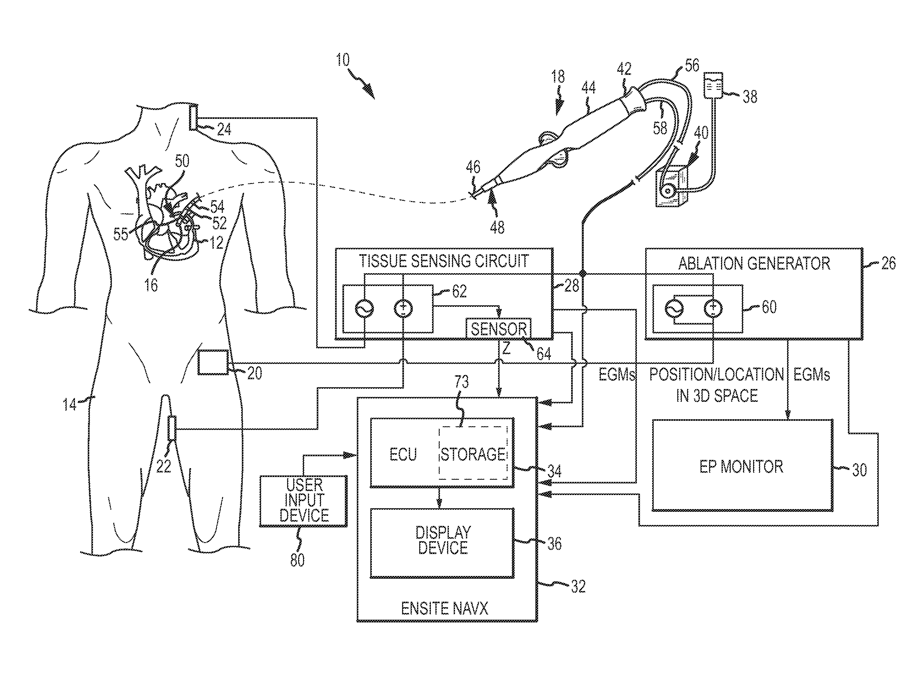
[0039]Referring now to the drawings wherein like reference numerals are used to identify identical components in the various views, FIG. 1 illustrates one exemplary embodiment of a system 10 configured, at least in part, for assessing the formation of a lesion in a tissue 12 of a body 14 as a result of an ablation procedure being performed on the tissue 12. In an exemplary embodiment wherein the tissue 12 is cardiac tissue, the system 10 is configured to assess the formation of a lesion in the tissue 12 being ablated by radio frequency (RF) energy or power delivered from an electrode 16 disposed on a catheter 18. For the sake of clarity and brevity alone, the description set forth below will be with respect to cardiac tissue only. It should be understood, however, that the present disclosure may find application in connection with assessing lesion depth in other types of tissue during ablation procedures. Accordingly, the present disclosure is not meant to be limited solely to cardi...


PUM


Abstract

A method and system for assessing lesion formation in tissue is provided. The system includes an electronic control unit (ECU) configured to acquire magnitudes for a component of a complex impedance between an electrode and tissue, and the power applied to the tissue during lesion formation. The ECU is configured to calculate a value responsive to the complex impedance component and the power. The value is indicative of a predicted lesion depth, a likelihood the lesion has reached a predetermined depth, or a predicted tissue temperature. The method includes acquiring magnitudes for a component of a complex impedance between an electrode and tissue and the power applied during lesion formation. The method includes calculating a value responsive to the complex impedance component and the power, the value being indicative of a predicted lesion depth, a likelihood the lesion has reached a predetermined depth, and/or a predicted tissue temperature.

Description

CROSS REFERENCE TO RELATED APPLICATIONS[0001]This application is a continuation-in-part of U.S. patent application Ser. No. 12 / 622,488 entitled “System and Method for Assessing Lesions in Tissue” filed Nov. 20, 2009 and currently pending, the entire disclosure of which is incorporated herein by reference. U.S. patent application Ser. No. 12 / 622,488 claimed the benefit of U.S. Provisional Application Ser. No. 61 / 177,876 entitled “System and Method for Assessing Lesions in Tissue” filed May 13, 2009 and now expired, and is a continuation-in-part of U.S. patent application Ser. No. 12 / 253,637 entitled “System and Method for Assessing Coupling Between an Electrode and Tissue” filed Oct. 17, 2008 and currently pending, the entire disclosures of which are incorporated herein by reference. U.S. patent application Ser. No. 12 / 253,637 is, in turn, a continuation-in-part of U.S. patent application Ser. No. 12 / 095,688 entitled “Assessment of Electrode Coupling of Tissue Ablation” filed May 30,...

Claims


Application Information

Patent Timeline
Patent Type & Authority Applications(United States)
IPC IPC(8): A61B18/12A61B18/18
CPCA61B5/0402A61B5/742A61B18/1233A61B18/14A61B18/1492A61B2018/00178A61B2018/00351A61B2018/00714A61B2018/00738A61B2018/00779A61B2018/00875A61B2018/0212A61B2019/2211A61B2019/465A61B2218/002A61B5/4836A61B5/1079A61B5/1076A61B5/0538A61B5/01A61B5/0084A61B5/042A61B2034/301A61B2090/065A61B5/0036A61B5/283A61B5/318
Inventor FISH, JEFFREY M.BYRD, ISRAEL A.CLARK, LYNN E.DANDO, JEREMY D.GEURKINK, CHRISTOPHER J.PURYEAR, HARRY A.PAUL, SAURAV
Owner ST JUDE MEDICAL ATRIAL FIBRILLATION DIV
Why Eureka
Try Eureka