Image display device and method, as well as program

a technology applied in the field of image display device and image display method, can solve the problems of difficult to find small abnormal shadows, requires a long time to interpret images, and becomes more likely to miss abnormal shadows, so as to reduce the burden on the user, perform the interpretation more efficiently, and narrow the range of objects to be interpreted with higher attention

Inactive Publication Date: 2011-06-16
FUJIFILM CORP
View PDF15 Cites 25 Cited by
  • Summary
  • Abstract
  • Description
  • Claims
  • Application Information

AI Technical Summary

Benefits of technology

[0008]In view of the above-described circumstances, the present invention is directed to enabling efficient interpretation of medical images containing abnormal shadow candidates.

Problems solved by technology

With any of the above-mentioned display methods, however, it is very difficult to find a small abnormal shadow, such as a small calcification, from an image displayed on a monitor without missing any abnormal shadow, and it requires a long time to interpret the images.
Further, although it is rare to miss an abnormal shadow immediately after the start of interpretation because the user is highly attentive, it will become more likely to miss an abnormal shadow in a repetition of the same work because of decreased attentiveness.

Method used


Image

  • Image display device and method, as well as program
  • Image display device and method, as well as program
  • Image display device and method, as well as program

Examples


first embodiment

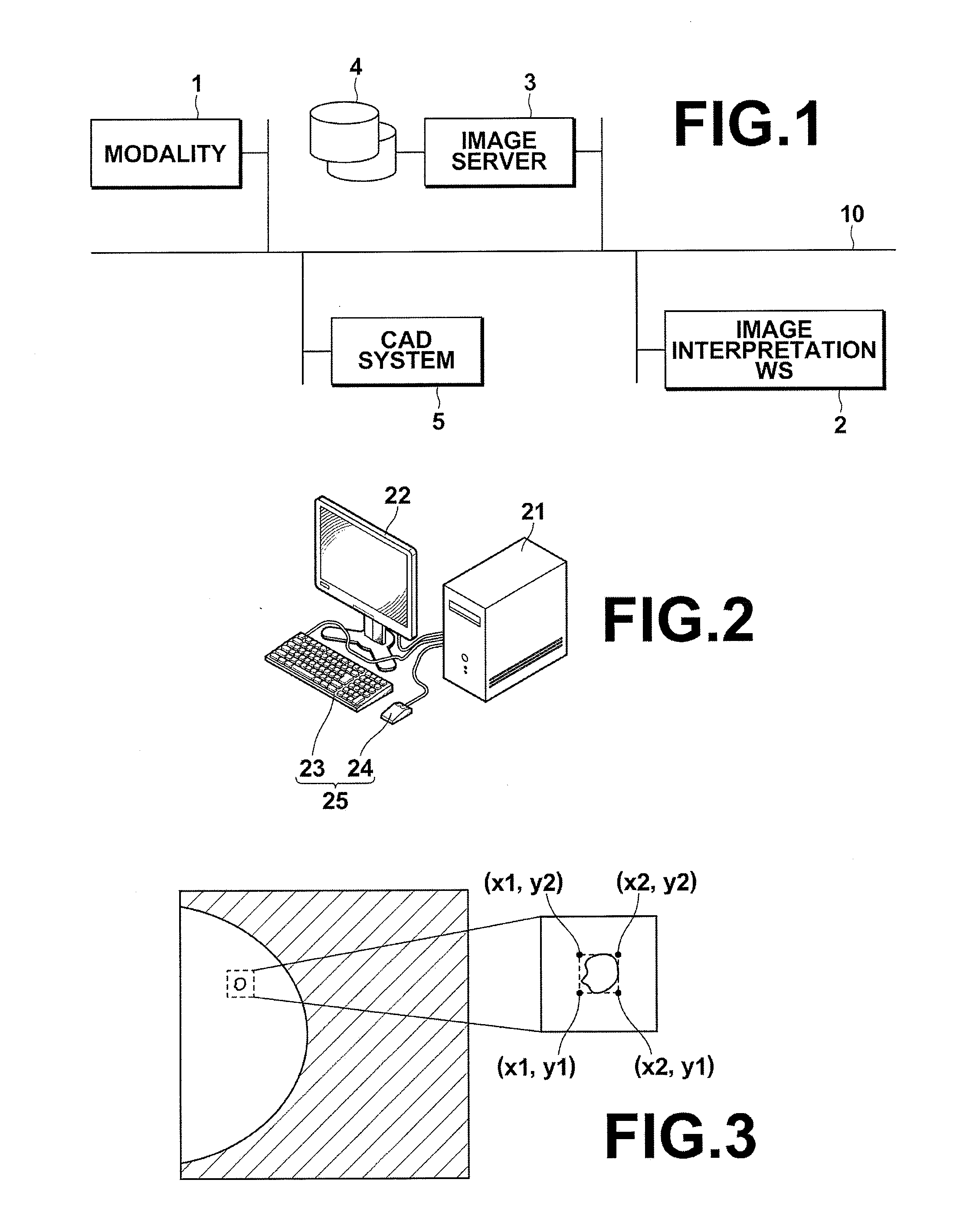
[0050]Hereinafter, embodiments of the present invention will be described with reference to the drawings. FIG. 1 is a diagram illustrating the schematic configuration of a medical information system including an image interpretation workstation, to which an image display device according to the present invention is applied. As shown in FIG. 1, the medical information system according to this embodiment includes: an imaging device for obtaining a medical image (modality) 1, an image interpretation workstation (WS) 2, an image server 3, an image database 4 and a CAD system (computer aided diagnosis system) 5, which are connected via a network 10 to be able to communicate with each other. Further, the components in this embodiment are controlled by a program which is installed from a recording medium, such as a CD-ROM. Alternatively, the program may be download from a server connected via a network, such as the Internet, before it is installed.

[0051]The modality 1 include a device that...

second embodiment

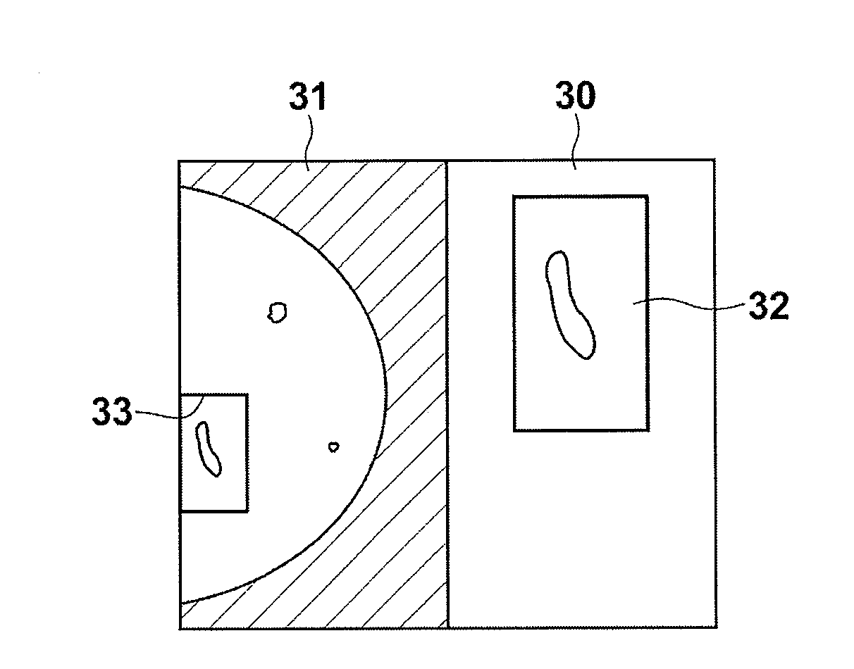
[0071]FIG. 9 is a diagram illustrating the regions of interest and the sub-regions of interest in the As shown in FIG. 9, the abnormal shadow candidates are contained in the regions of interest R6, R9 and R11 of the image containing the breast area R0, and further, sub-regions of interest R6-1, R9-1 and R11-1, which represent the ranges where the abnormal shadow candidates are present, are contained in the regions of interest R6, R9 and R11, respectively. It should be noted that each of the sub-regions of interest R6-1, R9-1 and R11-1 may be the range where the abnormal shadow candidate is present which is determined based on the detection information D, or may be a range provided with some margin by extending the range where the abnormal shadow candidate is present by, for example, about 50% in upward, downward, rightward and leftward directions (four directions).

[0072]If the descending order of the degree of malignancy of the abnormal shadow candidates contained in the sub-region...

third embodiment

[0079]In the third embodiment, if the descending order of the degree of malignancy of the abnormal shadow candidate contained in the sub-regions of interest R6-1, R9-1 and R11-1 is “R9-1, R11-1, R6-1”, the processing device 21 determines the display order of the sub-regions of interest as “R9-1, R11-1, R6-1”. After the sub-regions of interest have been displayed, the display order is determined to display the remaining regions of interest in order from the upper-left corner.

[0080]FIG. 12 is a diagram for explaining the display order the images of the regions of interest and the sub-regions of interest in the third embodiment. As shown in FIG. 12, in the third embodiment, first, the image of the sub-region of interest R9-1 is displayed. When the user instructs to switch the display, each of the images of the sub-regions of interest R11-1 and R6-1 is displayed in this order. When the user further instructs to switch the display, the remaining regions of interest are displayed in order...


PUM


Abstract

A medical image of a subject and detection information including positions of abnormal shadow candidates and degrees of malignancy of abnormal shadows detected from the medical image are acquired. The medical image is divided into a plurality of regions of interest, and images of the regions of interest are sequentially displayed with displaying images of the regions of interest containing the abnormal shadow candidates in descending order of the degree of malignancy based on the detection information prior to images of the regions of interest containing no abnormal shadow candidate.

Description

BACKGROUND OF THE INVENTION[0001]1. Field of the Invention[0002]The present invention relates to an image display device and an image display method for displaying medical images containing abnormal shadow candidates detected therefrom, as well as a program for causing a computer to carry out the image display method.[0003]2. Description of the Related Art[0004]A computer aided diagnosis (CAD) system, which automatically detects an abnormal shadow candidate in an image and performs enhanced display, etc., of the detected abnormal shadow candidate, has been known in the medical field. A doctor interprets the image containing the abnormal shadow candidate detected by the CAD system, and finally determines whether or not the abnormal shadow candidate in the image represents a lesion, such as a tumor mass or calcification.[0005]Examples of known techniques for detecting the abnormal shadow candidate include: a technique that involves applying image processing using an iris filter to a r...

Claims


Application Information

Patent Timeline
Patent Type & Authority Applications(United States)
IPC IPC(8): A61B6/02A61B8/08
CPCG06T7/0012G06T2207/30068G06T2207/20104G06T2207/20021
Inventor SENDAI, TOMONARIHASEGAWA, AKIRA
Owner FUJIFILM CORP
Why Eureka
Try Eureka